. 2013 Apr 11;50(1):104-15.
doi: 10.1016/j.molcel.2013.02.017. Epub 2013 Mar 21.

A mammalian pre-mRNA 5' end capping quality control mechanism and an unexpected link of capping to pre-mRNA processing

Affiliations

A mammalian pre-mRNA 5' end capping quality control mechanism and an unexpected link of capping to pre-mRNA processing

Xinfu Jiao et al. Mol Cell. .

Abstract

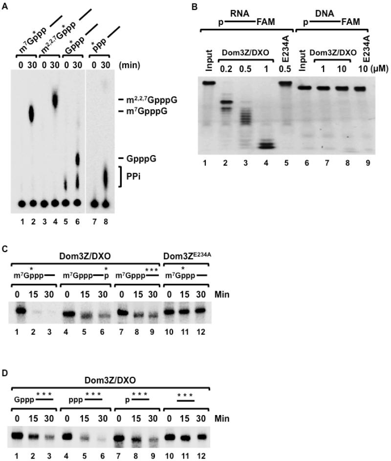
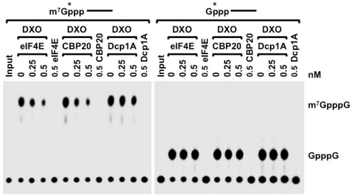
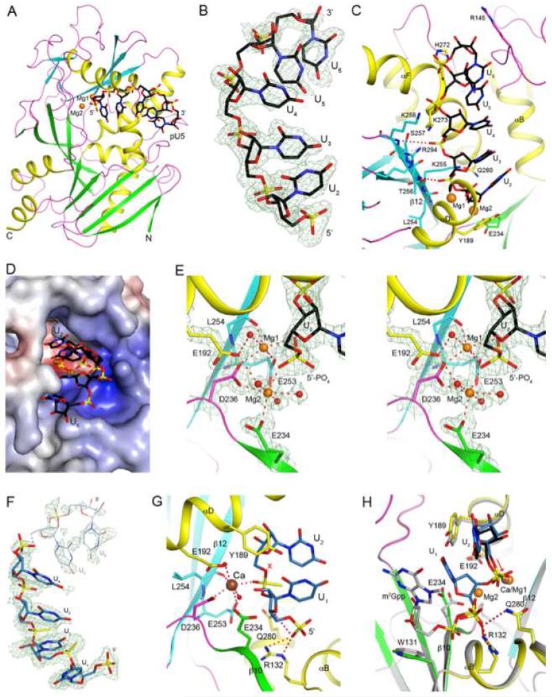
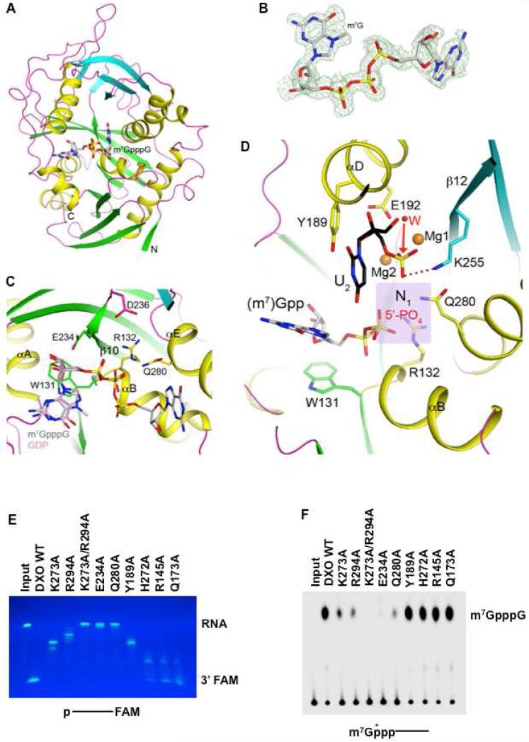
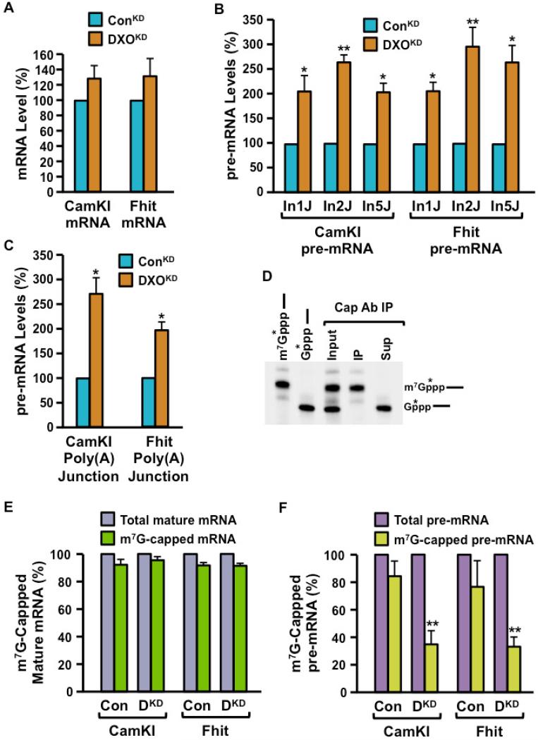
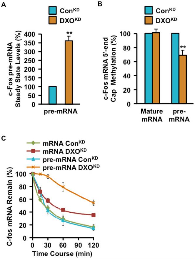
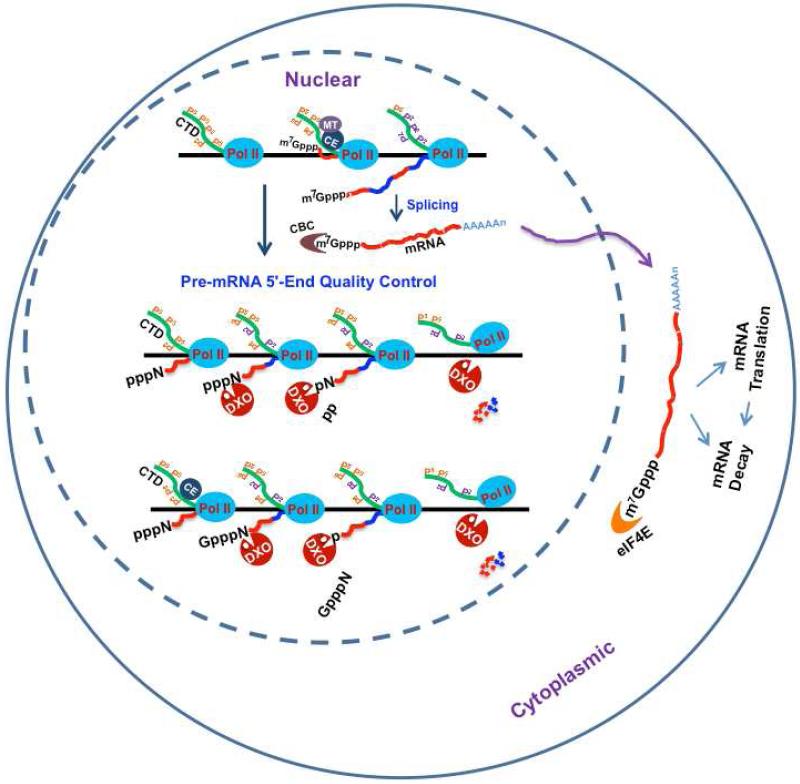
Recently, we reported that two homologous yeast proteins, Rai1 and Dxo1, function in a quality control mechanism to clear cells of incompletely 5' end-capped messenger RNAs (mRNAs). Here, we report that their mammalian homolog, Dom3Z (referred to as DXO), possesses pyrophosphohydrolase, decapping, and 5'-to-3' exoribonuclease activities. Surprisingly, we found that DXO preferentially degrades defectively capped pre-mRNAs in cells. Additional studies show that incompletely capped pre-mRNAs are inefficiently spliced at all introns, a fact that contrasts with current understanding, and are also poorly cleaved for polyadenylation. Crystal structures of DXO in complex with substrate mimic and products at a resolution of up to 1.5Å provide elegant insights into the catalytic mechanism and molecular basis for their three apparently distinct activities. Our data reveal a pre-mRNA 5' end capping quality control mechanism in mammalian cells, indicating DXO as the central player for this mechanism, and demonstrate an unexpected intimate link between proper 5' end capping and subsequent pre-mRNA processing.


Figures

Figure 1
Figure 1. Dom3Z/DXO possesses pyrophosphohydrolase, decapping and 5′-3′ exoribonuclease activities
(A) Decapping activity of Dom3Z/DXO toward RNAs with various 5′ ends depicted schematically at the top is shown where the RNA is represented by a line. The asterisk denotes the 32P position. Reactions were carried out with 25nM pcP RNA and 25nM recombinant His-tagged Dom3Z protein at 37°C for the indicated times. Decapping products were resolved on PEI-TLC developed in 0.45 M (NH4)2SO4 (lanes 1-6) or 0.75M KH2PO4 (pH 3.4; lanes 7-8)). Migrations of cap analoge markers are indicated. (B) Wild type and mutant Dom3Z/DXO proteins were incubated with 5′-end monophosphate 30-nt RNA or DNA substrates labeled at the 3′-end with the FAM (6-carboxyfluorescein) fluorophore. Remaining RNA or DNA fragments were resolved by 5% denaturing PAGE and visualized under UV light confirming the distributive, 5′-3′ exoribonuclease activity. The RNA or DNA is denoted by a line, the 5′ monophosphate is represented by the p at the beginning of the line. The catalytically inactive E234A mutant is shown. (C) In vitro decay reactions were carried out with 100nM recombinant His-tagged Dom3Z/DXO for the indicated times with methyl-capped RNAs (25 nM) represented schematically with the 32P labeling indicated by the asterisk. RNAs remaining were resolved by 5% denaturing PAGE. Catalytically inactive Dom3Z/DXO mutant (Dom3ZE234A) was used as a negative control. (D) The 5′ end substrate specificity of Dom3Z/DXO was tested as in (A) above with RNAs containing distinct 5′ ends as denoted schematically. RNAs with a 5′ hydroxyl were not degraded by Dom3Z/DXO.
Figure 2
Figure 2. Cap binding proteins compete for DXO decapping on methyl-capped RNA in vitro
Decapping activity of 20nM DXO on 32P 5′-end labeled methyl-capped or unmethyl-capped RNA in the presence of the indicated eIF4E, CBP20 or Dcp1A proteins are shown. Decapping assay was carried out as described in legend to Fig 1. Both eIF4E and CBP20 can compete DXO decapping on methyl-capped RNA but not unmethyl-capped RNA.
Figure 3
Figure 3. Crystal structure of wild-type murine DXO in complex with pU5 and two Mg2+ ions
(A). Schematic drawing of the structure of DXO (in green and cyan for the large and small β-sheets, respectively, yellow for the helices, and magenta for the loops) in complex with pU5 RNA oligonucleotide (in black for carbon atoms) and Mg2+ ions (orange). (B). Simulated-annealing omit Fo-Fc electron density at 1.8 Å resolution for pU5, contoured at 2.5σ. (C). Interactions between the pU5 RNA with the DXO active site. (D). Molecular surface of the active site region of DXO. The pU5 RNA is shown as sticks. (E). The coordination spheres of the two Mg2+ ions, and detailed interactions between the 5′-end phosphate group of pU5 and the two Mg2+ ions. (F). Simulated-annealing omit Fo-Fc electron density at 1.7 Å resolution for pU(S)6, contoured at 2.2σ. Very weak electron density is observed for the last two nucleotides, and they are not included in the atomic model. (G). Interactions between the first two nucleotides of pU(S)6 with DXO. The oligo is shown in light blue, and the Ca2+ ion is shown as a sphere in brown. The red X indicates the expected position of a water/hydroxide ion to initiate hydrolysis based on the observed conformation of the phosphorothioate group. (H). Overlay of the structures of the pU(S)6 and pU5 complexes in the active site region of DXO. The pU(S)6 oligo is shown in light blue, pU5 in black. DXO in the pU(S)6 complex is shown in color, and that in the pU5 complex in gray. The red arrow indicates the O5-P bond, and the two phosphate groups differ by ~60° rotation around this bond. The m7Gpp portion of the methylated cap (see Fig. 4 for more information) is also shown (gray). All the structure figures were produced with PyMOL (www.pymol.org).
Figure 4
Figure 4. Crystal structure of wild-type murine DXO in complex with the m7GpppG cap
(A) Schematic drawing of the structure of DXO (in green and cyan for the two β-sheets, yellow for the helices, and magenta for the loops) in complex with the m7GpppG cap (in gray for carbon atoms). (B) Simulated-annealing omit Fo-Fc electron density at 1.5 Å resolution for m7GpppG, contoured at 3σ. (C) Interactions between the m7GpppG cap with the DXO active site. (D) Overlay of the structures of DXO in complex with pU5 (in black) and two Mg2+ ions (orange) and m7Gpp (gray). (E) The indicated DXO wild type and mutant proteins were incubated with 5′-end monophosphate 30-nt RNA substrate labeled at the 3′-end with the fluorophore FAM. Remaining RNA fragments were resolved by 15% denaturing PAGE and visualized under UV light confirming the distributive, 5′-3′ exonuclease activity. (F) The indicated DXO wild type and mutant proteins were tested for decapping activity. Methyl-capped RNA labeled with 32P at the 5′-end was used in decapping reactions. Reaction conditions and labeling are as described in the legend to Figure 1.
Figure 5
Figure 5. DXO preferentially functions to clear incompletely capped pre-mRNAs in cells
(A) Total RNA isolated from control shRNA expressing (ConKD) or DXO-specific shRNA expressing (DXOKD) cells were subjected to qRT-PCR analysis with primers specific to the CamKI and Fhit mRNA. Values were normalized to the GAPDH mRNA and plotted relative to the control shRNA, which was arbitrarily set to 100. (B) The relative levels of CamKI and Fhit pre-mRNA in ConKD and DXOKD cells were quantitated with primers that span intron-exon junctions 1, 2 and 5 (In1J, In2J, In5J respectively). The level in ConKD cells was arbitrarily set to 100. (C) Total RNA isolated from ConKD and DXOKD cells subjected to qRT-PCR analysis with primers that span the CamKI and Fhit gene poly(A) addition site were used to determine the relative level of unpolyadenylated transcripts in DXOKD cells. Levels in the ConKD cells were arbitrarily set to 100. (D) Methyl-capped RNAs were purified with monoclonal anti-trimethylguanosine antibody column under conditions that resolve methyl-capped from incompletely capped RNAs as described (Chang et al., 2012; Jiao et al., 2010). 0.5μg of total 293T RNA was spiked with in vitro generated N7-methylated and unmethylated cap-labeled RNAs prior to capped RNA affinity purification. RNAs bound or unbound to the column were isolated and resolved by denaturing urea PAGE. Identity of the individual RNAs used, the input mixture and the resulting resolved RNAs are indicated. (E and F) Methyl-capped RNAs were purified with monoclonal anti-trimethylguanosine antibody column as in (D) (Chang et al., 2012; Jiao et al., 2010) and levels of methyl-capped CamKI and Fhit mRNA (E) or pre-mRNA (F) were quantitated by qRT-PCR and presented relative to the corresponding total level of mRNA or pre-mRNA respectively. Levels of the corresponding total mRNA was arbitrarily set to 100. Data in all four panels were derived from at least three independent experiments and the error bars represent +/− SD. * represents p < 0.05; ** represents p < 0.01.
Figure 6
Figure 6. Defectively capped c-fos pre-mRNAs were stabilized in the absence of DXO in cells
(A) Steady state levels of c-fos pre-mRNA in 293T cells expressing control shRNA (ConKD) or DXO-specific shRNA (DXOKD) were determined by qRT-PCR with primers that span intron 1 exon 2-junction and presented relative to GAPDH mRNA levels. Levels of c-fos pre-mRNA in the ConKD cells were arbitrarily set to 1. (B) Methyl capped RNAs were isolated as in Figure 5D-F from ConKD and DXOKD cells and levels of mature c-fos mRNA determined with primers that span exons 1 and 2 and c-fos pre-mRNA as in (A) above. A reduce level of methylated capped c-fos, pre-mRNA was detected in the DXOKD cells. (C) Stability of c-fos mRNA and pre-mRNA was determined in the indicated cells following actinomycin D transcriptional arrest. Data are presented relative to corresponding levels of GAPDH mRNA. Data in all three panels were derived from three independent assays with ±SD denoted by the error bars.
Figure 7
Figure 7. Model of 5′-end quality control in mammalian cells
Incompletely 5′-end capped pre-mRNA (unmethylated capped and uncapped 5′ triphosphate pre-mRNA) would be preferentially detected by DXO and subjected to 5′-end cleavage and degradation. The RNA Polymerase II (Pol II), the carboxyl terminal domain of RNAP II (CTD), the triphosphatase-guanylyltransferase capping enzyme (CE), the methyltransferase (MT), the nuclear cap binding complex (CBC) and the cytoplasmic cap binding protein, eIF4E, are as indicated.

Comment in

Similar articles

Cited by

References

    1. Benarroch D, Jankowska-Anyszka M, Stepinski J, Darzynkiewicz E, Shuman S. Cap analog substrates reveal three clades of cap guanine-N2 methyltransferases with distinct methyl acceptor specificities. RNA. 2010;16:211–220. - PMC - PubMed
    1. Brunger AT, Adams PD, Clore GM, DeLano WL, Gros P, Grosse-Kunstleve RW, Jiang JS, Kuszewski J, Nilges M, Pannu NS, et al. Crystallography & NMR system: A new software suite for macromolecular structure determination. Acta Crystallogr D Biol Crystallogr. 1998;54:905–921. - PubMed
    1. Calero G, Wilson KF, Ly T, Rios-Steiner JL, Clardy JC, Cerione RA. Structural basis of m7GpppG binding to the nuclear cap-binding protein complex. Nat Struct Biol. 2002;9:912–917. - PubMed
    1. Chang JH, Jiao X, Chiba K, Kiledjian M, Tong L. Dxo1, a novel eukaryotic enzyme with both decapping and 5′-3′ exoribonuclease activity. Nat Struct Mol Biol. 2012 in press. - PMC - PubMed
    1. Colot HV, Stutz F, Rosbash M. The yeast splicing factor Mud13p is a commitment complex component and corresponds to CBP20, the small subunit of the nuclear cap-binding complex. Genes Dev. 1996;10:1699–1708. - PubMed

Publication types

MeSH terms