. 2013;8(3):e58397.
doi: 10.1371/journal.pone.0058397. Epub 2013 Mar 8.

Graft-versus-host disease is enhanced by selective CD73 blockade in mice

Affiliations

Graft-versus-host disease is enhanced by selective CD73 blockade in mice

Long Wang et al. PLoS One. 2013.

Abstract

CD73 functions as an ecto-5'-nucleotidase to produce extracellular adenosine that has anti-inflammatory and immunosuppressive activity. We here demonstrate that CD73 helps control graft-versus-host disease (GVHD) in mouse models. Survival of wild-type (WT) recipients of either allogeneic donor naïve CD73 knock-out (KO) or WT T cells was similar suggesting that donor naïve T cell CD73 did not contribute to GVHD. By contrast, donor CD73 KO CD4(+)CD25(+) regulatory T cells (Treg) had significantly impaired ability to mitigate GVHD mortality compared to WT Treg, suggesting that CD73 on Treg is critical for GVHD protection. However, compared to donor CD73, recipient CD73 is more effective in limiting GVHD. Pharmacological blockade of A2A receptor exacerbated GVHD in WT recipients, but not in CD73 KO recipients, suggesting that A2 receptor signaling is primarily implicated in CD73-mediated GVHD protection. Moreover, pharmacological blockade of CD73 enzymatic activity induced stronger alloreactive T cell activity, worsened GVHD and enhanced the graft-versus-leukemia (GVL) effect. These findings suggest that both donor and recipient CD73 protects against GVHD but also limits GVL effects. Thus, either enhancing or blocking CD73 activity has great potential clinical application in allogeneic bone marrow transplants.


Conflict of interest statement

Competing Interests: The authors have declared that no competing interests exist.

Figures

Figure 1
Figure 1. CD73 affects GVHD development.
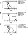
Lethally irradiated BALB/c CD73 KO (A) and WT (C) or B6D2F1 (BDF1) (D) mice were given i.v. injections of 5×10⁶ T cell depleted bone marrow (BM) cells from C57BL/6 (B6) mice donors alone (n = 5) or with 2×10⁷ splenocytes from WT (n = 10) or CD73 KO (n = 10) B6 donors. P values indicate the differences between recipients receiving WT versus CD73 KO cells. Results are representative of 2 independently performed experiments with similar results. (B) Ten days later, pathological analyses of lung, liver, skin, and colon of recipient mice (A) receiving WT or CD73 KO SP were performed by H&E staining. Combined results of pathology scores for 5 mice in each group. Error bars indicate the standard errors of the mean in the group. Representative micrographs from recipient livers (magnification 200X) are shown.
Figure 2
Figure 2. Naïve CD73 deficient (KO) T cells exhibit normal activation and proliferation in response to alloantigen.
B6 naïve WT or CD73 KO spleen T cells (CD25CD62L+) (5×10⁶) were injected i.v. into B6D2F1 (BDF1) mice. After 5 days, recipient spleens were harvested and stained with anti-H-2Kd antibody together with either anti-CD4 or anti-CD8 antibodies. (A) Percentage of donor (H-2Kd negative) CD4+ or CD8+ T cells in total spleen cells was determined by flow cytometry. Numbers in quadrants indicate percent positive cells in each. Absolute numbers of donor T cells in recipient spleen (n = 4) (B) and liver (n = 4) (C) were determined at 5 and 7 days after T cell transfer. (D) B6 WT or CD73 KO T cells (5 × 10⁵/well) were co-cultured with 1 × 10⁵ BALB/c mouse CD11c+ dendritic cells (DC) for 3 days. To determine the role of CD73 on DC, BALB/c WT T cells (5 × 10⁵/well) were co-cultured with 1 × 10⁵ B6 WT or CD73 KO CD11c+ DC for 3 days. Alloreactive T cell proliferation was measured by [3H]thymidine incorporation. CPM, counts per minute. (E) Culture supernatants were collected for IL-2 detection by ELISA. Data are given as means ± SEM. Results are representative of 2 independently performed experiments with similar results.
Figure 3
Figure 3. CD73 deficient (KO) CD4+CD25+ Tregs have reduced ability to inhibit GVHD.
(A) Lethally irradiated BALB/c mice were given i.v. injections of 5×10⁶ T cell depleted BM cells from B6 mice donors alone (n = 5) or with 1×10⁶ (1 M) or 0.5×10⁶ (0.5 M) splenic naïve T cells (CD25CD62L+) from WT (n = 10) or CD73 KO (n = 10) B6 donors. (B) Lethally irradiated BALB/c mice were given i.v. injections of 5×10⁶ T cell depleted BM cells from B6 mice donors alone (n = 5) or with 2×10⁷ total splenocytes or CD25 negative (CD25) splenocytes from WT (n = 10) or CD73 KO (n = 10) B6 donors. BM+WT-SP versus BM+CD73 KO-SP, p = 0.0399; BM+WT-SP versus BM+WT-CD25, p = 0.0305. (C) Suppression of the alloresponse of B6 CD4+CD25 T responder cells (Tres) to BABL/c cells by B6 WT or CD73 KO Tregs (ratio of Tres/Treg; 1∶1) in the MLR. WT Tregs were incubated with or without APCP at 100 µM before co-culture with indicated cells. NECA at 10 µM was added to the co-cultures with CD73 KO Tregs for the duration of the culture. Data are given as means ± SD of triplicates. (D) Lethally irradiated BALB/c mice were given i.v. injections of 5×10⁶ T cell depleted BM cells from B6 mice donors alone (n = 5) or plus 5×10⁵ purified B6 WT (n = 10) or CD73 KO (n = 10) CD4+CD25 T cells ±5×10⁵ CD4+CD25+ Tregs. BM+WT-T versus BM+WT-T+Treg, p<0.0001; BM+WT-T+WT-Treg versus BM+WT-T+CD73 KO-Treg, p = 0.024.
Figure 4
Figure 4. Recipient CD73 limits GVHD development.
(A) Lethally irradiated WT (n = 10) or CD73 KO (n = 10) BALB/c mice were given i.v. injections of 5×10⁶ T cell depleted BM cells from B6 mice donors alone (n = 5) or with 2×10⁷ splenocytes from WT B6 donors. Lethally irradiated WT (n = 10) or CD73 KO (n = 10) B6 mice were given i.v. injections of 5×10⁶ T cell depleted BM cells from BALB/c mice donors alone (n = 5) or with 2×10⁷ splenocytes from WT BALB/c donors (B) or CD73 KO BALB/c donors (C). (A,B) P values indicate the differences between WT versus CD73 KO recipients. (C) BALB WT→B6 CD73KO versus BALB CD73KO→B6 CD73KO, p = 0.0103; BALB WT→B6 CD73KO versus BALB CD73KO→B6 WT, p = 0.0029; BALB CD73KO→B6 WT versus BALB CD73KO→B6 CD73KO, p<0.0001; BALB WT→B6 WT versus BALB CD73KO→B6 WT; p = 0.0284. Results are representative of 2 independently performed experiments with similar results.
Figure 5
Figure 5. Enhanced proliferation but normal survival of allogeneic donor T cells in CD73 KO recipients.
(A) Sublethally irradiated WT or CD73 KO BALB/c mice were given i.v. injections of 2×10⁷ dye (CFSE)-labeled splenocytes from WT B6 donors. Division of host-reactive donor T cells was measured 2 and 5 days after cell transfer. CFSE intensity was gated on H-2Kd-negative (donor) CD4+ or CD8+ T cells. The percentage of donor T cells with more than one division is indicated in each panel and summarized in (B). (C) Annexin V staining of donor cells gated on H-2Kd-negative CD4+ or CD8+ T cells 5 days after cell transfer. The percentage of apoptotic donor T cells (annexin V+) is indicated in each panel (C) and summarized in (D). Data are given as means ± SEM. Results are representative of 3 independently performed experiments with similar results.
Figure 6
Figure 6. Blockade of CD73 enzymatic activity using the CD73-selective inhibitor APCP exacerbates GVHD but enhances GVL effects.
(A) Lethally irradiated BALB/c mice were given i.v. injections of 5×10⁶ T cell depleted BM cells from B6 mice donors alone (n = 5) or with 2×10⁷ splenocytes from WT B6 donors. In the groups receiving T cell transfer, the mice were treated with PBS or APCP 20 mg/kg i.v. twice weekly following donor BM and T cell transfer. (B) Enriched B6 WT T cells were injected i.v. into lethally irradiated BALB/c mice at 2×10⁶ per mouse, and cell expansion was determined 14 days after cell transfer. Mean of absolute number of CD4+ or CD8+ cells per spleen was shown in recipients given donor (H-2Kb+) T cells (n = 5). (C) Indicated cytokines were measured in recipient serum on day 14 after donor BM and T cell transfer as described above (n = 5). (D) Lethally irradiated B6 mice (8–10 per group) were given i.v. injections of 5×10⁶ T cell-depleted BM cells from BALB/c mice donors alone or with 106 BALB/c splenic T cells. In the groups receiving T cell transfer, the mice were treated with PBS or APCP 20 mg/kg i.v. twice weekly following donor BM and T cell transfer. Recipient mice were given 105 EL4-luc lymphoma cells as a separate i.v. injection at the same time of transfer. The recipient mice were monitored daily for survival. (E) The average of relative bioluminescence signal intensity of 5 mice per group at different time points as indicated was shown. P values indicate the differences between control PBS versus APCP treatment. Data are given as means ± SEM.
Figure 7
Figure 7. A2AR antagonist administration worsens GVHD.
(A) Lethally irradiated B6 mice were given i.v. injections of 5×10⁶ T cell depleted BALB/c BM cells with 2×10⁷ splenocytes. Recipients received daily i.p. injections of 2 mg/kg SCH58261, 2 mg/kg MRS1754 or vehicle (0.1% DMSO). Injections were initiated 48 h before cell transfer and continued for 14 days. SCH58261 versus vehicle, p = 0.0204. (B) Indicated cytokines were measured in recipient serum on day 14 after donor BM and T cell transfer as described above (n = 5). (C) Enriched BALB/c WT T cells were injected i.v. into lethally irradiated B6 WT or CD73 KO mice at 2×10⁶ per mouse, and cell expansion was determined 14 days after cell transfer. Recipients received daily i.p. injections of 2 mg/kg SCH58261 or vehicle (0.1% DMSO) as described above. Mean of absolute numbers of CD4+ or CD8+ cells per spleen was shown in recipients given donor T cells (n = 5). (D) Lethally irradiated B6 CD73 KO mice were given i.v. injections of 5×10⁶ T cell-depleted BALB/c BM cells with 2×10⁷ splenocytes. Recipients received daily i.p. injections of 2 mg/kg SCH58261 or vehicle (0.1% DMSO) as described above.

Similar articles

Cited by

References

    1. Shlomchik WD (2007) Graft-versus-host disease. Nat Rev Immunol 7: 340–352. - PubMed
    1. Ferrara JL, Levy R, Chao NJ (1999) Pathophysiologic mechanisms of acute graft-vs.-host disease. Biol Blood Marrow Transplant 5: 347–356. - PubMed
    1. Welniak LA, Blazar BR, Murphy WJ (2007) Immunobiology of allogeneic hematopoietic stem cell transplantation. Annu Rev Immunol 25: 139–170. - PubMed
    1. Holler E (2002) Cytokines, viruses, and graft-versus-host disease. Curr Opin Hematol 9: 479–484. - PubMed
    1. van den Brink MR, Burakoff SJ (2002) Cytolytic pathways in haematopoietic stem-cell transplantation. Nat Rev Immunol 2: 273–281. - PubMed

Publication types

MeSH terms

Substances