. 2013 Apr 26;288(17):12114-29.
doi: 10.1074/jbc.M112.409599. Epub 2013 Mar 21.

Shedding of discoidin domain receptor 1 by membrane-type matrix metalloproteinases

Affiliations

Shedding of discoidin domain receptor 1 by membrane-type matrix metalloproteinases

Hsueh-Liang Fu et al. J Biol Chem. .

Abstract

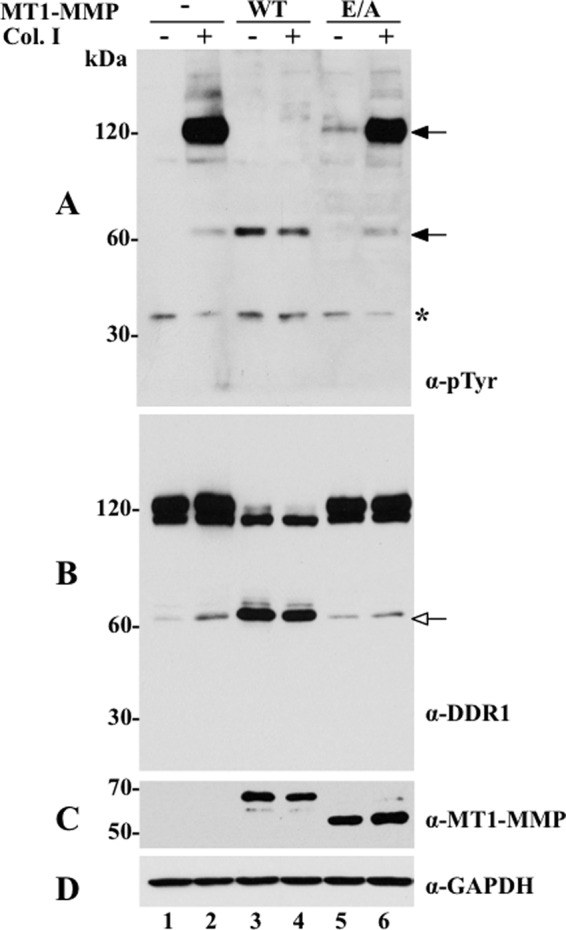
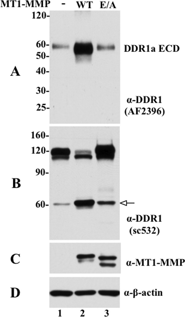
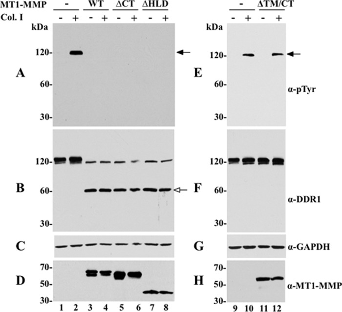
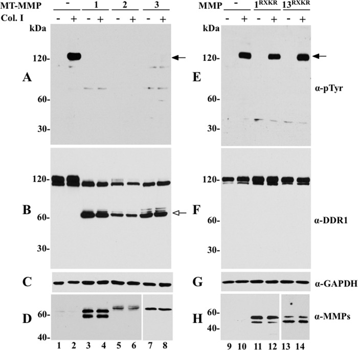
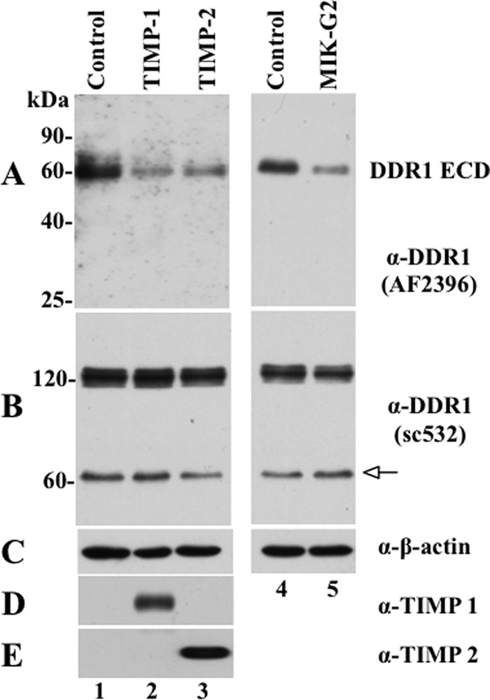
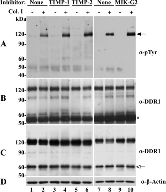
The discoidin domain receptors (DDRs) are receptor tyrosine kinases that upon binding to collagens undergo receptor phosphorylation, which in turn activates signal transduction pathways that regulate cell-collagen interactions. We report here that collagen-dependent DDR1 activation is partly regulated by the proteolytic activity of the membrane-anchored collagenases, MT1-, MT2-, and MT3-matrix metalloproteinase (MMP). These collagenases cleave DDR1 and attenuate collagen I- and IV-induced receptor phosphorylation. This effect is not due to ligand degradation, as it proceeds even when the receptor is stimulated with collagenase-resistant collagen I (r/r) or with a triple-helical peptide harboring the DDR recognition motif in collagens. Moreover, the secreted collagenases MMP-1 and MMP-13 and the glycosylphosphatidylinositol-anchored membrane-type MMPs (MT4- and MT6-MMP) have no effect on DDR1 cleavage or activation. N-terminal sequencing of the MT1-MMP-mediated cleaved products and mutational analyses show that cleavage of DDR1 takes place within the extracellular juxtamembrane region, generating a membrane-anchored C-terminal fragment. Metalloproteinase inhibitor studies show that constitutive shedding of endogenous DDR1 in breast cancer HCC1806 cells is partly mediated by MT1-MMP, which also regulates collagen-induced receptor activation. Taken together, these data suggest a role for the collagenase of membrane-type MMPs in regulation of DDR1 cleavage and activation at the cell-matrix interface.


Figures

FIGURE 1.
FIGURE 1.
MT1-MMP cleaves DDR1 and inhibits collagen I-induced receptor activation. COS1 cells were transiently transfected to co-express DDR1a without (−) or with (+) wild type (WT) or catalytically inactive (E/A) MT1-MMP and serum-starved (18 h) before stimulation (2 h) with (+) 10 μg/ml of rat tail collagen I (Col. I) or vehicle control (−), as described under “Experimental Procedures.” After stimulation, the cells were lysed in RIPA buffer, and the lysates from each experimental condition were divided in two fractions. Equal amounts of the two fractions were then resolved by reducing 8% SDS-PAGE in two identical separate gels followed by immunoblot analyses. One blot was probed with Tyr(P) (α-pTyr) (4G10®) antibody (A) and the other with DDR1 antibody (sc-532) (B). The blot in B was then reprobed with antibodies to MT1-MMP (Lem2/15) (C) or GAPDH, as loading control (D). Black arrows in A indicate phosphorylated DDR1 forms, and white arrow in B indicates the CTF of DDR1. Asterisk indicates a nonspecific band.
FIGURE 2.
FIGURE 2.
MT1-MMP enhances DDR1 ectodomain shedding. COS1 cells were transfected to co-express DDR1a without (−) or with wild type (WT) or catalytically inactive (E/A) MT1-MMP, and the conditioned media were collected, as described under “Experimental Procedures.” Equal volumes of the conditioned media were TCA-precipitated and resolved by reducing 12% SDS-PAGE followed by immunoblot analyses using a DDR1 antibody against the N-terminal ectodomain from R&D Systems (AF2396). A single immunoreactive fragments of ∼60 kDa was detected, indicated as soluble DDR1 (A). B and C, cells from the same experiment were lysed in RIPA buffer, and equal amounts of protein from each sample was resolved by reducing 7.5% SDS-PAGE followed by immunoblot analyses. The blot was probed with DDR1 antibody (sc-532) (B). The blot in B was then reprobed with antibodies to MT1-MMP (Lem2/15) (C) or β-actin, as loading control (D). White arrow in B indicates the CTF of DDR1.
FIGURE 3.
FIGURE 3.
Structural requirements for MT1-MMP-mediated cleavage of DDR1. COS1 cells were transiently transfected to express DDR1a without (−) or with (+) wild type MT1-MMP (WT), MT1-MMPΔCT (ΔCT), MT1-MMPΔHLD (ΔHLD) (A–D), or MT1-MMPΔTM/CT (ΔTM/CT) (E–H). The cells were then serum-starved (18 h) before stimulation (2 h) with (+) 10 μg/ml of rat tail collagen I (Col. I) or vehicle control (−). After stimulation, the cells were lysed, and the lysates were examined for DDR1 phosphorylation (A and E) and cleavage (B and F), as described in the legend of Fig. 1. The blot in B was then reprobed with antibodies to MT1-MMP (Lem2/15) (D) or GAPDH, as loading control (C). The blot in F was reprobed with anti-GAPDH antibodies (G). The blot in H shows serum-free conditioned media collected from COS1 cells expressing secreted MT1-MMPΔTM/CT (lanes 11 and 12) or vector control (lanes 9 and 10). Black arrows in A and E indicate phosphorylated DDR1 and white arrow in B indicates the CTF of DDR1. α-pTyr, Tyr(P).
FIGURE 4.
FIGURE 4.
Effects of membrane-anchored and secreted collagenases on DDR1 phosphorylation and cleavage. COS1 cells were transiently transfected to co-express DDR1a without (−) or with (+) MT1-MMP, MT2-MMP, MT3-MMP, MMP-1RXKR, or MMP-1RXKR. Serum-starved cells were then stimulated (2 h) with (+) 10 μg/ml rat tail collagen I (Col. I) or vehicle control (−). After stimulation, the cells were lysed, and the lysates were examined for DDR1 phosphorylation (A and E) and cleavage (B and F), as described in the legend of Fig. 1. The blots in B and F were then reprobed with antibodies to the respective MMPs (D and H) or GAPDH, as loading control (C and G). Black arrows in A and E indicate phosphorylated DDR1 and white arrow in B indicates the CTF of DDR1. α-pTyr, Tyr(P).
FIGURE 5.
FIGURE 5.
MT1-MMP dampens DDR1a phosphorylation in response to r/r collagen I and triple-helical GVMGFO peptide. COS1 cells were transiently transfected to co-express DDR1a without (−) or with (+) wild type (WT) or catalytically inactive (E/A) MT1-MMP, and serum-starved (18 h) before stimulation (2 h) with (+) either 20 μg/ml wild type (WT) or r/r (r/r) mouse tail type I collagen (A and C) or with 200 μg/ml triple-helical GVMGFO peptide. After stimulation, the cells were lysed, and the lysates were examined for DDR1 phosphorylation (A and B) and cleavage (C and D), as described in the legend of Fig. 1. The blots in C and D were also probed with anti-GAPDH antibodies, without stripping. Black arrows in A and C indicate phosphorylated DDR1 and white arrow in B and D indicate the CTF of DDR1. α-pTyr, Tyr(P).
FIGURE 6.
FIGURE 6.
A, alignment of the EJXM region of DDR1 and DDR2. B, close-up stereo view of the docked peptide FSSLEL (as capped sticks; nitrogen in blue, oxygen in red, carbon in gray; hydrogens not shown) into the active site of MT1-MMP. The protein is depicted as a translucent green Connolly surface. The Zn2+ ion is shown as a gray sphere, and the histidines coordinated to it are shown as capped sticks. C, close-up stereo view of the docked peptide QQPVAK shown in the same representation as in B.
FIGURE 7.
FIGURE 7.
Effects of deletions at the EJXM region of DDR1 on receptor activation and cleavage. A, schematic depicting the structural domain organization of DDR1 and the sequence of the deletions generated at the EJXM region of DDR1. B–G, COS1 cells were transiently transfected to express wild type or EJXM deletion mutants without (−) or with (+) wild type MT1-MMP (B–E) or with wild type (WT) or catalytically inactive (E/A) MT1-MMP (F and G). Serum-starved cells were then stimulated (2 h) with (+) 10 μg/ml rat tail collagen I (Col. I) or vehicle control (−). After stimulation, the cells were lysed, and the lysates were examined for DDR1 cleavage (B, D, and F) and phosphorylation (C, E and G), with antibodies to DDR1 (sc-532) or Tyr(P) (α-pTyr) (4G10®). The blots in B, D, and F were also reprobed with anti-GAPDH antibodies without stripping. White arrows in B, D, and F indicate the CTF of DDR1, and black arrows in C, E, and G indicate phosphorylated DDR1.
FIGURE 8.
FIGURE 8.
TIMPs and MIK-G2 inhibit DDR1 shedding in HCC1806 cells. HCC1806 cells seeded in 6-well plates were treated overnight without (Control) or with 100 nm of purified TIMP-1 or TIMP-2 or 10 μm MIK-G2. The serum-free media (400 μl/well) were then collected and TCA-precipitated, and the resultant pellets were resolved by reducing 12% SDS-polyacrylamide gel followed by immunoblot analyses. The blot was probed with N-terminal DDR1 antibody, AF2396 (A) and then reprobed with antibodies to TIMP-1 (D) and TIMP-2 (E). For total protein analysis, 30 μg of protein from each lysate were resolved by reducing 7.5% SDS-polyacrylamide gel followed by immunoblot analyses. The blot was probed with anti-DDR1 antibody (sc-532) (B), and then reprobed with antibodies to anti-β-actin antibody as loading control (C). White arrow indicates the CTF of DDR1.
FIGURE 9.
FIGURE 9.
Effects of TIMPs and MIK-G2 on collagen I-induced DDR1 activation in HCC1806 cells. HCC1806 cells were incubated with the inhibitors as described in the legend of Fig. 8, and 20 h later the cells were treated (2 h) with or without 20 μg/ml of rat tail collagen I. The cells were then lysed with RIPA buffer (200 μl per well), and equal amounts of protein lysates were immunoprecipitated with DDR1 antibody sc-532. The immunoprecipitates were resolved by reducing 7.5% SDS-PAGE followed by immunoblot analyses with Tyr(P) (α-pTyr) (4G10®) antibody (A). The membrane was then stripped and probed with DDR1 (sc-532) for total DDR1 immunoprecipitated (B). Another fraction of the lysates was subjected to immunoblot analyses for total DDR1 expression level using DDR1 antibody (sc-532) antibody (C). The blot in C was then reprobed with antibodies to β-actin, as loading control (D). Black arrow in A indicates phosphorylated DDR1, and white arrow in C indicates the CTF of DDR1. Asterisk in B indicates the IgG fraction.

Similar articles

Cited by

References

    1. Leitinger B. (2011) Transmembrane collagen receptors. Annu. Rev. Cell Dev. Biol. 27, 265–290 - PubMed
    1. Valiathan R. R., Marco M., Leitinger B., Kleer C. G., Fridman R. (2012) Discoidin domain receptor tyrosine kinases: new players in cancer progression. Cancer Metastasis Rev. 31, 295–321 - PMC - PubMed
    1. Vogel W. F., Abdulhussein R., Ford C. E. (2006) Sensing extracellular matrix: an update on discoidin domain receptor function. Cell. Signal. 18, 1108–1116 - PubMed
    1. Vogel W., Gish G. D., Alves F., Pawson T. (1997) The discoidin domain receptor tyrosine kinases are activated by collagen. Mol. Cell 1, 13–23 - PubMed
    1. Shrivastava A., Radziejewski C., Campbell E., Kovac L., McGlynn M., Ryan T. E., Davis S., Goldfarb M. P., Glass D. J., Lemke G., Yancopoulos G. D. (1997) An orphan receptor tyrosine kinase family whose members serve as nonintegrin collagen receptors. Mol. Cell 1, 25–34 - PubMed

Publication types

LinkOut - more resources