. 2013 Mar 20;33(12):5138-51.
doi: 10.1523/JNEUROSCI.5321-12.2013.

Secretory leukocyte protease inhibitor reverses inhibition by CNS myelin, promotes regeneration in the optic nerve, and suppresses expression of the transforming growth factor-β signaling protein Smad2

Affiliations

Secretory leukocyte protease inhibitor reverses inhibition by CNS myelin, promotes regeneration in the optic nerve, and suppresses expression of the transforming growth factor-β signaling protein Smad2

Sari S Hannila et al. J Neurosci. .

Abstract

After CNS injury, axonal regeneration is limited by myelin-associated inhibitors; however, this can be overcome through elevation of intracellular cyclic AMP (cAMP), as occurs with conditioning lesions of the sciatic nerve. This study reports that expression of secretory leukocyte protease inhibitor (SLPI) is strongly upregulated in response to elevation of cAMP. We also show that SLPI can overcome inhibition by CNS myelin and significantly enhance regeneration of transected retinal ganglion cell axons in rats. Furthermore, regeneration of dorsal column axons does not occur after a conditioning lesion in SLPI null mutant mice, indicating that expression of SLPI is required for the conditioning lesion effect. Mechanistically, we demonstrate that SLPI localizes to the nuclei of neurons, binds to the Smad2 promoter, and reduces levels of Smad2 protein. Adenoviral overexpression of Smad2 also blocked SLPI-induced axonal regeneration. SLPI and Smad2 may therefore represent new targets for therapeutic intervention in CNS injury.


Figures
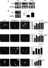
Figure 1.
Figure 1.
SLPI mRNA expression is increased in response to elevation of intracellular cAMP, and exogenous SLPI overcomes inhibition by MAG and myelin in vitro. A, Reverse transcription (RT)-PCR analysis of P5–P6 CGNs, P5–P6 DRG neurons, and P1 cortical neurons treated with 1 mm dbcAMP for 18 h. B, RT-PCR analysis of lesioned and unlesioned lumbar DRGs isolated from P28 rats 24 h after a unilateral sciatic nerve lesion. Graph represents the average densitometric measurements for lesioned and unlesioned DRGs from three individual animals (measured in arbitrary units) ± SEM. C–E, Untreated P5–P6 DRG neurons plated on monolayers of control (C) or MAG-expressing (D) CHO cells and SLPI-treated DRG neurons plated on MAG-expressing CHO cells (E). F–H, Untreated P1 cortical neurons plated on monolayers of control (F) or MAG-expressing (G) CHO cells and SLPI-treated cortical neurons plated on MAG-expressing CHO cells (H). I–K, Untreated P1 cortical neurons (I) and neurons treated with either 1 mm dbcAMP (J) or 10 μg/ml SLPI (K) plated on CNS myelin. L–N, DRG neurons from P28 rats that received intrathecal delivery of sterile saline plated on monolayers of control (L) or MAG-expressing CHO cells (M) and DRG neurons from P28 rats that received intrathecal delivery of SLPI plated on monolayers of MAG-expressing CHO cells (N). Neurite outgrowth was measured from a minimum of 200 neurons for each treatment. Graphs represent the average length of the longest neurite per neuron (depicted as percentage of control) ± SEM (*p < 0.05; **p < 0.01, ***p < 0.001).
Figure 2.
Figure 2.
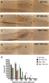
Transected dorsal column axons fail to regenerate after a conditioning lesion in Slpi−/− mice. Representative images of spinal cords from wild-type mice that received a dorsal column lesion only (A, WT DCL), wild-type mice that received a conditioning lesion before the dorsal column lesion (B, WT DCL + CL), Slpi−/− mice that received a dorsal column lesion only (C, Slpi−/− DCL), and Slpi−/− mice that received a conditioning lesion before the dorsal column lesion (D, Slpi−/− DCL + CL). Axons that have regenerated beyond the lesion site (*) are indicated by the arrowheads. E, Quantification of axonal density at points 100, 200, and 300 μm rostral and caudal to the lesion site. Graph depicts average axonal density (square micrometers) ± SEM (**p < 0.01, ***p < 0.001). Scale bar, 100 μm.
Figure 3.
Figure 3.
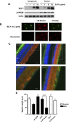
Nuclear localization of SLPI is required to overcome inhibition by myelin. A, Western blot of P5–P6 CGN treated with 1, 5, or 10 μg/ml SLPI for 1 h and subjected to subcellular fractionation. Membranes were probed for pCREB to demonstrate extraction of nuclear proteins. B, Representative images of P5–P6 DRG neurons treated with 10 μg/ml fSLPI or fluorescein alone for 1 h and counterstained for βIII tubulin. C, Representative images of retinas from adult Fischer rats that were killed 4 h after receiving intravitreal injections of 10 μg of fSLPI or unconjugated fluorescein. Sections were immunostained for βIII tubulin (red) and counterstained with DAPI (blue). PR, Photoreceptor layer; ONL, outer nuclear layer; INL, inner nuclear layer; IPL, inner plexiform layer; GCL, ganglion cell layer. Scale bars: 20×, 100 μm; 40×, 100 μm. D, Quantification of neurite outgrowth for P5–P6 DRG neurons treated with SLPI-conjugated beads and plated on CHO cell monolayers. Neurite outgrowth was quantified from a minimum of 200 neurons per condition. Graph depicts average neurite length ± SEM (***p < 0.001).
Figure 4.
Figure 4.
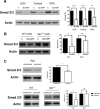
Smad2 knockdown overcomes inhibition by CNS myelin. A, Western blot of P6 CGNs treated with Smad2 siRNA or nontargeting (NT) siRNA. B, Quantification of neurite outgrowth for P6 CGNs plated on CNS myelin and treated with Smad2 siRNA. C, Representative images of control (C) and siRNA-treated P6 CGNs at 24, 48, and 72 h after the addition of Smad2 siRNA. Neurite outgrowth was quantified from a minimum of 200 neurons per condition. Graph depicts average neurite length ± SEM (***p < 0.001).
Figure 5.
Figure 5.
SLPI binds to the Smad2 promoter. P5–P6 CGNs were treated with 10 μg/ml SLPI for 1 h and crosslinked with 1% PFA. Chromatin was isolated and immunoprecipitated (IP) using antibodies to histone H3 (positive control), SLPI, and biotin (negative control). DNA was then amplified using primers for the TNFα promoter, the FASN promoter, and the Smad2 promoter.
Figure 6.
Figure 6.
Elevation of intracellular cAMP suppresses Smad2 in an SLPI-dependent manner. A, Western blots of P6 CGNs, P1 cortical neurons, and P6 DRG neurons treated with 1 mm dbcAMP for 18 h. B, Western blots of P6 CGNs isolated from wild-type (WT) and Slpi−/− mouse pups and treated with 1 mm dbcAMP for 18 h. C, Western blots of lysates prepared from lesioned and unlesioned lumbar DRGs isolated 24 h after a unilateral sciatic nerve lesion in P28 rats, wild-type mice, and Slpi−/− mice. Graphs depict densitometric measurements expressed as percentage of control ± SEM (*p < 0.05, **p < 0.01, ****p < 0.0001). All membranes were probed successively for Smad2/3 and actin. C, Control.
Figure 7.
Figure 7.
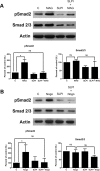
Myelin-associated inhibitors induce phosphorylation of Smad2, and SLPI reduces pSmad2 levels in the presence of myelin-associated inhibitors. A, Western blots of P6 CGNs treated with 10 μg/ml SLPI for 1 h and then exposed to 20 μg/ml MAG-Fc for 30 min. B, Western blots of P6 CGNs treated with 10 μg/ml SLPI for 1 h and then exposed to 1 μg/ml Nogo-AP for 30 min. Graphs depict densitometric measurements expressed as percentage of control ± SEM (*p < 0.05). Membranes were probed successively for pSmad2, Smad2/3, and actin. C, Control.
Figure 8.
Figure 8.
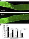
SLPI promotes regeneration of retinal ganglion cell axons. A, Representative images of optic nerves from rats that received intravitreal injections of sterile saline or 10 μg of SLPI after crush injury. Axons that have regenerated beyond the lesion site (*) are indicated by the arrows. B, Quantification of axonal density in regions 0–500, 500–1000, 1000–1500, and 1500–2000 μm distal to the lesion site. Graph depicts average axonal density (square micrometers) ± SEM (**p < 0.01, ***p < 0.001). Scale bar, 100 μm.
Figure 9.
Figure 9.
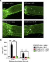
SLPI-mediated regeneration of retinal ganglion cell axons is abolished by ectopic overexpression of Smad2. A, Representative images of optic nerves from rats that received intravitreal injections of RFP or Smad2 adenovirus, followed by crush injury and intravitreal injection of sterile saline or 10 μg of SLPI. Axons that have regenerated beyond the lesion site (*) are indicated by the arrows. B, Quantification of axonal density in regions 0–500 and 500–1000 μm distal to the lesion site. Graph depicts average axonal density (square micrometers) ± SEM (****p < 0.0001). Scale bar, 100 μm.
Figure 10.
Figure 10.
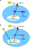
Schematic of myelin-mediated activation of the TGFβ signaling pathway and downregulation of Smad2 by SLPI. A, Myelin-associated inhibitors activate the TGFβ receptor complex through as yet undefined means. The activated receptor complex phosphorylates Smad2, which is essential for inhibition of neurite outgrowth by myelin. B, SLPI, either exogenously applied or produced in response to elevation of cAMP, localizes to the nucleus of the neuron and binds to the Smad2 promoter, inhibiting transcription and reducing the amount of Smad2 protein within the neuron. Thus, less Smad2 is available for phosphorylation, which allows the neuron to extend neurites in the presence of myelin.

Similar articles

Cited by

References

    1. Abdollah S, Macías-Silva M, Tsukazaki T, Hayashi H, Attisano L, Wrana JL. TbetaRI phosphorylation of Smad2 on Ser465 and Ser467 is required for Smad2-Smad4 complex formation and signaling. J Biol Chem. 1997;272:27678–27685. - PubMed
    1. Ashcroft GS, Lei K, Jin W, Longenecker G, Kulkarni AB, Greenwell-Wild T, Hale-Donze H, McGrady G, Song XY, Wahl SM. Secretory leukocyte protease inhibitor mediates non-redundant functions necessary for normal wound healing. Nat Med. 2000;6:1147–1153. - PubMed
    1. Asher RA, Morgenstern DA, Fidler PS, Adcock KH, Oohira A, Braistead JE, Levine JM, Margolis RU, Rogers JH, Fawcett JW. Neurocan is upregulated in injured brain and in cytokine-treated astrocytes. J Neurosci. 2000;20:2427–2438. - PMC - PubMed
    1. Buss A, Pech K, Kakulas BA, Martin D, Schoenen J, Noth J, Brook GA. TGF-beta1 and TGF-beta2 expression after traumatic human spinal cord injury. Spinal Cord. 2008;46:364–371. - PubMed
    1. Cafferty WB, Gardiner NJ, Das P, Qiu J, McMahon SB, Thompson SW. Conditioning injury-induced spinal axon regeneration fails in interleukin-6 knock-out mice. J Neurosci. 2004;24:4432–4443. - PMC - PubMed

Publication types

MeSH terms

LinkOut - more resources