. 2013 May 3;288(18):12791-804.
doi: 10.1074/jbc.M113.465443. Epub 2013 Mar 20.

Sumoylation of Krüppel-like factor 4 inhibits pluripotency induction but promotes adipocyte differentiation

Affiliations

Sumoylation of Krüppel-like factor 4 inhibits pluripotency induction but promotes adipocyte differentiation

Soroush Tahmasebi et al. J Biol Chem. .

Abstract

Ectopic expression of transcription factors has been shown to reprogram somatic cells into induced pluripotent stem (iPS) cells. It remains largely unexplored how this process is regulated by post-translational modifications. Several reprogramming factors possess conserved sumoylation sites, so we investigated whether and how this modification regulates reprogramming of fibroblasts into iPS cells. Substitution of the sole sumoylation site of the Krüppel-like factor (KLF4), a well known reprogramming factor, promoted iPS cell formation. In comparison, much smaller effects on reprogramming were observed for sumoylation-deficient mutants of SOX2 and OCT4, two other classical reprogramming factors. We also analyzed KLF2, a KLF4 homolog and a member of the KLF family of transcription factors with a known role in reprogramming. KLF2 was sumoylated at two conserved neighboring motifs, but substitution of the key lysine residues only stimulated reprogramming slightly. KLF5 is another KLF member with an established link to embryonic stem cell pluripotency. Interestingly, although it was much more efficiently sumoylated than either KLF2 or KLF4, KLF5 was inactive in reprogramming, and its sumoylation was not responsible for this deficiency. Furthermore, sumoylation of KLF4 but not KLF2 or KLF5 stimulated adipocyte differentiation. These results thus demonstrate the importance KLF4 sumoylation in regulating pluripotency and adipocyte differentiation.

Keywords: Adipocyte; Cell Differentiation; KLF2; KLF4; KLF5; Reprogramming; Sumoylation; Transcription Factors; iPS Cells.


Figures

FIGURE 1.
FIGURE 1.
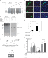
Sumoylation represses transcriptional activity of KLF4. A, domain organization of KLF4 illustrated with sequence alignment of a conserved sumoylation motif and adjacent residues in KLF4 proteins from zebrafish to human. AD, transcriptional activation domain; RD, repression domain; ZF, zinc finger; ψ, bulky hydrophobic residues such as Ile, Leu, or Val; x, any residue. B, sumoylation assays. Expression plasmids for HA-SUMO2 and FLAG-tagged wild-type and mutant KLF4 were co-transfected into HEK293 cells as indicated. 48 h post-transfection, cells were lysed in buffer S, and soluble extracts were prepared for immunoprecipitation (IP) on anti-FLAG M2-agarose and Western blotting (WB) with anti-HA and -FLAG antibodies. HA-SUMO1 was difficult to express, so SUMO2 was used instead. C, subcellular localization of GFP-KLF4 and -K269R after transient transfection of the corresponding expression plasmids into HEK293 cells. Cells were fixed and incubated with Hoechst 33258 for subsequent fluorescence microscopy to detect GFP expression (green) and nuclei (blue). D, reporter gene assays. The Gal4-tk-Luc construct contains five tandem binding sites for the yeast transcription factor Gal4 upstream from the thymidine kinase (tk) core promoters and the coding sequence of luciferase. This construct was transfected into HEK293 cells along with an expression plasmid for expression of fusion proteins containing the Gal4 DNA-binding domain (residues 1–147) fused to KLF4 or its point mutants as indicated. Luciferase activities were normalized to β-galactosidase activities expressed from the CMV-β-Gal expression plasmid that was co-transfected as the internal control. The results were based on three independent assays. *, p = 0.0006 when compared with the empty vector-transfected control. E, Western blotting analysis of Gal4 fusion protein expression. Expression plasmids for the indicated Gal4 fusion proteins (wild-type and mutant KLF4 proteins fused to the C-terminal end of the N-terminal 147 residues of yeast Gal4) were transfected into HEK293 cells, and 2 days later, soluble extracts were prepared for immunoblotting with anti-Gal4 antibody (top) and anti-α-tubulin (bottom) antibodies.
FIGURE 2.
FIGURE 2.
Sumoylation inhibits transcriptional activity of SOX2. A, domain organization of mouse Sox2 shown with sequence alignment of a conserved sumoylation motif and adjacent residues in Sox2 proteins from Xenopus to humans. HMG, high mobility group DNA binding domain; AD1 and AD2, transcriptional activation domains 1 and 2, respectively. B, sumoylation assays. Expression plasmids for HA-SUMO2 and FLAG-tagged wild-type human SOX2 and mutants were co-transfected into HEK293 cells. Extracts were prepared as in Fig. 1B for IP on anti-FLAG M2-agarose and subsequent immunoblotting with anti-HA and -FLAG antibodies as indicated. C, subcellular localization of GFP-tagged wild-type and mutant SOX2 proteins. After transient transfection with the corresponding expression plasmids, HEK293 cells were fixed and incubated with Hoechst 33258 for fluorescence microscopy to detect GFP expression (green) and nuclei (blue). D, reporter gene assays. The assays were performed as for Fig. 1D except that expression plasmids for Sox2 and mutants were analyzed. The results were calculated from three independent assays. *, p < 0.05 when compared with the empty vector-transfected control. E, ChIP analysis. Formaldehyde was used to fix mouse ES cells, and after brief sonication, soluble chromatin was prepared for IP with the α-KLF4 antibody (lane 3) or control IgG (lane 2). The immunoprecipitates were used for PCR to amplify the fragment corresponding to nucleotides −450 to −191 of the mouse Nanog promoter, indicated by arrows (top). F, reporter gene assays. The assays were performed as for Fig. 1D except that the reporter construct contains a mouse Nanog promoter fragment (−2340 to +150) controlling luciferase expression. The expression plasmids for FLAG-tagged KLF4 and SOX2 were transfected as specified. The results were calculated from five different sets of data, with p values indicated.
FIGURE 3.
FIGURE 3.
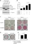
Sumoylation of KLF4 and SOX2 down-regulates iPS cell induction. A, Western blotting analysis. HEK293 cell lysates were analyzed 48 h after transduction with lentivirus expressing FLAG-tagged KLF4, SOX2, or OCT4 as indicated. B, quantification of alkaline phosphatase (ALP)-positive colonies 9 days after MEFs were transduced with lentiviruses expressing OCT4, N-MYC (both untagged), and FLAG-tagged (f-) KLF4 and SOX2 proteins as indicated. The p value is shown (n = 3). C, alkaline phosphatase staining of primary iPS colonies 6 or 9 days after infection of MEFs with a mixture of lentiviruses expressing untagged OCT4 and N-MYC, along with lentiviruses for FLAG-tagged KLF4 and SOX2 as indicated. D, alkaline phosphatase staining of primary iPS colonies 6 days after MEFs were infected with the lentivirus expressing FLAG-tagged OCT4 or mutant K123R, along with lentiviruses expressing N-MYC and FLAG-tagged sumoylation-deficient mutants of KLF4 and SOX2. E, quantification of experiments performed as in D.
FIGURE 4.
FIGURE 4.
Characterization of iPS cell clones expressing wild-type or mutant KLF4. A, phase contrast, alkaline phosphatase staining, and immunostaining of a representative iPS cell clone derived from MEFs infected with lentiviruses expressing KLF4 (FLAG-tagged), untagged OCT4, SOX2, and N-MYC. B, phase contrast, alkaline phosphatase staining, and immunostaining of a representative iPS cell clone derived from MEFs infected with lentivirus expressing K269R (FLAG-tagged), untagged OCT4, SOX2, and N-MYC. C, RT-PCR analysis of different ES cell markers in the two representative clones described in A and B. Mouse ES cells and MEFs were used as positive and negative controls, respectively. D, Western blotting analysis of the two representative iPS cell clones described in A and B. Top blot, soluble extracts were used for IP on anti-FLAG M2-agarose, and the eluted proteins were analyzed by immunoblotting with the anti-FLAG antibody. Perhaps due to different sites of integration, mutant K269R was expressed to a slightly lower level than that of the wild type. The doublet on lane 1 is perhaps due to phosphorylation. A similar doublet was observed elsewhere (e.g. Fig. 1B, top right). Lower three blots, soluble extracts were directly used for immunoblotting with the indicated antibodies. On the anti-KLF4 blot, the single asterisk labels a lower mysterious band perhaps related to endogenous KLF4, and the double asterisks mark the position of nonspecific bands. E, embryoid body formation of cells from the two representative iPS cell clones described in A and B.
FIGURE 5.
FIGURE 5.
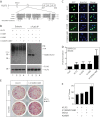
Sumoylation of KLF2 inhibits transcription and reprogramming. A, domain organization of KLF2 shown with sequence alignment of conserved sumoylation motifs and adjacent residues from human, mouse, and Xenopus KLF2 proteins. See supplemental Fig. S4 for the entire sequence alignment. PPXY, a motif for interaction with WW domains such as those in two nuclear targets in hippo signaling and organ size control (37, 47, 48); ZF, zinc finger. B, sumoylation assays. Expression plasmids for HA-SUMO2 and FLAG-tagged wild-type and mutant KLF2 were co-transfected into HEK293 cells as indicated. Extracts were prepared 48 h later for IP on anti-FLAG M2-agarose and subsequent immunoblotting analysis with anti-HA and -FLAG antibodies. WB, Western blot. C, subcellular localization of GFP-tagged wild-type and mutants of KLF2 after transient transfection of the corresponding expression plasmids into HEK293 cells. Cells were fixed and incubated with Hoechst 33258 for fluorescence microscopy to detect GFP expression (green) and nuclei (blue). D, reporter gene assays. The Gal4-tk-Luc construct was transfected into HEK293 cells along with expression plasmids for expression of the Gal4 fusion protein containing the Gal4 DNA-binding domain fused to the wild-type and point mutants of KLF2 as indicated. Luciferase activities were normalized to β-galactosidase activities expressed from CMV-β-Gal co-transfected as the internal control. E, alkaline phosphatase staining of primary iPS colonies 9 days after infection of MEFs with a mixture of lentiviruses expressing untagged Oct4 and N-MYC, along with lentiviruses for FLAG-tagged KLF2 and Sox2 as indicated. F, quantification of alkaline phosphatase staining as performed in E. The quantification was based on two sets of independent experiments; *, p = 0.0048.
FIGURE 6.
FIGURE 6.
KLF5 sumoylation and the effects on subcellular localization and transcription. A, domain organization of KLF5 illustrated with sequence alignment of two conserved sumoylation motifs and adjacent residues in human, mouse, and Xenopus KLF5 proteins. ZF, zinc finger. B, sumoylation assays. Expression plasmids for HA-SUMO2 and FLAG-tagged wild-type and mutant KLF5 were co-transfected into HEK293 cells as indicated. 48 h later, extracts were prepared for IP on anti-FLAG M2-agarose and immunoblotting with anti-HA and -FLAG antibodies. Asterisks denote nonspecific bands. WB, Western blot. C, subcellular localization of GFP-tagged wild-type and mutants of KLF5 and after transient transfection of the corresponding expression plasmids into HEK293 cells. Cells were fixed and incubated with Hoechst 33258 for fluorescence microscopy to detect GFP expression (green) and nuclei (blue). D, reporter gene assays. The Gal4-tk-Luc construct was transfected into HEK293 cells along with expression plasmids for expression of the Gal4 fusion proteins containing the Gal4 DNA-binding domain fused to the wild-type and point mutants of KLF5 as indicated. Luciferase activities were normalized to β-galactosidase activities expressed from CMV-β-Gal co-transfected as the internal control.
FIGURE 7.
FIGURE 7.
Defects of the KLF4 mutant K269R in promoting adipocyte differentiation. A, adipocyte differentiation from MEFs. The cells were infected with lentivirus expressing GFP, FLAG-KLF4, or FLAG-K269R with (bottom) or without (top) lentivirus for N-MYC. Two days after infection, adipocyte differentiation was initiated with the differentiation medium containing 5 μg/ml insulin, 1 μm dexamethasone, and 0.1 μm rosiglitazone. After 2 days, cells were switched to the medium containing 5 μg/ml insulin only and maintained there for the rest of the experiment. Oil Red O staining was performed on day 14; stained dishes were examined under a light microscope, and images of representative areas were taken. B, quantification of Oil Red O staining assays performed in A. The quantification was based on two sets of independent experiments. *, p < 0.05; ***, p < 0.001. C, adipocyte differentiation from 3T3-L1 preadipocytes. The cells were infected with lentivirus expressing GFP, FLAG-KLF4, or FLAG-K269R. Two days after infection, the cells were switched to the differentiation medium containing 5 μg/ml insulin, 1 μm dexamethasone in the absence (top) or presence (bottom) of 0.1 μm rosiglitazone. Oil Red O staining was performed 8 days later, and images of stained dishes were taken afterward. D, quantification of adipocyte differentiation. Isopropyl alcohol was used to extract Oil-Red O stain from stained cells in the dishes (C) for measurement of absorbance at 520 nm. The quantification was based on two sets of independent experiments, with the p values shown. E, adipocyte differentiation from 3T3-L1 preadipocytes. The cells were infected with lentivirus as in C. Two days later, adipocyte differentiation was initiated with the medium containing 5 μg/ml insulin, 1 μm dexamethasone, and isobutyl-1-methylxanthine at the indicated concentrations. Oil Red O staining was performed on day 8, and images of stained dishes were taken afterward.

Similar articles

Cited by

References

    1. Gill G. (2004) SUMO and ubiquitin in the nucleus: different functions, similar mechanisms? Genes Dev. 18, 2046–2059 - PubMed
    1. Hochstrasser M. (2009) Origin and function of ubiquitin-like proteins. Nature 458, 422–429 - PMC - PubMed
    1. Geiss-Friedlander R., Melchior F. (2007) Concepts in sumoylation: a decade on. Nat. Rev. Mol. Cell Biol. 8, 947–956 - PubMed
    1. Nacerddine K., Lehembre F., Bhaumik M., Artus J., Cohen-Tannoudji M., Babinet C., Pandolfi P. P., Dejean A. (2005) The SUMO pathway is essential for nuclear integrity and chromosome segregation in mice. Dev. Cell 9, 769–779 - PubMed
    1. Hay R. T. (2005) SUMO: a history of modification. Mol. Cell 18, 1–12 - PubMed

Publication types

MeSH terms