. 2013 Jun;11(6):651-64.
doi: 10.1158/1541-7786.MCR-12-0578. Epub 2013 Mar 19.

The tyrosine phosphatase SHP2 regulates focal adhesion kinase to promote EGF-induced lamellipodia persistence and cell migration

Affiliations

The tyrosine phosphatase SHP2 regulates focal adhesion kinase to promote EGF-induced lamellipodia persistence and cell migration

Zachary R Hartman et al. Mol Cancer Res. 2013 Jun.

Abstract

The Src homology phosphotyrosyl phosphatase 2 (SHP2) is a positive effector of receptor tyrosine kinases (RTK) signaling. Furthermore, SHP2 is known to promote cell migration and invasiveness, key steps in cancer metastasis. To date, however, the mechanism by which SHP2 regulates cell movement is not fully understood. In the current report, a new role for SHP2 in regulating cell migration has been suggested. We show that SHP2 mediates lamellipodia persistence and cell polarity to promote directional cell migration in the MDA-MB231 and the MDA-MB468 basal-like and triple-negative breast cancer cell lines. We further show that SHP2 modulates the activity of focal adhesion kinase (FAK) by dephosphorylating pTyr397, the autophosphorylation site that primes FAK function. Because hyperactivation of FAK is known to counter the maturation of nascent focal complexes to focal adhesions, we propose that one of the mechanisms by which SHP2 promotes lamellipodia persistence is by downregulating FAK activity through dephosphorylation of pTyr397. The finding that inhibition of FAK activity partially restores EGF-induced lamellipodia persistence and cell migration in SHP2-silenced cells supports our proposition that SHP2 promotes growth factor-induced cell movement by acting, at least in part, on FAK. However, the effect of SHP2 inhibition in nonstimulated cells seems FAK independent as there was no significant difference between the control and the SHP2-silenced cells in pY397-FAK levels. Also, FAK inhibition did not rescue Golgi orientation defects in SHP2-silenced cells, suggesting that SHP2 acts through other mechanisms to promote cell polarity.


Conflict of interest statement

Conflict of interest: The authors do not have any conflict of interest to disclose.

Figures

Figure 1
Figure 1
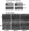
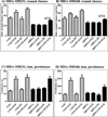
A) Silencing SHP2 protein expression in the MDA-MB231 and the MDA-MB468 cells using two specific shRNA constructs designated as shRNA-1 and shRNA-2. Anti-β-actin immunostaining was used as a loading control. Control and shRNA cells derived from the MDA-MB231 (B) and the MDA-MB468 (C) were grown to confluence followed by wounding with a P200 pipette tip. Images of wounds were taken at 0and 24 hour time points. Rate of wound closure per hour in 8 hours in the MDA-MB231 (D) and the MDA-MB468 (E) was estimated by dividing the overall change in wound space similar to that described in Table 1. Data presented is mean ± SEM. *** indicates p < 0.001 and **indicates p<0.01. Representative images of control and shRNA cells derived from the MDA-MB231 (F) and MDA-MB468 (G) stained with DAPI and GM130 after 6 hours of wound healing are shown. Arrowheads represent cells that were considered as oriented to the wound. H) Golgi orientation was calculated as a percentage of cells at the wound edge with stained golgi facing the wound space. Ten fields of cells were gathered per independent experiment, totaling more than 500 cells for each cell line. Data is presented as mean± SEM.
Figure 1
Figure 1
A) Silencing SHP2 protein expression in the MDA-MB231 and the MDA-MB468 cells using two specific shRNA constructs designated as shRNA-1 and shRNA-2. Anti-β-actin immunostaining was used as a loading control. Control and shRNA cells derived from the MDA-MB231 (B) and the MDA-MB468 (C) were grown to confluence followed by wounding with a P200 pipette tip. Images of wounds were taken at 0and 24 hour time points. Rate of wound closure per hour in 8 hours in the MDA-MB231 (D) and the MDA-MB468 (E) was estimated by dividing the overall change in wound space similar to that described in Table 1. Data presented is mean ± SEM. *** indicates p < 0.001 and **indicates p<0.01. Representative images of control and shRNA cells derived from the MDA-MB231 (F) and MDA-MB468 (G) stained with DAPI and GM130 after 6 hours of wound healing are shown. Arrowheads represent cells that were considered as oriented to the wound. H) Golgi orientation was calculated as a percentage of cells at the wound edge with stained golgi facing the wound space. Ten fields of cells were gathered per independent experiment, totaling more than 500 cells for each cell line. Data is presented as mean± SEM.
Figure 2
Figure 2
SHP2 localizes to the leading edge of BTBC cells. The control and the shRNA cells derived from the MDA-MB231 (A) and the MDA-MB468 (B) lines were seeded on fibronectin-coated coverslips and were allowed to attach overnight. Cells were then fixed, permeablized, and stained with anti-cortactin and anti-SHP2 antibodies. Exposure times to acquire images of SHP2 staining were held constant between control and shRNA slides. Arrows indicate leading edges of cells.
Figure 2
Figure 2
SHP2 localizes to the leading edge of BTBC cells. The control and the shRNA cells derived from the MDA-MB231 (A) and the MDA-MB468 (B) lines were seeded on fibronectin-coated coverslips and were allowed to attach overnight. Cells were then fixed, permeablized, and stained with anti-cortactin and anti-SHP2 antibodies. Exposure times to acquire images of SHP2 staining were held constant between control and shRNA slides. Arrows indicate leading edges of cells.
Figure 3
Figure 3
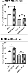
Bar graphs showing the impact of SHP2 silencing on EGF-induced wound healing over a period of 2 hours in the MDA-MB231 (A) and the MDA-MB468 (B) cells. SHP2 depletion alters EGF-stimulated membrane protrusions in BTBC cells. C) Kymographs of movies collected from live-cell imaging experiments for both the MDA-MB231 and MDA-MB468 cells are shown. Kymographs were used to calculate the persistence of the lamellipodia in the MDA-MB231 (D) and the MDA-MB468 (E) cells in a manner described by Kelley, et al (48). Data presented are mean ± SEM of protrusion persistence time in seconds collected from at least 10 cells per cell line. * indicates p< 0.05, and *** indicates p < 0.001.
Figure 3
Figure 3
Bar graphs showing the impact of SHP2 silencing on EGF-induced wound healing over a period of 2 hours in the MDA-MB231 (A) and the MDA-MB468 (B) cells. SHP2 depletion alters EGF-stimulated membrane protrusions in BTBC cells. C) Kymographs of movies collected from live-cell imaging experiments for both the MDA-MB231 and MDA-MB468 cells are shown. Kymographs were used to calculate the persistence of the lamellipodia in the MDA-MB231 (D) and the MDA-MB468 (E) cells in a manner described by Kelley, et al (48). Data presented are mean ± SEM of protrusion persistence time in seconds collected from at least 10 cells per cell line. * indicates p< 0.05, and *** indicates p < 0.001.
Figure 4
Figure 4
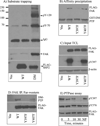
SHP2 regulates FAK activity in BTBC cells. A) Substrate-trapping studies in mouse embryo fibroblasts (MEFs) expressing vector alone, FLAG-WT-SHP2 or FLAG-DM-SHP2. Cells were seeded on fibronectin for 30 minutes, and lysates prepared from these cells were subjected to anti-FLAG immunoprecipitation followed by immunostaining analysis with anti-pTyr, anti-FAK and anti-FLAG (for SHP2) antibodies. B) In vitro affinity precipitation with GST-DM-PTP of cell lysates prepared from FAK-null MEFs expressing vector alone or FLAG-tagged FAK proteins. The precipitates were analyzed by immunostaining with ant-FLAG antibody for FAK proteins and with anti-GST for GST-DM-PTP. C) Input total cell lysates used for affinity precipitation were stained with anti-FLAG, anti-pY397 and anti-β-actin antibodies. D) Far-Western analysis with GST-DM-PTP to show that the presence of pY397 is needed for binding. E) In vitro phosphatase assay using wild-type FAK as a substrate and purified wild-type SHP2 as an enzyme. Immunostaining results for pY397, pY576 and FAK (FLAG-FAK) are shown. NP: no PTP.
Figure 5
Figure 5
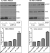
Analysis of impact of SHP2 silencing on the pY397-FAK levels. The control and the shRNA cells derived from the MDA-MB231 (A) and MDA-MB468 (B) were grown to about 90% confluence, serum-starved for approximately 4 hours, and then left unstimulated or stimulated with 100 ng/ml EGF for 20 minutes. Lysates prepared from these cells were analyzed by immunostaining with anti-pY397 antibody. Bar graphs show band density measurements of the anti-pY397 staining from three independent experiments. The MDA-MB231 (C) and the MDA-MB468 (D) cells grown on coverslips were stimulated with the same concentration of EGF and then analyzed by immunofluorescence microscopy after staining with anti-pY397 antibody. The DAPI staining also was performed to show the nucleus.
Figure 5
Figure 5
Analysis of impact of SHP2 silencing on the pY397-FAK levels. The control and the shRNA cells derived from the MDA-MB231 (A) and MDA-MB468 (B) were grown to about 90% confluence, serum-starved for approximately 4 hours, and then left unstimulated or stimulated with 100 ng/ml EGF for 20 minutes. Lysates prepared from these cells were analyzed by immunostaining with anti-pY397 antibody. Bar graphs show band density measurements of the anti-pY397 staining from three independent experiments. The MDA-MB231 (C) and the MDA-MB468 (D) cells grown on coverslips were stimulated with the same concentration of EGF and then analyzed by immunofluorescence microscopy after staining with anti-pY397 antibody. The DAPI staining also was performed to show the nucleus.
Figure 6
Figure 6
Effect of FAK inhibition on EGF-induced wound healing, lamellipodia persistence, and golgi orientation. SHP2-depleted MDA-MB231 (A) and MDA-MB468 (B) cells were grown to confluence before scratching with a pipette tip. Cells were then fed with serum-free medium and left untreated or treated with 10 ng/ml FAK inhibitor (PF) and 100 ng/ml EGF. Rate of wound healing was assessed in the same manner as in Fig. 3A and B. Live-cell imaging was used to assess EGF-induced lamellipodia persistence in the presence and absence of PF (10 nM) in theMDA-MB231 (C) and the MDA-MB468 (D) cells. Data presented is mean ± SEM of protrusion persistence time in seconds collected from at least 10 cells per cell line. E) Effect of the FAK inhibitor on golgi orientation. Analysis of pY397-FAK levels in the MDA-MB231 (F) and MDA-MB468 (G) cells in the presence and absence of FAK inhibitor and EGF.
Figure 6
Figure 6
Effect of FAK inhibition on EGF-induced wound healing, lamellipodia persistence, and golgi orientation. SHP2-depleted MDA-MB231 (A) and MDA-MB468 (B) cells were grown to confluence before scratching with a pipette tip. Cells were then fed with serum-free medium and left untreated or treated with 10 ng/ml FAK inhibitor (PF) and 100 ng/ml EGF. Rate of wound healing was assessed in the same manner as in Fig. 3A and B. Live-cell imaging was used to assess EGF-induced lamellipodia persistence in the presence and absence of PF (10 nM) in theMDA-MB231 (C) and the MDA-MB468 (D) cells. Data presented is mean ± SEM of protrusion persistence time in seconds collected from at least 10 cells per cell line. E) Effect of the FAK inhibitor on golgi orientation. Analysis of pY397-FAK levels in the MDA-MB231 (F) and MDA-MB468 (G) cells in the presence and absence of FAK inhibitor and EGF.

Similar articles

Cited by

References

    1. Freeman RM, Plutzky J, Neel BG. Identification of a human src homology 2-containing protein-tyrosine-phosphatase: a putative homolog of Drosophila corkscrew. Proceedings of the National Academy of Sciences. 1992;89(23) 11239-. - PMC - PubMed
    1. Sugimoto S, Lechleider RJ, Shoelson SE, Neel BG, Walsh CT. Expression, purification, and characterization of SH2-containing protein tyrosine phosphatase, SH-PTP2. Journal of Biological Chemistry. 1993;268(30) 22771-. - PubMed
    1. Hof P, Pluskey S, Dhe-Paganon S, Eck MJ, Shoelson SE. Crystal structure of the tyrosine phosphatase SHP-2. Cell. 1998 Feb 20;92(4):441–450. - PubMed
    1. Qu CK. The SHP-2 tyrosine phosphatase: signaling mechanisms and biological functions. Cell Research. 2000;10(4):279–288. - PubMed
    1. Higashi H, Nakaya A, Tsutsumi R, Yokoyama K, Fujii Y, Ishikawa S, et al. Helicobacter pylori CagA induces Ras-independent morphogenetic response through SHP-2 recruitment and activation. The Journal of biological chemistry. 2004;279(17):17205–17216. - PubMed

Publication types

MeSH terms

Substances