. 2013 Mar 13;33(11):5040-52.
doi: 10.1523/JNEUROSCI.2896-12.2013.

Postsynaptic density scaffold SAP102 regulates cortical synapse development through EphB and PAK signaling pathway

Affiliations

Postsynaptic density scaffold SAP102 regulates cortical synapse development through EphB and PAK signaling pathway

Yasunobu Murata et al. J Neurosci. .

Abstract

Membrane-associated guanylate kinases (MAGUKs), including SAP102, PSD-95, PSD-93, and SAP97, are scaffolding proteins for ionotropic glutamate receptors at excitatory synapses. MAGUKs play critical roles in synaptic plasticity; however, details of signaling roles for each MAGUK remain largely unknown. Here we report that SAP102 regulates cortical synapse development through the EphB and PAK signaling pathways. Using lentivirus-delivered shRNAs, we found that SAP102 and PSD-95, but not PSD-93, are necessary for excitatory synapse formation and synaptic AMPA receptor (AMPAR) localization in developing mouse cortical neurons. SAP102 knockdown (KD) increased numbers of elongated dendritic filopodia, which is often observed in mouse models and human patients with mental retardation. Further analysis revealed that SAP102 coimmunoprecipitated the receptor tyrosine kinase EphB2 and RacGEF Kalirin-7 in neonatal cortex, and SAP102 KD reduced surface expression and dendritic localization of EphB. Moreover, SAP102 KD prevented reorganization of actin filaments, synapse formation, and synaptic AMPAR trafficking in response to EphB activation triggered by its ligand ephrinB. Last, p21-activated kinases (PAKs) were downregulated in SAP102 KD neurons. These results demonstrate that SAP102 has unique roles in cortical synapse development by mediating EphB and its downstream PAK signaling pathway. Both SAP102 and PAKs are associated with X-linked mental retardation in humans; thus, synapse formation mediated by EphB/SAP102/PAK signaling in the early postnatal brain may be crucial for cognitive development.


Figures

Figure 1.
Figure 1.
SAP102 and PSD-95 are the two major postsynaptic MAGUKs in developing cortical neurons. A, B, Different developmental expression profiles of SAP102, PSD-95, and PSD-93 in cultured cortical neurons. Homogenates (1 μg) from cultured cortical neurons at DIV 10, 14, 18, 22, and 26 were analyzed by quantitative Western blotting. These blots were quantified and plotted as a percentage of each MAGUK's level at DIV 26. Total MAGUK levels were analyzed with an anti-pan-MAGUK antibody recognizing SAP102, PSD-95, PSD-93, and SAP97. SAP102 was expressed at relatively high levels from the earliest developmental stage examined, and PSD-95 levels were gradually increased whereas PSD-93 remained at relatively low levels until DIV 18 and increased very rapidly between DIV 18 and 22. No clear band was detected with the anti-SAP97 antibodies (see Materials and Methods). (n = 6 immunoblots from three separate sets of cultures, data are presented as mean ± SEM.) C, Lentivirus-delivered shRNAs effectively knocked down specific MAGUKs in DIV 14 cultured cortical neurons. Neurons were infected with lentiviruses expressing shRNA or the corresponding control scrambled shRNA (scr.sh) at DIV 2. For each MAGUK two different shRNA and scrambled shRNA lentiviruses were used. The shRNA specifically reduced the target protein to a point that it was undetectable or barely detectable on immunoblots, and the knockdown lasted for at least four weeks in the cultures (data not shown). As illustrated for SAP102, the GFP lentivirus control was not different from the SAP102 scr.sh. This was tested in one-way ANOVAs (data not shown). The same ANOVA test was used for GFP lentivirus or tdTomato lentivirus and the scrambled shRNAs for each MAGUK in all subsequent experiments and no significant differences were found. Therefore, GFP or tdTomato lentivirus is shown as the control in most experiments. D, E, SAP102 shRNA and PSD-95 shRNA, but not PSD-93 shRNA, significantly decreased the amount of total MAGUK expression levels in developing cultured cortical neurons. Total MAGUK levels were analyzed with an anti-pan-MAGUK antibody recognizing SAP102, PSD-95, PSD-93, and SAP97. Knockdown of SAP102 or PSD-95, but not PSD-93, significantly reduced the total MAGUK levels, suggesting SAP102 and PSD-95 are predominant MAGUKs in these developing neurons. Triple knockdown of SAP102, PSD-95, and PSD-93, was achieved by coinfection with a lentivirus against each MAGUK. Triple KD decreased the total MAGUK levels by 95%, indicating that the last member of MAGUKs, SAP97, is far less abundant than SAP102, PSD-95 or PSD-93 in young cortical neurons. (n = 6 immunoblots from three separate sets of cultures.) One-way ANOVA tests were used in this and all subsequent data presented. Data are presented as mean ± SEM, and asterisks are p values comparing to the shRNA lentivirus infected neurons to the GFP lentivirus infected control neurons; *p < 0.05, **p < 0.01.
Figure 2.
Figure 2.
SAP102 and PSD-95 play important roles in excitatory synapse formation and synaptic AMPAR localization in developing cortical neurons. A, B, SAP102 shRNAs and PSD-95 shRNAs, but not PSD-93 shRNAs, reduced puncta of the presynaptic marker Bassoon. Cortical neurons were infected with lentiviruses containing shRNA or scrambled shRNA (scr.sh) at DIV 2 and immunostained with an anti-Bassoon antibody at DIV 14. The density of Bassoon puncta on dendrites showed a 40% decrease in SAP102 KD and PSD-95 KD neurons compared with their appropriate scrambled control, but remained constant in PSD-93 KD neurons. Note that similar results were obtained from two different shRNA lentiviruses for each MAGUK, which ensure the validity of the knockdown effects. Since two different shRNA sequences showed very similar results, we did not show the results of second shRNA in some subsequent experiments to avoid repetition. Scrambled shRNAs had no significant effect on the densities of Bassoon puncta. Scale bar, 5 μm. (n = >15 dendrites from 8 neurons). C, D, SAP102 shRNA and PSD-95 shRNA reduced the excitatory presynaptic marker VGLUT1. DIV 14 neurons were doubly immunostained with anti-VGLUT1 (excitatory presynaptic marker, red) and anti-VGAT (inhibitory presynaptic marker, blue). The densities of VGLUT1 puncta were significantly reduced in SAP102 KD and PSD-95 KD neurons whereas the density of VGAT puncta was not significantly altered in any of MAGUK KDs. Scale bar, 5 μm. (n = >15 dendrites from 8 neurons). E–G, SAP102 and PSD-95 play major roles in maintaining synaptic AMPAR levels in developing cortical neurons. The PSD fraction was prepared from DIV 14 cortical cultures and analyzed by Western blotting. The levels of NMDAR and AMPAR subunits in homogenate were not altered by any of MAGUK KDs (E). The levels of glutamate receptor subunits in the PSD fraction (F) were plotted as a histogram (G). SAP102 shRNAs and PSD-95 shRNAs, but not PSD-93 shRNAs, significantly reduced the levels of the GluA1 and GluA2 AMPAR subunits in the PSD fraction. None of MAGUK KDs changed the levels of NMDAR subunits in the PSD fraction, thereby indicating a greater effect of MAGUK KDs on AMPARs than NMDARs at the PSD. (n = 6 immunoblots from 3 independent sets of cultures.) H, The subcellular fractions from DIV 14 cultured cortical neurons, including homogenate, cytosol, crude membrane, synaptosome, and PSD fraction, were analyzed by Western blotting. SAP102, PSD-95, GluN1, and GluA2 were strongly enriched in the PSD fraction. The absence of Synaptophysin, presynaptic vesicle related protein, in the PSD fraction validates the isolation of PSD fraction.
Figure 3.
Figure 3.
SAP102 shRNA increases dendritic filopodia length. A, Representative confocal images of dendritic protrusions in control GFP lentivirus, SAP102 KD, and PSD-95 KD neurons at DIV 14. Cultured cortical neurons were infected with GFP control, SAP102 shRNA1, SAP102 shRNA2, PSD-95 shRNA1, or PSD-95 shRNA2 lentivirus at DIV 2. Neurons were fixed and imaged at DIV 14. Scale bar, 5 μm. B, The density of dendritic protrusions was not significantly altered in SAP102 KD and PSD-95 KD neurons (n = 10 dendrites from 5 neurons. The number of dendritic protrusions per micrometer dendrite is presented as mean ± SEM.) C, The average length of dendritic protrusions was significantly increased in SAP102 KD neurons reflecting the prevalence of long dendritic filopodia caused by SAP102 shRNAs. The average length of dendritic protrusions in PSD-95 KD was slightly shorter compared with GFP lentivirus-infected neurons because of the prevalence of small stubby protrusions in PSD-95 KD neurons, but the difference did not reach statistical significance. (n = 10 dendrites from 5 neurons.)
Figure 4.
Figure 4.
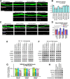
SAP102 is in a complex with EphB2, and SAP102 KD reduces surface expression and dendritic localization of EphB. A, B, EphB2 and SAP102 were reciprocally coimmunoprecipitated from developing (P14; 1 d after completion of eye-lid opening) rat visual cortex. IP from visual cortical homogenates showed that (A) EphB2 was immunoprecipitated with the anti-SAP102 antibody, but not with the anti-PSD-95 antibody, and that (B) SAP102, but not PSD-95, was immunoprecipitated with the anti-EphB2 antibody. Synaptophysin and PSD-95 were used as negative controls. Normal rabbit IgG and normal mouse IgG were used as additional negative controls of IP. C, D, Association between EphB2 and SAP102 is not mediated by their direct binding. C, HEK cells were cotransfected with EphB2 and SAP102-GFP expression plasmids, and coimmunoprecipitated with the anti-GFP antibody. EphB2 was not pulled down with SAP102-GFP, suggesting that these two proteins do not directly bind to each other. D, GluN2B was coimmunoprecipitated with SAP102-GFP from HEK cells cotransfected with GluN2B and SAP102-GFP, validating the reliability of coimmunoprecipitation assay in HEK cells. E–G, SAP102 shRNA reduced the surface expression level of EphB2. Cultured neurons were infected with either the GFP control, SAP102 shRNA, PSD-95 shRNA, or PSD-93 shRNA lentivirus at DIV 2. Surface proteins on DIV 14 cultured cortical neurons were biotinylated, pulled down with streptavidin agarose resin, and analyzed by Western blotting (G). The proteins unbound to streptavidin agarose resin were collected as cytosolic proteins and also analyzed (F). The surface or cytosolic expression levels of EphB2, GluN1, GluN2A, GluN2B, and GluA2 for each MAGUK KD were quantified and plotted as a percentage of the GFP lentivirus control. The surface expression level of EphB2 was significantly decreased in SAP102 KD neurons. The surface expression levels of GluN1 and GluN2A were not significantly altered in any MAGUK KD neurons. However, in PSD-95 KD neurons, the surface expression level of GluN2B was increased (G) and the cytosolic level of GluN2B was decreased (F), suggesting that SAP102 compensates for the absence of PSD-95 by holding more GluN2B on the surface of PSD-95 KD neurons. The GluA2 AMPAR subunit was significantly decreased in the surface fraction but increased in the cytosolic fraction of SAP102 KD or PSD-95 KD neurons, suggesting both SAP102 and PSD-95 play a key role in surface AMPAR expression. Synaptophysin was used as a negative control of surface biotinylation assay. Transferrin receptor (TfR) was used as a loading control to normalize protein levels. (n = 6 immunoblots from 3 separate sets of cultures; data were presented as mean ± SEM.) H, I, SAP102 shRNA reduced the number of EphB puncta on dendrites. Cortical culture neurons were infected with lentiviruses expressing tdTomato only, SAP102 shRNA or PSD-95 shRNA at DIV 2, and fixed at DIV 14. EphBs were visualized by incubation with ephrinB2-Fc followed by anti-Fc and secondary antibodies (see Materials and Methods). The number of EphB puncta on dendrites was quantified and plotted as a percentage of control. These data revealed a significant decrease of EphB puncta in SAP102 KD neurons. (n = 10 dendrites from 5 neurons.) The density of EphB puncta per micrometer of dendrite is normalized to the tdTomato lentivirus control and presented as mean ± SEM.
Figure 5.
Figure 5.
SAP102 shRNAs impaired ephrinB-induced actin reorganization, synapse formation, and AMPAR trafficking. A, B, SAP102 shRNA impaired ephrinB-induced reorganization of actin filaments. DIV 14 cortical neuron cultures were treated with dimerized Fc (control) or ephrinB2-Fc for 1 h, and stained with phalloidin conjugated with Alexa 543 to visualize the F-actin. In the Fc-treated (control) conditions, phalloidin signals within GFP-positive lentivirus-infected dendrites and dendritic protrusions were comparable among neurons infected with lentivirus expressing GFP only, SAP102 shRNA1, SAP102 shRNA2, PSD-95 shRNA1, and PSD-95 shRNA2. After ephrinB2-Fc treatment, GFP lentivirus as well as PSD-95 KD neurons showed >2-fold increases in phalloidin signal intensity. However, ephrinB treatment failed to increase phalloidin signal in both SAP102 shRNA1 and SAP102 shRNA2 neurons. The phalloidin signal intensity per micrometer of dendrite is normalized to Fc-treated GFP lentivirus neurons and presented as mean 0± SEM (n = >10 dendrites from 5 neurons). C, D, SAP102 KD impaired ephrinB-induced synapse formation. DIV 14 cortical neurons were treated with dimerized Fc (control) or ephrinB2-Fc for 1 h, and immunostained for the presynaptic marker Bassoon. EphrinB2-Fc treatment significantly increased Bassoon puncta density by >30% in GFP lentivirus neurons as well as PSD-95 KD neurons, but did not cause a significant increase in SAP102 KD neurons. Bassoon puncta associated with GFP-positive lentivirus-infected dendrites and dendritic protrusions were analyzed, and the density of Bassoon puncta is normalized to Fc-treated GFP lentivirus-infected neurons and presented as mean ± SEM (n = >10 dendrites from 5 neurons). E, F, SAP102 KD impaired ephrinB-induced synaptic AMPAR trafficking. DIV 14 cortical culture neurons were treated with dimerized Fc (control) or ephrinB2-Fc for 1 h, and PSD fractions were prepared to analyze the amount of AMPAR GluA2 subunit and of NMDAR GluN1subunit by Western blotting. In Fc-treated (control) conditions, the amounts of GluA2 subunit in PSD fractions were significantly lower in both SAP102 KD and PSD-95 KD neurons compared with GFP lentivirus-infected neurons. EphrinB2-Fc treatment increased the amounts of GluA2 subunit in PSD fractions of the GFP lentivirus as well as the PSD-95 KD neurons. However, in SAP102 KD neurons, this EphrinB2-Fc-induced increase in synaptic AMPARs was abolished. The levels of GluA2 in the PSD fraction is normalized to Fc-treated GFP lentivirus infected neurons and presented as mean ± SEM (n = 3 independently prepared and analyzed sets of cultures).
Figure 6.
Figure 6.
Rac1-GEF Kalirin-7 and the GluN2B NMDAR subunit are preferentially immunoprecipitated with SAP102 from the developing rat visual cortex. A, Proteins immunoprecipitated with an anti-SAP102 or an anti-PSD-95 antibody from P14 visual cortex were analyzed by Western blotting. Anti-pan Kalirin antibody detects several isoforms: Kalirin-7 around 190 kDa and Kalirin-9 and Kalirin-12 in the higher molecular weight region. Among Kalirin isoforms, only Kalirin-7 was preferentially pulled down with SAP102 as indicated by an asterisk. Both SAP102 and PSD-95 pulled down a similar amount of the GluN1 NMDAR subunit. However, SAP102 pulled down more GluN2B subunit than PSD-95, in contrast, PSD-95 pulled down higher levels of the GluN2A subunit than SAP102, showing preferential interactions between SAP102 and GluN2B and between PSD-95 and GluN2A in intact developing visual cortex. Asterisks indicate Kalirin-7 at 190 kDa pulled down with SAP102, GluN2A pulled down PSD-95, and GluN2B pulled down with SAP102. Synaptophysin, a presynaptic vesicle associated protein, was shown as a negative control. Normal rabbit IgG and normal mouse IgG were used as negative controls of IP. B, Proteins immunoprecipitated from adult visual cortex were analyzed by Western blotting. Kalirin-7 was pulled down by both anti-SAP102 and anti-PSD-95 antibodies showing a difference in protein interaction between developing and mature visual cortex.
Figure 7.
Figure 7.
SAP102 shRNA downregulates group I PAK activity. A, B, Group I PAK activity was analyzed with an anti-phospho PAK antibody recognizing phosphorylated serines at 144 of PAK1, 141 of PAK2, and 139 of PAK3; these phosphorylations are necessary for activation and maintenance of PAK activity. DIV 14 cultured cortical neurons were treated with dimerized control Fc or ephrinB2-Fc for 1 h, and whole lysates were prepared and analyzed by Western blotting. Under the control condition (Fc-treated), SAP102 KD neurons showed a significantly reduced level of PAK phosphorylation compared with GFP lentivirus or PSD-95 KD neurons. After a 1 h treatment with ephrinB2-Fc, the PAK phosphorylation level was increased by >50% in GFP lentivirus as well as PSD-95 KD neurons. However, in SAP102 KD neurons, the PAK phosphorylation levels remained unchanged and were significantly lower than GFP lentivirus or PSD-95 KD neurons, suggesting PAK activity is downregulated in SAP102 KD neurons. There were no significant differences in the expression levels of either PAK1 or PAK3 between GFP lentivirus, SAP102 KD, and PSD-95 KD neurons. (n = 3 independently prepared and analyzed sets of cultures, data is normalized to Fc-treated GFP lentivirus infected neurons and presented as mean ± SEM.)

Similar articles

Cited by

References

    1. Allen KM, Gleeson JG, Bagrodia S, Partington MW, MacMillan JC, Cerione RA, Mulley JC, Walsh CA. PAK3 mutation in nonsyndromic X-linked mental retardation. Nat Genet. 1998;20:25–30. - PubMed
    1. Banker G, Goslin K. Culturing nerve cells. Cambridge, MA: MIT; 1991.
    1. Béïque JC, Lin D-T, Kang MG, Aizawa H, Takamiya K, Huganir RL. Synapse-specific regulation of AMPA receptor function by PSD-95. Proc Natl Acad Sci U S A. 2006;103:19535–19540. - PMC - PubMed
    1. Bienvenu T, des Portes V, McDonell N, Carrié A, Zemni R, Couvert P, Ropers HH, Moraine C, van Bokhoven H, Fryns JP, Allen K, Walsh CA, Boué J, Kahn A, Chelly J, Beldjord C. Missense mutation in PAK3, R67C, causes X-linked nonspecific mental retardation. Am J Med Genet. 2000;93:294–298. - PubMed
    1. Boda B, Alberi S, Nikonenko I, Node-Langlois R, Jourdain P, Moosmayer M, Parisi-Jourdain L, Muller D. The mental retardation protein PAK3 contributes to synapse formation and plasticity in hippocampus. J Neurosci. 2004;24:10816–10825. - PMC - PubMed

Publication types

MeSH terms

LinkOut - more resources