. 2013;8(3):e58311.
doi: 10.1371/journal.pone.0058311. Epub 2013 Mar 6.

HOXC9 regulates formation of parachordal lymphangioplasts and the thoracic duct in zebrafish via stabilin 2

Affiliations

HOXC9 regulates formation of parachordal lymphangioplasts and the thoracic duct in zebrafish via stabilin 2

Sandra J Stoll et al. PLoS One. 2013.

Abstract

HOXC9 belongs to the family of homeobox transcription factors, which are regulators of body patterning and development. HOXC9 acts as a negative regulator on blood endothelial cells but its function on lymphatic vessel development has not been studied. The hyaluronan receptor homologs stabilin 1 and stabilin 2 are expressed in endothelial cells but their role in vascular development is poorly understood. This study was aimed at investigating the function of HOXC9, stabilin 2 and stabilin 1 in lymphatic vessel development in zebrafish and in endothelial cells. Morpholino-based expression silencing of HOXC9 repressed parachordal lymphangioblast assembly and thoracic duct formation in zebrafish. HOXC9 positively regulated stabilin 2 expression in zebrafish and in HUVECs and expression silencing of stabilin 2 phenocopied the HOXC9 morphant vascular phenotype. This effect could be compensated by HOXC9 mRNA injection in stabilin 2 morphant zebrafish embryos. Stabilin 1 also regulated parachordal lymphangioblast and thoracic duct formation in zebrafish but acts independently of HOXC9. On a cellular level stabilin 1 and stabilin 2 regulated endothelial cell migration and in-gel sprouting angiogenesis in endothelial cells. HOXC9 was identified as novel transcriptional regulator of parachordal lymphangioblast assembly and thoracic duct formation in zebrafish that acts via stabilin 2. Stabilin 1, which acts independently of HOXC9, has a similar function in zebrafish and both receptors control important cellular processes in endothelial cells.


Conflict of interest statement

Competing Interests: The authors have declared that no competing interests exist.

Figures

Figure 1
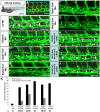
Figure 1. Silencing of HOXC9 expression in zebrafish inhibits assembly of parachordal lymphangioplasts (PLs).
(A) Overall morphology of 48 hpf zebrafish embryo after control morpholino injection. Red box shows region displayed in (B) and (C). (B) Normal formation of the PLs (arrows) in 48 hpf tg(fli1:EGFP) zebrafish embryo after injection of 4 ng control morpholino. (C) Silencing of HOXC9 expression using 2 ng translational-blocking morpholino disrupted the formation of the PLs (asterisks) in 48 hpf tg(fli1:EGFP) zebrafish embryo. (D) Quantification of 48 hpf tg(fli1:EGFP) zebrafish embryos showing a disturbed PL formation. Embryos were divided in three groups depending on the PL appearance being completely absent, partially formed or completely present. (E) RT-PCR analysis for increased expression of Stab2 in zebrafish driven by mRNA (50 pg) mediated overexpression of HOXC9. (F) Quantification of (E), n = 3 per group. *p<0.05 vs.Orange-mRNA. (G) RT-PCR analysis for reduced expression of Stab2 in zebrafish driven by morpholino (2 ng) mediated silencing of HOXC9. (H) Quantification of (G), n = 4 per group. *p<0.05 vs.Co-Mo. Black scale bar: 500 µm. White scale bar: 50 µm.
Figure 2
Figure 2. Silencing of Stab2 expression in zebrafish inhibits assembly of parachordal lymphangioplasts (PLs).
(A) Overall morphology of 48 hpf zebrafish embryo after control morpholino injection. Red box shows region displayed in (B–D). (B) Normal formation of the PLs (arrows) in 48 hpf tg(fli1:EGFP) zebrafish embryo after injection of 4 ng control morpholino. (C,D) Silencing of Stab2 expression using 4 ng splice-blocking morpholino targeting exon 2 (C) or 2 ng splice-blocking morpholino targeting exon 9 (D) disrupted the formation of the PLs (asterisks) in 48 hpf tg(fli1:EGFP) zebrafish embryos. (E) Quantification of 48 hpf tg(fli1:EGFP) zebrafish embryos showing a disturbed PL formation. Embryos were divided in three groups depending on the PL appearance being completely absent, partially formed or completely present. (F) Functionality of the splice blocking morpholino Stab2-Ex2-Mo. RT-PCR of control morpholino (4 ng) and Stab2-Ex2-Mo (4 ng) injected zebrafish embryos at 24 hpf. (G) Functionality of the splice blocking morpholino Stab2-Ex9-Mo. RT-PCR of control morpholino (4 ng) and Stab2-Ex9-Mo (2 ng) injected zebrafish embryos at 24 hpf. WT: wild type, morph: morphant. Black scale bar: 500 µm. White scale bar: 50 µm.
Figure 3
Figure 3. HOXC9 overexpression rescues the defects in parachordal lymphangioplast (PL) formation in Stab2 morphants.
(A) Overall morphology of 48 hpf zebrafish embryo after control morpholino injection. Red box shows region displayed in (B–D). (B) Normal formation of the PLs (arrows) in 48 hpf tg(fli1:EGFP) zebrafish embryo after injection of 4 ng control morpholino. (C) Silencing of Stab2 expression using 4 ng splice-blocking morpholino disrupted the formation of the PLs (asterisks) in 48 hpf tg(fli1:EGFP) zebrafish embryo. (D) Injection of HOXC9 mRNA (50 pg) rescued the Stab2 loss-of-function phenotype in 48 hpf tg(fli1:EGFP) zebrafish embryo. (E) Quantification of 48 hpf tg(fli1:EGFP) fish embryos showing a disturbed PL formation including rescue experiments using HOXC9 mRNA (50 pg). Embryos were divided in three groups depending on the PL appearance being completely absent, partially formed or completely present. (F) Functionality of the mRNA injection. Western Blot of Orange-mRNA (50 pg) and HOXC9-mRNA (50 pg) injected zebrafish embryos at 24 hpf. (G) Quantification of (F), n = 3 per group. *p<0.05 vs. Orange-mRNA. Black scale bar: 500 µm. White scale bar: 50 µm.
Figure 4
Figure 4. Silencing of Stab1 expression in zebrafish inhibits assembly of parachordal lymphangioplasts (PLs).
(A) Overall morphology of 48 hpf zebrafish embryo after control morpholino injection. Red box shows region displayed in (B–D). (B) Normal formation of the PLs (arrows) in 48 hpf tg(fli1:EGFP) zebrafish embryo after injection of 4 ng control morpholino. (C,D) Silencing of Stab1 expression using 4 ng splice-blocking morpholino targeting exon 3 (C) or 12 ng splice-blocking morpholino targeting exon 4 (D) disrupted the formation of the PLs (asterisks) in 48 hpf tg(fli1:EGFP) fish embryos. (E) Quantification of 48 hpf tg(fli1:EGFP) fish embryos showing a disturbed PL formation. Embryos were divided in three groups depending on the PL appearance being completely absent, partially formed or completely present. (F) Functionality of the splice blocking morpholinos Stab1-Ex3-Mo and Stab1-Ex4-Mo. RT-PCR of control morpholino (4 ng), Stab1-Ex3-Mo (4 ng) and Stab1-Ex4-Mo (12 ng) injected zebrafish embryos at 24 hpf. WT: wild type, morph: morphant. Black scale bar: 500 µm. White scale bar: 50 µm.
Figure 5
Figure 5. HOXC9 overexpression rescues the defects in parachordal lymphangioplast (PL) formation in Stab1 morphants.
(A) Overall morphology of 48 hpf zebrafish embryo after control morpholino injection. Red box shows region displayed in (B–D). (B) Normal formation of the PLs (arrows) in 48 hpf tg(fli1:EGFP) fish embryo after injection of 4 ng control morpholino. (C) Silencing of Stab1 expression using 4 ng splice-blocking morpholino disrupted the formation of the PLs (asterisks) in 48 hpf tg(fli1:EGFP) zebrafish embryo. (D) Injection of HOXC9 mRNA (50 pg) rescued the Stab1 loss-of-function phenotype in 48 hpf tg(fli1:EGFP) zebrafish embryo. (E) Quantification of 48 hpf tg(fli1:EGFP) zebrafish embryos showing a disturbed PL formation including rescue experiments using HOXC9 mRNA (50 pg). Embryos were divided in three groups depending on the PL appearance being completely absent, partially formed or completely present. (F) RT-PCR analysis for increased expression of Stab2 in zebrafish injected with the Stab1-Ex3-Mo (4 ng) and HOXC9 mRNA (50 pg). (G) Quantification of (F), n = 3 per group. *p<0.05 vs. Co-Mo. Black scale bar: 500 µm. White scale bar: 50 µm.
Figure 6
Figure 6. Silencing of HOXC9, Stab1 and Stab2 expression in zebrafish inhibits formation of the thoracic duct (TD).
(A) 120 hpf tg(fli1:EGFP) zebrafish embryo. Blue box marks the region magnified in (A′). (A′) Trunk vasculature of the embryo shown in (A). Red box marks the region magnified in (B–J). (B) Normal formation of the TD (arrows and dotted line) in 120 hpf tg(fli1:EGFP) zebrafish embryos after injection of control morpholino. Blue bar marks dorsal aorta and red bar marks cardinal vein. (C–G) Silencing of HOXC9, Stab1 and Stab2 expression using the indicated morpholinos disrupted the formation of the TD (asterisks) in 120 hpf tg(fli1:EGFP) zebrafish embryos. (H–J) Co-injection of 50 pg HOXC9 mRNA rescued the defects in TD formation caused by silencing of HOXC9, Stab1 and Stab2 using the indicated morpholinos. (K) Quantification of defects in TD formation of embryos shown in (A–J) including rescue experiments with 50 pg HOXC9-mRNA. Black scale bar: 500 µm. White scale bar: 50 µm.
Figure 7
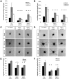
Figure 7. Silencing of Stab2 or Stab1 expression in endothelial cells inhibits endothelial sprouting and migration.
(A,B) siRNA mediated silencing of Stab2 (A) or Stab1 (B) inhibited basal, VEGF-A and VEGF-C driven in-gel sprouting in HUVECs. (C,D) Representative spheroids from the sprouting assay shown in (A,B). (E,F) Silencing of Stab2 (E) or Stab1 (F) in HUVECs inhibited basal and VEGF-A driven endothelial migration in the modified Boyden chamber assay. HUVECs were transfected with two different Stab2 or Stab1 siRNAs and allowed to migrate through a membrane for 3 h using 25 ng/ml VEGF-A. Migrated cells were stained and counted under the microscope. n = 3 per group. *p<0.05 vs. control siRNA.

Similar articles

Cited by

References

    1. Adams RH, Alitalo K (2007) Molecular regulation of angiogenesis and lymphangiogenesis. Nat Rev Mol Cell Biol 8: 464–478. - PubMed
    1. Tammela T, Alitalo K (2010) Lymphangiogenesis: Molecular mechanisms and future promise. Cell 140: 460–476. - PubMed
    1. Butler MG, Isogai S, Weinstein BM (2009) Lymphatic development. Birth Defects Res C Embryo Today 87: 222–231. - PMC - PubMed
    1. Shah N, Sukumar S (2010) The Hox genes and their roles in oncogenesis. Nat Rev Cancer 10: 361–371. - PubMed
    1. Cantile M, Schiavo G, Terracciano L, Cillo C (2008) Homeobox genes in normal and abnormal vasculogenesis. Nutr Metab Cardiovasc Dis 18: 651–658. - PubMed

Publication types

MeSH terms

Grants and funding

This work was supported by grants from the Deutsche Forschungsgemeinschaft (www.dfg.de) (SFB/TR23, project Z5, KR1887/5-1 to J.K.) and by the German Cardiac Society (http://dgk.org/)(to S.J.S.). The funders had no role in study design, data collection and analysis, decision to publish, or preparation of the manuscript.

LinkOut - more resources