Clinical Trial
. 2013 Mar 26;110(13):5091-6.
doi: 10.1073/pnas.1217197110. Epub 2013 Mar 11.

Inhibition of TFG function causes hereditary axon degeneration by impairing endoplasmic reticulum structure

Affiliations
Clinical Trial

Inhibition of TFG function causes hereditary axon degeneration by impairing endoplasmic reticulum structure

Christian Beetz et al. Proc Natl Acad Sci U S A. .

Abstract

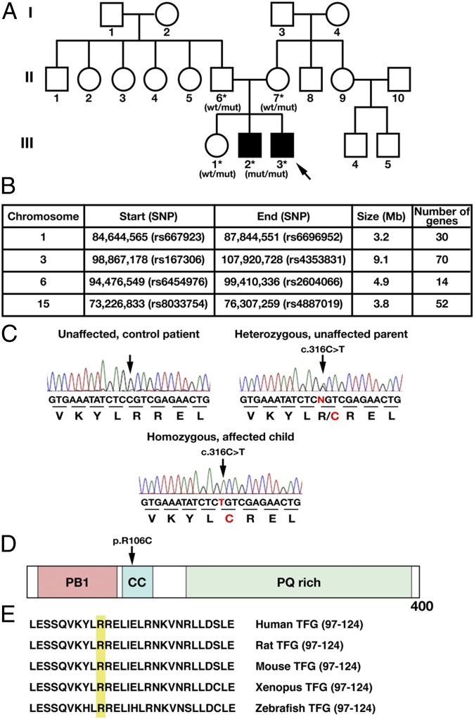
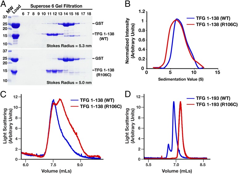
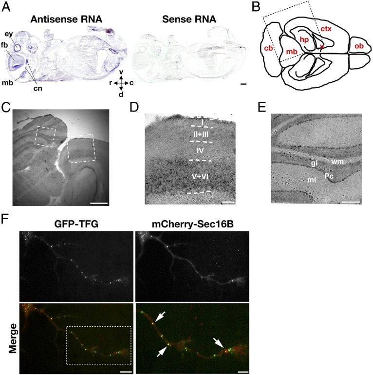
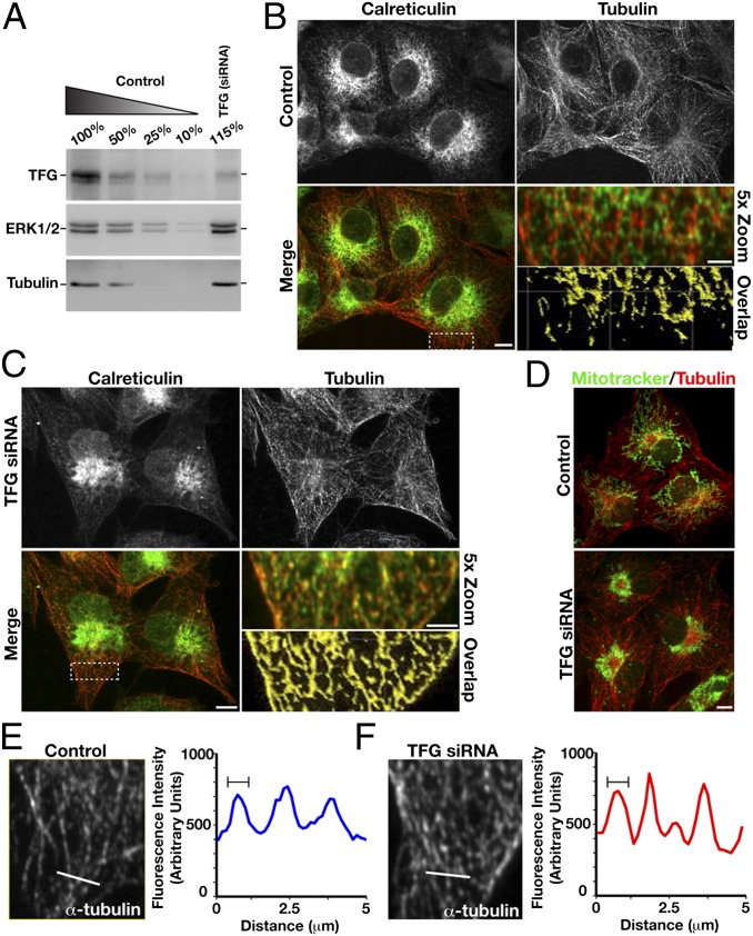
Hereditary spastic paraplegias are a clinically and genetically heterogeneous group of gait disorders. Their pathological hallmark is a length-dependent distal axonopathy of nerve fibers in the corticospinal tract. Involvement of other neurons can cause additional neurological symptoms, which define a diverse set of complex hereditary spastic paraplegias. We present two siblings who have the unusual combination of early-onset spastic paraplegia, optic atrophy, and neuropathy. Genome-wide SNP-typing, linkage analysis, and exome sequencing revealed a homozygous c.316C>T (p.R106C) variant in the Trk-fused gene (TFG) as the only plausible mutation. Biochemical characterization of the mutant protein demonstrated a defect in its ability to self-assemble into an oligomeric complex, which is critical for normal TFG function. In cell lines, TFG inhibition slows protein secretion from the endoplasmic reticulum (ER) and alters ER morphology, disrupting organization of peripheral ER tubules and causing collapse of the ER network onto the underlying microtubule cytoskeleton. The present study provides a unique link between altered ER architecture and neurodegeneration.


Conflict of interest statement

The authors declare no conflict of interest.

Figures

Fig. 1.
Fig. 1.
Association of a complicated form of hereditary spastic paraplegia with a homozygous missense alteration affecting a conserved residue of TFG. (A) Pedigree chart of a family affected by HSP. Filled and unfilled symbols indicate affected and unaffected individuals, respectively. Asterisks mark family members for whom DNA was available and who were applied in linkage analysis. Indicated below the identifiers are the results of TFG Sanger sequencing (mut, mutant allele; wt, WT allele). Arrow denotes the index case. (B) Runs of homozygosity >3 Mb and specific to the patients. (C) Exemplary results of Sanger sequencing of TFG exon 4 in an unaffected control patient, one of the affected parents, and the index case. (D) Schematic illustrating the domain organization of TFG. The alteration (p.R106C) detected in this study is highlighted. CC, coiled-coil domain; PB1, Phox and Bem1p domain; PQ rich, proline and glutamine-rich domain. (E) Amino acid sequence alignment of the TFG coiled-coil domain, showing the high level of conservation among different species.
Fig. 2.
Fig. 2.
Mutant p.R106C isoforms of TFG exhibit defects in normal oligomerization in vitro. (A) Purified GST-fused forms of WT and p.R106C TFG (amino acids 1–138) were cleaved in solution to remove the tag and separated by size-exclusion chromatography. Eluted fractions were separated by SDS/PAGE and stained with Coomassie stain. The Stokes radius of each protein was determined based on the elution profile of characterized standards. (B) Purified proteins described in A were applied to a glycerol density gradient (10–30%) and fractionated by hand. Individual fractions recovered were separated by SDS/PAGE and stained by using Coomassie stain. Densitometry measurements were made to quantify the amount of protein in each fraction, which is presented in a graphical format. (C and D) WT and p.R106C TFG (amino acids 1–138 and 1–193) were purified and examined by light scattering following size-exclusion chromatography. The lack of overlap between the peaks for each pair indicates that the oligomerization of TFG is altered in the presence of the R106C mutation.
Fig. 3.
Fig. 3.
TFG is highly expressed in the brain cortex and accumulates at ER exit sites in cultured murine neurons and human epithelial cells. (A) Sagittal section of a murine embryo (embryonic day 16) hybridized in situ with TFG-specific antisense (Left) and sense (Right) RNA probes. c, caudal; cn, cranial nerve ganglia; d, dorsal; ey, eye; fb, forebrain; mb, midbrain; r, rostral; v, ventral. (Scale bar, 1 mm.) (B) Cartoon illustrating a horizontal brain section from an adult mouse at Bregma − 3.4 mm. cb, cerebellum; ctx, cortex; hp, hippocampus; mb, midbrain; ob, olfactory bulb; v, ventricle. (C) TFG-specific in situ hybridization of an area corresponding to the region boxed in B. (Scale bar, 1 mm.) (D) Magnification of the cortical area marked by the right box in C. Roman numerals indicate cortical layers. (Scale bar, 200 µm.) (E) Magnification of the cerebellar area marked by left box in C. gl, granular layer; ml, molecular layer; Pc, Purkinje cell layer; wm, white matter. (Scale bar, 200 µm.) (F) Murine neurons transfected with plasmids encoding GFP-TFG and mCherry-Sec16B were imaged by using total internal reflection fluorescence microscopy. (Scale bar, 10 μm.) (Lower Right) A 1.8× zoom of the boxed region. Arrows highlight ER exit sites at which TFG and Sec16B colocalize. (Scale bar, 5 μm.)
Fig. 4.
Fig. 4.
Depletion of TFG disrupts ER architecture and distribution of mitochondria in mammalian cells. (A) Immunoblots of extracts prepared from COS7 cells depleted of TFG by siRNA. Serial dilutions of extracts prepared from control cells were loaded to quantify depletion levels. Blotting with antibodies directed against ERK1/2 and tubulin were performed to control loading. (BD) COS7 cells that were transfected with siRNAs targeting TFG or mock-transfected were fixed and stained by using antibodies directed against calreticulin and tubulin and imaged by using swept-field confocal optics. Cells were incubated with MitoTracker before fixation to stain mitochondria (D). (Scale bars, 5 μm.) Additionally, a 5× zoom of a boxed region of the peripheral ER (B and C) is provided. Color overlays show the ER (green) relative to microtubules (red). (Scale bars, 2 μm.) By using Imaris software, we specifically identified the areas of overlap in the boxed regions, which highlight differences in ER–microtubule associations in control cells compared with cells treated with TFG siRNA (B and C, Bottom Right). (E and F) Cropped images of microtubules in control (E) and TFG-depleted (F) cells. Line scan analysis (examined region highlighted with a 5-μm white line) was used to demonstrate that the thickness of peripheral microtubules, as determined by their fluorescence intensity, does not vary between the conditions. Data shown are representative of the population (n = 23 cells analyzed per condition).

Similar articles

Cited by

  • Autophagy in major human diseases.
    Klionsky DJ, Petroni G, Amaravadi RK, Baehrecke EH, Ballabio A, Boya P, Bravo-San Pedro JM, Cadwell K, Cecconi F, Choi AMK, Choi ME, Chu CT, Codogno P, Colombo MI, Cuervo AM, Deretic V, Dikic I, Elazar Z, Eskelinen EL, Fimia GM, Gewirtz DA, Green DR, Hansen M, Jäättelä M, Johansen T, Juhász G, Karantza V, Kraft C, Kroemer G, Ktistakis NT, Kumar S, Lopez-Otin C, Macleod KF, Madeo F, Martinez J, Meléndez A, Mizushima N, Münz C, Penninger JM, Perera RM, Piacentini M, Reggiori F, Rubinsztein DC, Ryan KM, Sadoshima J, Santambrogio L, Scorrano L, Simon HU, Simon AK, Simonsen A, Stolz A, Tavernarakis N, Tooze SA, Yoshimori T, Yuan J, Yue Z, Zhong Q, Galluzzi L, Pietrocola F. Klionsky DJ, et al. EMBO J. 2021 Oct 1;40(19):e108863. doi: 10.15252/embj.2021108863. Epub 2021 Aug 30. EMBO J. 2021. PMID: 34459017 Free PMC article. Review.
  • STIM1 Is Required for Remodeling of the Endoplasmic Reticulum and Microtubule Cytoskeleton in Steering Growth Cones.
    Pavez M, Thompson AC, Arnott HJ, Mitchell CB, D'Atri I, Don EK, Chilton JK, Scott EK, Lin JY, Young KM, Gasperini RJ, Foa L. Pavez M, et al. J Neurosci. 2019 Jun 26;39(26):5095-5114. doi: 10.1523/JNEUROSCI.2496-18.2019. Epub 2019 Apr 25. J Neurosci. 2019. PMID: 31023836 Free PMC article.
  • Unraveling the Role of Heme in Neurodegeneration.
    Chiabrando D, Fiorito V, Petrillo S, Tolosano E. Chiabrando D, et al. Front Neurosci. 2018 Oct 9;12:712. doi: 10.3389/fnins.2018.00712. eCollection 2018. Front Neurosci. 2018. PMID: 30356807 Free PMC article. Review.
  • Involvement of neuronal and muscular Trk-fused gene (TFG) defects in the development of neurodegenerative diseases.
    Yamamotoya T, Hasei S, Akasaka Y, Ohata Y, Nakatsu Y, Kanna M, Fujishiro M, Sakoda H, Ono H, Kushiyama A, Misawa H, Asano T. Yamamotoya T, et al. Sci Rep. 2022 Feb 4;12(1):1966. doi: 10.1038/s41598-022-05884-7. Sci Rep. 2022. PMID: 35121777 Free PMC article.
  • Spatial and temporal immunoreactivity in the rat brain using an affinity purified polyclonal antibody to DNSP-11.
    Sonne JWH, Groshong JS, Seavey C, Gash DM. Sonne JWH, et al. J Chem Neuroanat. 2019 Oct;100:101664. doi: 10.1016/j.jchemneu.2019.101664. Epub 2019 Aug 5. J Chem Neuroanat. 2019. PMID: 31394198 Free PMC article.

References

    1. Blackstone C, O’Kane CJ, Reid E. Hereditary spastic paraplegias: Membrane traffic and the motor pathway. Nat Rev Neurosci. 2011;12(1):31–42. - PMC - PubMed
    1. Schüle R, Schöls L. Genetics of hereditary spastic paraplegias. Semin Neurol. 2011;31(5):484–493. - PubMed
    1. Fink JK. Hereditary spastic paraplegia. Curr Neurol Neurosci Rep. 2006;6(1):65–76. - PubMed
    1. Salinas S, Proukakis C, Crosby A, Warner TT. Hereditary spastic paraplegia: clinical features and pathogenetic mechanisms. Lancet Neurol. 2008;7(12):1127–1138. - PubMed
    1. Ikenaka K, et al. Disruption of axonal transport in motor neuron diseases. Int J Mol Sci. 2012;13(1):1225–1238. - PMC - PubMed

Publication types

MeSH terms

LinkOut - more resources