. 2013 Apr 11;50(1):5-15.
doi: 10.1016/j.molcel.2013.01.039. Epub 2013 Mar 7.

STING recognition of cytoplasmic DNA instigates cellular defense

Affiliations

STING recognition of cytoplasmic DNA instigates cellular defense

Takayuki Abe et al. Mol Cell. .

Abstract

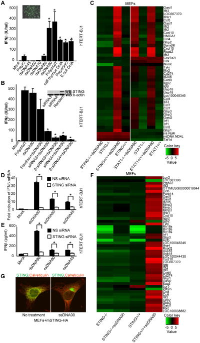
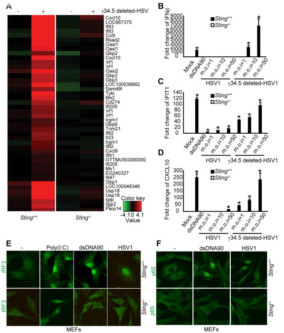
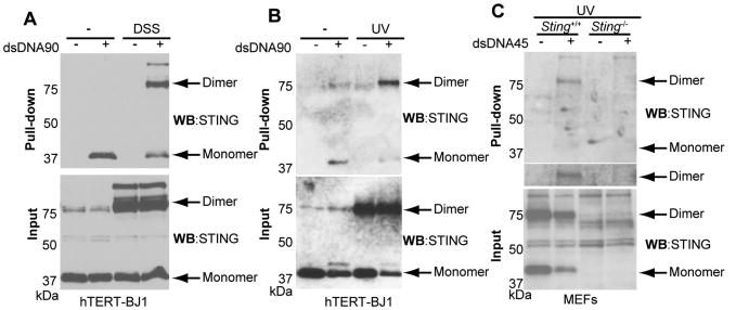
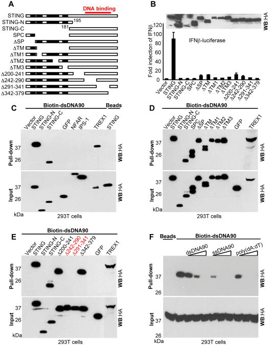
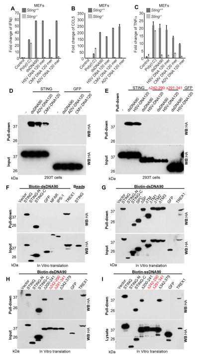
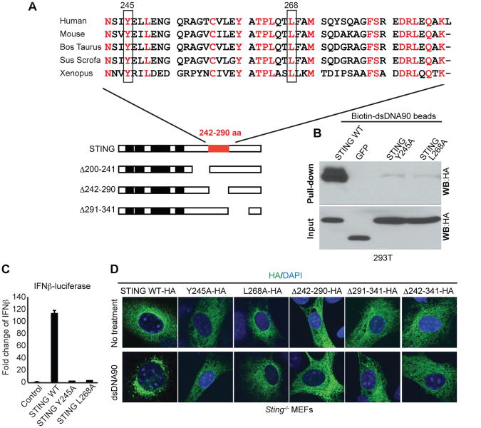
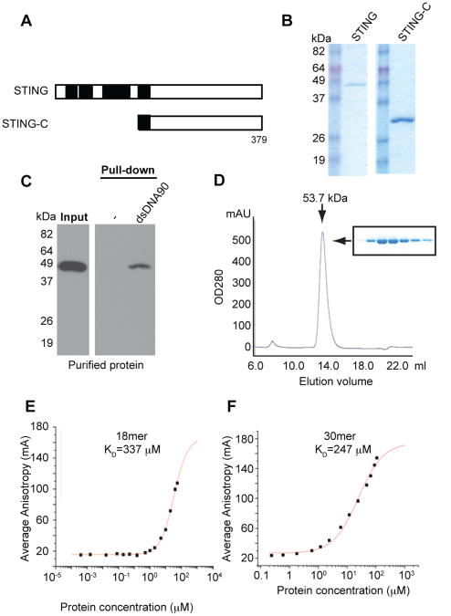
How the cell recognizes cytosolic DNA including DNA-based microbes to trigger host-defense-related gene activation remains to be fully resolved. Here, we demonstrate that STING (stimulator of interferon genes), an endoplasmic reticulum translocon-associated transmembrane protein, acts to detect cytoplasmic DNA species. STING homodimers were able to complex with self- (apoptotic, necrotic) or pathogen-related ssDNA and dsDNA and were indispensible for HSV-1-mediated transcriptional activation of a wide array of innate immune and proinflammatory genes in addition to type I IFN. Our data indicate that STING instigates cytoplasmic DNA-mediated cellular defense gene transcription and facilitates adoptive responses that are required for protection of the host. In contrast, chronic STING activation may manifest inflammatory responses and possibly autoimmune disease triggered by self-DNA.


Figures

Figure 1
Figure 1. STING controls cytosolic ssDNA and dsDNA innate signaling. See also Figure S1
(A) Human Telomerase Fibroblasts (hTERT-BJ1) were transfected with various nucleotides (3 μg/ml) for 16h. Endogenous IFNβ levels were measured. hTERT-BJ1 cells were transfected with FITC conjugated dsDNA90 and was examined by fluorescent microscopy to ensure efficient transfection. (B) hTERT-BJ1 cells were transfected with mock, random or two independent human STING siRNAs (siRNA 3 or 4) for 3 days followed by dsDNA90 transfection (3 μg/ml) for 16 hours. Endogenous IFNβ levels were measured. Silencing of hSTING protein was demonstrated by immunoblotting, with β-actin serving as a loading control. (C) Primary Sting+/+, Sting-/-, Stat1+/+ or Stat1-/- MEFs were transfected with or without dsDNA90 (3 μg/ml) for 3 hours. Total RNA was purified and examined for gene expression by Illumina Sentrix BeadChip Array (Mouse WG6 version 2). Most variable genes were selected. Rows represent individual genes; columns represent individual samples. Pseudo-colors indicate transcript levels below, equal to, or above the mean (green, black, and red, respectively). The scale represents the intensity of gene expression (log10 scale ranges between -5 and 5). (D) hTERT-BJ1 cells were treated with NS or STING siRNA. After 3 days, cells were treated with dsDNA90, ssDNA90 or ssDNA45 (3 μg/ml). IFNβ mRNA levels were measured by real time RT-PCR after 16 hours. (E) hTERT-BJ1 cells were treated with NS or STING siRNA. At 3 days, cells were treated with dsDNA90, ssDNA90 or ssDNA45 (3 μg/ml). IFNβ mRNA levels were measured after 16 hours. (F) Primary Sting+/+ or Sting-/- MEFs were transfected with or without ssDNA90 (3 μg/ml). After 3h, the same as c. (G) Primary MEFs with mSTING-HA were treated with or without ssDNA90 (3 μg/ml) for 3 hours and stained with anti-HA antibody (green) and calreticulin (red) as an ER marker. *P<0.05, Student's t-test. Error bars indicated s.d. Data are representative of at least two independent experiments.
Figure 2
Figure 2. STING is essential for HSV1-mediated innate immune signaling. See also Figure S6
(A) MEFs were infected with γ34.5 deleted-HSV1 (m.o.i=1) for 3h. Total RNA was purified and examined for gene expression using Illumina Sentrix BeadChip Array (Mouse WG6 version2). Most variable genes were selected. Rows represent individual genes; columns represent individual samples. Pseudo-colors indicate transcript levels below, equal to, or above the mean (green, black, and red, respectively). The scale represents the intensity of gene expression (log10 scale ranges between -4.1 and 4.1). (B) Sting+/+ or Sting-/- MEFs were treated with or without dsDNA, HSV1 or γ34.5 deleted-HSV1 for 3h. Total RNAs were purified and examined by real time PCR for IFNβ(B), IFIT1(C) or CXCL10(D). Error bars indicate s.d. (E) Sting+/+ or Sting-/- MEFs were treated with poly(I:C), dsDNA90 or HSV1 and cells were stained by anti-IRF3 antibody. PolyIC is RIG-I/MDA5 dependent and STING independent. (F) Sting+/+ or Sting-/- MEFs were treated with dsDNA90 or HSV1 and cells were stained by anit-p65 antibody at 3 hrs PI.
Figure 3
Figure 3. STING binds to DNA in vivo. See also Figure S2 and S3
(A) hTERT-BJ1 cells were transfected with biotin conjugated dsDNA90 (3 μg/ml) for 6h and treated with DSS. Lysates were precipitated using streptavidin agarose beads and analyzed by immunoblotting using anti-HA antibody. (B) hTERT-BJ1 cells were transfected with biotin conjugated dsDNA90 (3 μg/ml) for 6h and treated with UV. Same as A. (C) Sting+/+ or Sting-/- MEFs were transfected with biotin conjugated dsDNA45 and crosslinked by UV. Lysates were precipitated by streptavidin agarose beads and analyzed by immunoblotting.
Figure 4
Figure 4. STING binds to DNA in vitro. See also Figure S4
(A) Schematic of STING variants. (B) 293T cells were transfected with IFNβ-luciferase and STING variants and luciferase activity were measured. (C-E) 293T cells were transfected with the indicated plasmids. Cell lysates were precipitated with biotin conjugated dsDNA90 agarose beads and analyzed by immunoblotting using anti-HA antibody. (F) Full length STING-HA was expressed in 293T cells and lysates were incubated with biotin conjugated dsDNA90 agarose beads in the presence of dsDNA90, ssDNA90 or Poly(dA:dT) and analyzed by immunoblotting using anti-HA antibody.
Figure 5
Figure 5. STING binds to DNA. See also Figure S5
(A-C) Sting+/+ or Sting-/- MEFs were treated with poly(I:C), dsDNA90, HSV DNA 120mer, CMV DNA 120mer or ADV DNA 120mer for 3 hours. Total RNA was purified and examined by real time PCR for gene expression of IFNβ (A), CCL5(B) or TNFα(C). Error bars indicate s.d. (D-E) 293T cells were transfected with indicated plasmids. Cell lysates were precipitated with biotin-dsDNA90, biotin-ADV DNA 120mer, biotin-CMV DNA 120mer (D) or biotin-HSV DNA 120mer (E) agarose beads and analyzed by immunoblotting using anti-HA antibody. (F-H) In vitro translation products were incubated with biotin-dsDNA90 agarose beads and analyzed by immunoblotting using anti-HA antibody. (I) In vitro translation products were incubated with biotin-ssDNA90 agarose beads and analyzed by immunoblotting using anti-HA antibody.
Figure 6
Figure 6. Y245 and L268 are important for STING binding DNA. See also Figure S7
(A) Alignment of amino acid sequence of STING in 242-290aa. (B) 293T cells were transfected with STING WT-HA, GFP-HA, STING Y245A-HA or STING L268A-HA for 24 hours. Cell lysates were precipitated with biotin-dsDNA90 beads and analyzed by immunoblotting with anti HA antibody. (C) 293T cells were transfected with IFNβ-luciferase plasmid and plasmid encoding STING or mutants. After 24 hours, luciferase activity was measured. Error bars indicate s.d. (D) STING-/- MEFs were transfected with plasmid encoding STING or mutants using an Amaxa nucleofector apparatus (program A-023) with Amaxa MEF nucleofector kit 1 according to the manufacturer' s instructions. After 24 hours, the cells were treated with dsDNA90 for 6 hours and then stained using anti-HA (green) antibody and DAPI (blue).
Figure 7
Figure 7. STING-DNA binding analysis. See also Figure S5
(A) Schematic of STING variants. (B) Coomassie brilliant blue staining of purified hSTING-HA protein from 293T cells or purified hSTING-C-6xHis (aa152-379) protein from E.coli. (C) Purified hSTING-HA proteins were precipitated with biotin-conjugated dsDNA90 and analyzed by immunoblotting using STING antibody. (D) hSTING-C was highly purified by N-affinity chromatography and subjected to Superdex 200 GL 10/30 size-exclusion column. Our analysis indicated that purified hSTING-C has a molecular weight of 53.7 kDa, which is consistent with that predicted size of a dimer. (E-F) Direct binding of purified hSTING-C with 18mer (E) or 30mer (F) FAM labeled dsDNA oligonucleotide was determined using Fluorescence Polarization (FP) assays. The raw data of average fluorescence anisotropy is shown in a sigmoidal binding curve, with a calculated dissociation constant (KD) of 337 ± 26 μM and 247 ± 38 μM respectively.

Comment in

Similar articles

Cited by

References

    1. Ahn J, Gutman D, Saijo S, Barber GN. STING manifests self DNA-dependent inflammatory disease. Proceedings of the National Academy of Sciences of the United States of America. 2012;109:19386–19391. - PMC - PubMed
    1. Alnemri ES. Sensing cytoplasmic danger signals by the inflammasome. J Clin Immunol. 2010;30:512–519. - PMC - PubMed
    1. Brunette RL, Young JM, Whitley DG, Brodsky IE, Malik HS, Stetson DB. Extensive evolutionary and functional diversity among mammalian AIM2-like receptors. J Exp Med. 2012;209:1969–1983. - PMC - PubMed
    1. Burdette DL, Monroe KM, Sotelo-Troha K, Iwig JS, Eckert B, Hyodo M, Hayakawa Y, Vance RE. STING is a direct innate immune sensor of cyclic di-GMP. Nature. 2011;478:515–518. - PMC - PubMed
    1. Gall A, Treuting P, Elkon KB, Loo YM, Gale M, Jr, Barber GN, Stetson DB. Autoimmunity Initiates in Nonhematopoietic Cells and Progresses via Lymphocytes in an Interferon-Dependent Autoimmune Disease. Immunity. 2012;36:120–131. - PMC - PubMed

Publication types

MeSH terms

Associated data