. 2013 Apr;31(4):350-6.
doi: 10.1038/nbt.2537. Epub 2013 Mar 10.

Efficient and specific gene knockdown by small interfering RNAs produced in bacteria

Affiliations

Efficient and specific gene knockdown by small interfering RNAs produced in bacteria

Linfeng Huang et al. Nat Biotechnol. 2013 Apr.

Abstract

Synthetic small interfering RNAs (siRNAs) are an indispensable tool to investigate gene function in eukaryotic cells and may be used for therapeutic purposes to knock down genes implicated in disease. Thus far, most synthetic siRNAs have been produced by chemical synthesis. Here we present a method to produce highly potent siRNAs in Escherichia coli. This method relies on ectopic expression of p19, an siRNA-binding protein found in a plant RNA virus. When expressed in E. coli, p19 stabilizes an ∼21-nt siRNA-like species produced by bacterial RNase III. When mammalian cells are transfected by them, siRNAs that were generated in bacteria expressing p19 and a hairpin RNA encoding 200 or more nucleotides of a target gene reproducibly knock down target gene expression by ∼90% without immunogenicity or off-target effects. Because bacterially produced siRNAs contain multiple sequences against a target gene, they may be especially useful for suppressing polymorphic cellular or viral genes.


Figures
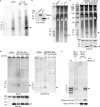
Figure 1
Figure 1
Ectopic p19 expression captures small RNAs in E. coli. (a) p19-coupled magnetic beads were incubated with total RNA isolated from mammalian ACH2 cells, or from E. coli cells that were either wild-type (WT) or transformed with a pcDNA3.1-p19 expression plasmid. Captured RNAs were 5' 32P-labeled, separated on a native polyacrylamide gel and detected by autoradiography. (b) FLAG-tagged p19 or TREX1, or empty vector (V), were expressed in E. coli. Left, anti-FLAG immunoblot. Right, total RNAs were separated on a denaturing polyacrylamide gel and stained with SYBR Gold. (c) Top, total RNAs purified from E. coli expressing an empty vector (V), or WT or mutant (Mut1: W39G, W42G and Mut2: K71A, R72G; mutants were defective in RNA binding) His-tagged p19 proteins were separated on a denaturing polyacrylamide gel and stained with SYBR Gold. Bottom, anti-His immunoblot. (d) p19-coupled magnetic beads were incubated with total RNA extracted from WT E. coli (DH5α or MG1655 Δlac) or RNase III mutant strains (rnc14 and rnc38 in MG1655 Δlac background) expressing or not His-tagged p19. p19-captured RNAs were separated on native (left) or denaturing (right) gels and stained with SYBR Gold. Bottom, anti-His immunoblot. The asterisk (*) indicates equal loading of a background band. (e) p19-coupled magnetic beads were incubated with total RNA extracted from E. coli WT BL21(DE3) cells or rnc14 mutant HT115(DE3) cells that were co-transfected with p19 and a vector encoding Flag-tagged E. coli RNase III (or empty vector). p19-bound RNAs were separated on a native polyacrylamide gel and stained with SYBR Gold. Bottom, anti-FLAG and ant-His immunoblots. Arrows indicate the ~21 nt small RNA band. M, markers. Data are representative of at least 2 independent experiments.
Figure 2
Figure 2
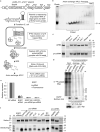
pro-siRNAs knockdown target gene expression. (a) Schematic of pGEX-4T-1-p19-T7 plasmid and the method to produce pro-siRNAs from E. coli. (b) Anion exchange HPLC fractions of SDS-eluted RNAs (isolated from E. coli engineered as in (a) to express pro-siRNAs) were separated on a native polyacrylamide gel and stained with SYBR Gold. HPLC fractions containing only ~21 nt small RNAs were combined (“Pooled”) for use in subsequent experiments. (c) Synthetic siRNAs (top) and HPLC purified pro-siRNAs (bottom) were incubated with indicated nucleases and separated on a native polyacrylamide gel stained with SYBR Gold. -, no nuclease. (d) Anti-Ago mAb 2A8 or mouse total IgG were used to immunoprecipitate RNAs from HeLa-d1EGFP cells transfected with negative control (NC) siRNA or EGFPFL pro-siRNAs. Immunoprecipitated RNAs were analyzed by 5' 32P end-labeling (top) or by Northern blot using a probe complementary to the full length EGFP coding sequence (middle). Bottom, anti-Ago immunoblot. (e) HeLa-d1EGFP cells were transfected with 4 nM of synthetic EGFP siRNA or with pro-siRNAs. EGFP expression was measured by qRTPCR (mRNA) and flow cytometry (fluorescence). Data are normalized to cells treated with negative control (NC) siRNA. mRNA level is relative to GAPDH. Data are mean±SD of 2 (qRT-PCR) and 3 (EGFP fluorescence) independent experiments. (f) HPLC-purified pro-siRNAs or synthetic siRNAs (EGFP and NC) were separated on native (top) or denaturing (bottom) polyacrylamide gels stained with SYBR Gold. M, markers. Data are representative of at least 2 independent experiments.
Figure 3
Figure 3
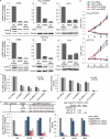
pro-siRNA-mediated knockdown of endogenous and viral gene expression in human cells. (a) Synthetic siRNAs or pro-siRNAs specific for the indicated target genes or negative control (NC) siRNA were transfected (4 nM) into HeLa-d1EGFP (top) or HCT116 (bottom) cells. Target gene expression was measured by qRT-PCR (bar graphs) or immunoblots (tubulin is loading control). (b) HeLa-d1EGFP (top) or HCT116 (bottom) cells were transfected with PLK1 siRNA or pro-siRNA (4 nM) or NC siRNA or EGFP pro-siRNA as nontargeting controls, respectively. Cells were counted at indicated time points. (c) HeLa-CD4 cells were transfected with vif siRNAs (vif siRNA-1 and vif siRNA-2, alone or together) or vif pro-siRNA 24 hr before HIV-1 infection. Left, vif mRNA was measured by qRT-PCR 36 hr after infection. Right, infectivity of culture supernatants from transfected HeLa-CD4 cells was measured by HIV-1 tat-driven expression of a luciferase reporter gene in TZM-bl assay. (d) Top, sequence of the gag siRNA (complementary to HIV-1 clade B) used here; corresponding sequences in clade A UG29 and clade C IN22 strains are shown together (left) with the proportion of identical bases in the entire gag coding region for the 3 isolates tested (right). Bottom, HeLa-CD4 cells were infected with HIV-1 clade B (IIIB), U87.CD4.CXCR4 cells were infected with HIV-1 clade A (UG29) and U87.CD4.CCR5 cells were infected with HIV-1 clade C (IN22). Cells were transfected with 20 nM of the indicated synthetic or pro-siRNA 12 hr before HIV-1 infection. Left, gag mRNA was measured by qRT-PCR 36 hr after infection. Right, infectivity of culture supernatants was measured by TZM-bl assay. Data are mean±SD of 3 (a–c) and 2 (d) independent experiments. mRNA expression and TZM-bl luciferase data are normalized to cells transfected with NC siRNA.
Figure 4
Figure 4
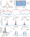
pro-siRNA sequences and assessment of off-target effects. (a) Length distribution of EGFPFL, EGFP100 and LMNA pro-siRNAs assessed by deep sequencing. (b) Percentage of deep sequencing reads aligning to the target gene hairpin, the E. coli genome or the plasmid backbone. (c) pro-siRNA deep sequencing read alignment to indicated positions of the EGFPFL, EGFP100 and LMNA target genes (sense and antisense). (d) Volcano plots of expression changes versus p value of all annotated transcripts detected by RNA deep sequencing in HeLa-d1EGFP cells transfected with EGFP synthetic siRNA or EGFP100 or EGFPFL pro-siRNAs relative to expression in cells transfected with a negative control (NC) siRNA. Arrows indicate EGFP and the numbers indicate its fold change (log2). Cut-off for significance is q value<0.05 (the default in Cufflinks). (e) Percent of significantly changed transcripts in (d and f). Numbers on top of bars indicate exact percentages. Significantly changed genes are listed in Supplementary Table 4. (f) Volcano plots of expression changes (1.2 fold less or more) versus p value detected by microarray in HeLa-d1EGFP cells transfected with LMNA synthetic siRNA or pro-siRNAs relative to expression in cells transfected with a NC siRNA. Arrows indicate LMNA and the numbers indicate its fold change (log2). Cut-off for significance is p<0.05 (paired T-test by dChip).

Comment in

  • Renewable RNAi.
    Blau JA, McManus MT. Blau JA, et al. Nat Biotechnol. 2013 Apr;31(4):319-20. doi: 10.1038/nbt.2547. Nat Biotechnol. 2013. PMID: 23563428 No abstract available.

Similar articles

Cited by

References

    1. Elbashir SM, et al. Duplexes of 21-nucleotide RNAs mediate RNA interference in cultured mammalian cells. Nature. 2001;411:494–498. - PubMed
    1. Caplen NJ, Parrish S, Imani F, Fire A, Morgan RA. Specific inhibition of gene expression by small double-stranded RNAs in invertebrate and vertebrate systems. Proc Natl Acad Sci U S A. 2001;98:9742–9747. - PMC - PubMed
    1. Rettig GR, Behlke MA. Progress toward in vivo use of siRNAs-II. Mol Ther. 2012;20:483–512. - PMC - PubMed
    1. Voinnet O, Pinto YM, Baulcombe DC. Suppression of gene silencing: a general strategy used by diverse DNA and RNA viruses of plants. Proc Natl Acad Sci U S A. 1999;96:14147–14152. - PMC - PubMed
    1. Silhavy D, et al. A viral protein suppresses RNA silencing and binds silencing-generated, 21- to 25-nucleotide double-stranded RNAs. Embo J. 2002;21:3070–3080. - PMC - PubMed

Publication types

Associated data