

Met is the most frequently amplified gene in endometriosis-associated ovarian clear cell adenocarcinoma and correlates with worsened prognosis
- PMID: 23469222
- PMCID: PMC3587638
- DOI: 10.1371/journal.pone.0057724
Met is the most frequently amplified gene in endometriosis-associated ovarian clear cell adenocarcinoma and correlates with worsened prognosis
Abstract
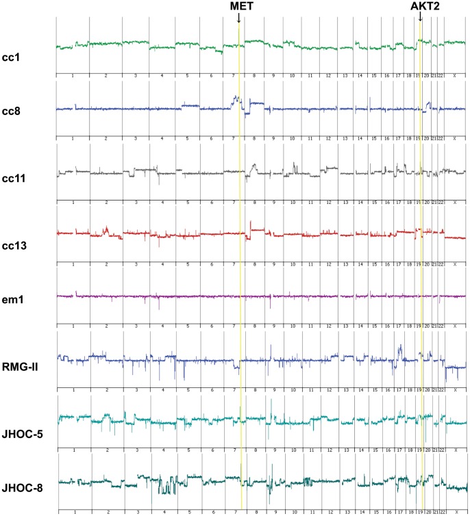
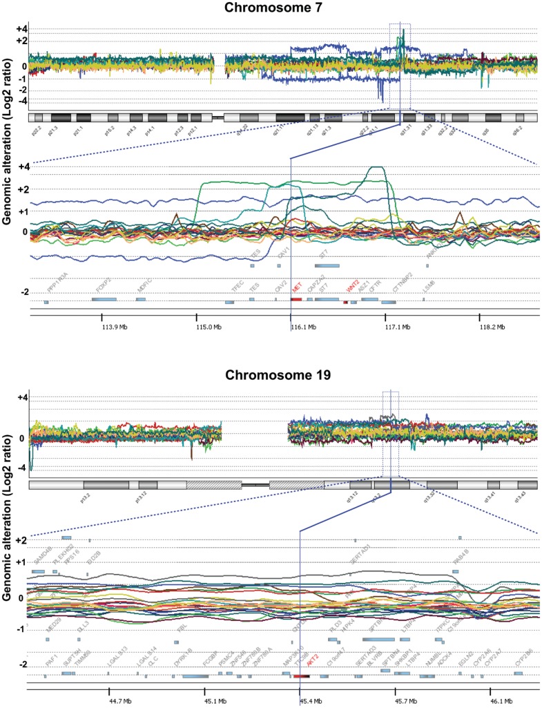
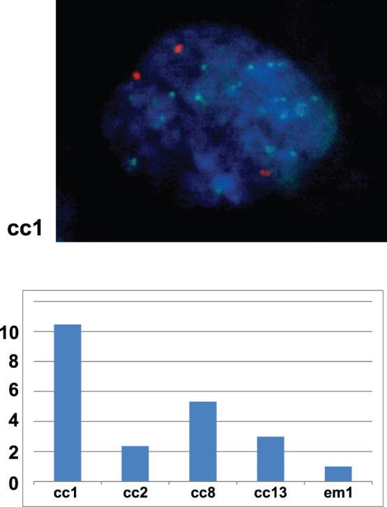
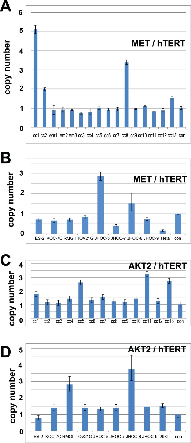
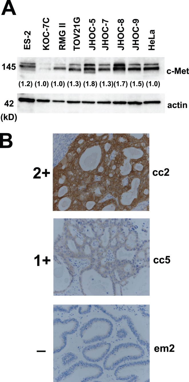
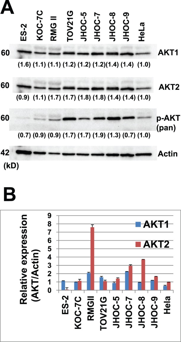
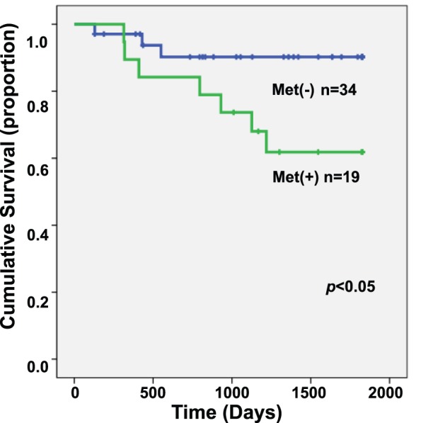
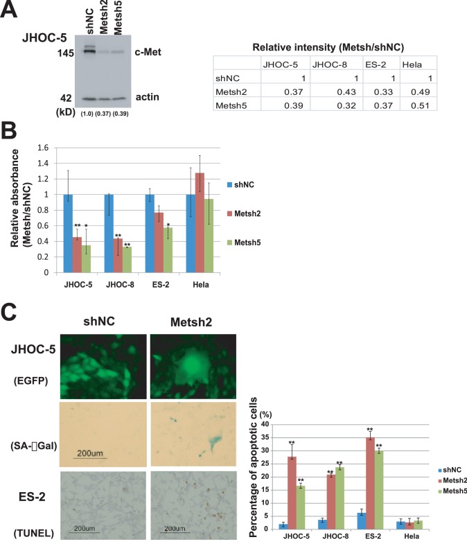
Clear cell adenocarcinoma of the ovary (OCC) is a chemo-resistant tumor with a relatively poor prognosis and is frequently associated with endometriosis. Although it is assumed that oxidative stress plays some role in the malignant transformation of this tumor, the characteristic molecular events leading to carcinogenesis remain unknown. In this study, an array-based comparative genomic hybridization (CGH) analysis revealed Met gene amplification in 4/13 OCC primary tumors and 2/8 OCC cell lines. Amplification of the AKT2 gene, which is a downstream component of the Met/PI3K signaling pathway, was also observed in 5/21 samples by array-based CGH analysis. In one patient, both the Met and AKT2 genes were amplified. These findings were confirmed using fluorescence in situ hybridization, real-time quantitative PCR, immunoblotting, and immunohistochemistry. In total, 73 OCC cases were evaluated using real-time quantitative PCR; 37.0% demonstrated Met gene amplification (>4 copies), and 8.2% had AKT2 amplification. Furthermore, stage 1 and 2 patients with Met gene amplification had significantly worse survival than patients without Met gene amplification (p<0.05). Met knockdown by shRNA resulted in reduced viability of OCC cells with Met amplification due to increased apoptosis and cellular senescence, suggesting that the Met signaling pathway plays an important role in OCC carcinogenesis. Thus, we believe that targeted inhibition of the Met pathway may be a promising treatment for OCC.
Conflict of interest statement
Figures
Similar articles
-
Accumulative copy number increase of MET drives tumor development and histological progression in a subset of ovarian clear-cell adenocarcinomas.Mod Pathol. 2012 Jan;25(1):122-30. doi: 10.1038/modpathol.2011.143. Epub 2011 Oct 7. Mod Pathol. 2012. PMID: 21983935
-
Gene amplification and protein overexpression of MET are common events in ovarian clear-cell adenocarcinoma: their roles in tumor progression and prognostication of the patient.Mod Pathol. 2011 Aug;24(8):1146-55. doi: 10.1038/modpathol.2011.70. Epub 2011 Apr 8. Mod Pathol. 2011. PMID: 21478826
-
MicroRNA-21 is a candidate driver gene for 17q23-25 amplification in ovarian clear cell carcinoma.BMC Cancer. 2014 Nov 3;14:799. doi: 10.1186/1471-2407-14-799. BMC Cancer. 2014. PMID: 25366985 Free PMC article.
-
Ovarian cancer: new developments in clear cell carcinoma and hopes for targeted therapy.Jpn J Clin Oncol. 2015 May;45(5):405-7. doi: 10.1093/jjco/hyu221. Epub 2015 Jan 12. Jpn J Clin Oncol. 2015. PMID: 25583423 Free PMC article. Review.
-
ARID1A mutations and PI3K/AKT pathway alterations in endometriosis and endometriosis-associated ovarian carcinomas.Int J Mol Sci. 2013 Sep 12;14(9):18824-49. doi: 10.3390/ijms140918824. Int J Mol Sci. 2013. PMID: 24036443 Free PMC article. Review.
Cited by
-
Cigarette smoke enhances oncogene addiction to c-MET and desensitizes EGFR-expressing non-small cell lung cancer to EGFR TKIs.Mol Oncol. 2018 May;12(5):705-723. doi: 10.1002/1878-0261.12193. Epub 2018 Apr 14. Mol Oncol. 2018. PMID: 29570930 Free PMC article.
-
Iron and thiols as two major players in carcinogenesis: friends or foes?Front Pharmacol. 2014 Aug 28;5:200. doi: 10.3389/fphar.2014.00200. eCollection 2014. Front Pharmacol. 2014. PMID: 25221514 Free PMC article. Review.
-
c-Met inhibitors attenuate tumor growth of small cell hypercalcemic ovarian carcinoma (SCCOHT) populations.Oncotarget. 2015 Oct 13;6(31):31640-58. doi: 10.18632/oncotarget.5151. Oncotarget. 2015. PMID: 26436697 Free PMC article.
-
Interplay Between MicroRNAs and Oxidative Stress in Ovarian Conditions with a Focus on Ovarian Cancer and Endometriosis.Int J Mol Sci. 2019 Oct 25;20(21):5322. doi: 10.3390/ijms20215322. Int J Mol Sci. 2019. PMID: 31731537 Free PMC article. Review.
-
The target therapy of ovarian clear cell carcinoma.Onco Targets Ther. 2014 Sep 23;7:1647-52. doi: 10.2147/OTT.S49993. eCollection 2014. Onco Targets Ther. 2014. PMID: 25285014 Free PMC article. Review.
References
-
- Mandai M, Yamaguchi K, Matsumura N, Baba T, Konishi I (2009) Ovarian cancer in endometriosis: molecular biology, pathology, and clinical management. Int J Clin Oncol 14: 383–391. - PubMed
-
- Yamaguchi K, Mandai M, Toyokuni S, Hamanishi J, Higuchi T, et al. (2008) Contents of endometriotic cysts, especially the high concentration of free iron, are a possible cause of carcinogenesis in the cysts through the iron-induced persistent oxidative stress. Clin Cancer Res 14: 32–40. - PubMed
Publication types
MeSH terms
Substances
Grants and funding
LinkOut - more resources
Full Text Sources
Other Literature Sources
Medical
Research Materials
Miscellaneous
