Shaping organisms with apoptosis
- PMID: 23449394
- PMCID: PMC3619238
- DOI: 10.1038/cdd.2013.11
Shaping organisms with apoptosis
Abstract
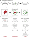
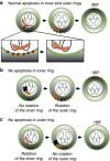
Programmed cell death is an important process during development that serves to remove superfluous cells and tissues, such as larval organs during metamorphosis, supernumerary cells during nervous system development, muscle patterning and cardiac morphogenesis. Different kinds of cell death have been observed and were originally classified based on distinct morphological features: (1) type I programmed cell death (PCD) or apoptosis is recognized by cell rounding, DNA fragmentation, externalization of phosphatidyl serine, caspase activation and the absence of inflammatory reaction, (2) type II PCD or autophagy is characterized by the presence of large vacuoles and the fact that cells can recover until very late in the process and (3) necrosis is associated with an uncontrolled release of the intracellular content after cell swelling and rupture of the membrane, which commonly induces an inflammatory response. In this review, we will focus exclusively on developmental cell death by apoptosis and its role in tissue remodeling.
Figures


Similar articles
-
Programmed cell death and cancer.Postgrad Med J. 2009 Mar;85(1001):134-40. doi: 10.1136/pgmj.2008.072629. Postgrad Med J. 2009. PMID: 19351640 Review.
-
Spreading the word: non-autonomous effects of apoptosis during development, regeneration and disease.Development. 2015 Oct 1;142(19):3253-62. doi: 10.1242/dev.127878. Development. 2015. PMID: 26443630 Free PMC article. Review.
-
Ecdysone-mediated programmed cell death in Drosophila.Int J Dev Biol. 2015;59(1-3):23-32. doi: 10.1387/ijdb.150055sk. Int J Dev Biol. 2015. PMID: 26374522 Review.
-
Distinct requirements of Autophagy-related genes in programmed cell death.Cell Death Differ. 2015 Nov;22(11):1792-802. doi: 10.1038/cdd.2015.28. Epub 2015 Apr 17. Cell Death Differ. 2015. PMID: 25882046 Free PMC article.
-
Autophagy: paying Charon's toll.Cell. 2007 Mar 9;128(5):833-6. doi: 10.1016/j.cell.2007.02.023. Cell. 2007. PMID: 17350570 Review.
Cited by
-
Caspase-Independent Regulated Necrosis Pathways as Potential Targets in Cancer Management.Front Oncol. 2021 Feb 16;10:616952. doi: 10.3389/fonc.2020.616952. eCollection 2020. Front Oncol. 2021. PMID: 33665167 Free PMC article. Review.
-
Large-scale death of retinal astrocytes during normal development is non-apoptotic and implemented by microglia.PLoS Biol. 2019 Oct 18;17(10):e3000492. doi: 10.1371/journal.pbio.3000492. eCollection 2019 Oct. PLoS Biol. 2019. PMID: 31626642 Free PMC article.
-
Apoptosis in Living Animals Is Assisted by Scavenger Cells and Thus May Not Mainly Go through the Cytochrome C-Caspase Pathway.J Cancer. 2013 Nov 15;4(9):716-23. doi: 10.7150/jca.7577. eCollection 2013. J Cancer. 2013. PMID: 24312141 Free PMC article.
-
K45A mutation of RIPK1 results in poor necroptosis and cytokine signaling in macrophages, which impacts inflammatory responses in vivo.Cell Death Differ. 2016 Oct;23(10):1628-37. doi: 10.1038/cdd.2016.51. Epub 2016 Jun 3. Cell Death Differ. 2016. PMID: 27258786 Free PMC article.
-
Preparation and characterization of nanoclay-hydrogel composite support-bath for bioprinting of complex structures.Sci Rep. 2020 Mar 24;10(1):5257. doi: 10.1038/s41598-020-61606-x. Sci Rep. 2020. PMID: 32210259 Free PMC article.
References
-
- Potts MB, Cameron S. Cell lineage and cell death: Caenorhabditis elegans and cancer research. Nat Rev Cancer. 2010;11:50–58. - PubMed
-
- Zakeri Z, Quaglino D, Ahuja HS. Apoptotic cell death in the mouse limb and its suppression in the hammertoe mutant. Dev Biol. 1994;165:294–297. - PubMed
-
- Coleman ML, Sahai EA, Yeo M, Bosch M, Dewar A, Olson MF. Membrane blebbing during apoptosis results from caspase-mediated activation of ROCK I. Nat Cell Biol. 2001;3:339–345. - PubMed
-
- Crawford ED, Wells JA. Caspase substrates and cellular remodeling. Ann Rev Biochem. 2011;80:1055–1087. - PubMed
Publication types
MeSH terms
Grants and funding
LinkOut - more resources
Full Text Sources
Other Literature Sources
Molecular Biology Databases

