. 2013;8(2):e56336.
doi: 10.1371/journal.pone.0056336. Epub 2013 Feb 18.

Identification of a novel protein-protein interaction motif mediating interaction of GPCR-associated sorting proteins with G protein-coupled receptors

Affiliations

Identification of a novel protein-protein interaction motif mediating interaction of GPCR-associated sorting proteins with G protein-coupled receptors

Olivier Bornert et al. PLoS One. 2013.

Abstract

GPCR desensitization and down-regulation are considered key molecular events underlying the development of tolerance in vivo. Among the many regulatory proteins that are involved in these complex processes, GASP-1 have been shown to participate to the sorting of several receptors toward the degradation pathway. This protein belongs to the recently identified GPCR-associated sorting proteins (GASPs) family that comprises ten members for which structural and functional details are poorly documented. We present here a detailed structure-function relationship analysis of the molecular interaction between GASPs and a panel of GPCRs. In a first step, GST-pull down experiments revealed that all the tested GASPs display significant interactions with a wide range of GPCRs. Importantly, the different GASP members exhibiting the strongest interaction properties were also characterized by the presence of a small, highly conserved and repeated "GASP motif" of 15 amino acids. We further showed using GST-pull down, surface plasmon resonance and co-immunoprecipitation experiments that the central domain of GASP-1, which contains 22 GASP motifs, is essential for the interaction with GPCRs. We then used site directed mutagenesis and competition experiments with synthetic peptides to demonstrate that the GASP motif, and particularly its highly conserved core sequence SWFW, is critically involved in the interaction with GPCRs. Overall, our data show that several members of the GASP family interact with GPCRs and highlight the presence within GASPs of a novel protein-protein interaction motif that might represent a new target to investigate the involvement of GASPs in the modulation of the activity of GPCRs.


Conflict of interest statement

Competing Interests: O. Bornert is an employee of the company BioXtal. This does not alter the authors’ adherence to all the PLOS ONE policies on sharing data and materials.

Figures

Figure 1
Figure 1. Schematic comparison of GASP family members.
Black boxes represent the conserved carboxyl-terminal domain of 250 amino acids. The percentage of identical amino acids shared with GASP-1 is indicated within each box. Small grey boxes represent a highly conserved motif of 15 amino acids that is repeated 22 times in GASP-1 and two times in GASP-2 to -5. The consensus sequence of this motif is: (E/D/G) (E/D) E X (I/L/V/S/T) (I/V/A/F) (G/N) (S/T) W F W (A/V/T/S/D/E) (G/E/R) (E/D/K) (E/D/K/A/Q). For GASP-2, two regions showing significant sequence homology with GASP-1 are separated by a gap represented by dotted lines. GASPs accession numbers from SPtrEMBL database are indicated on the left of the figure.
Figure 2
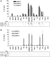
Figure 2. GST-pull down experiments with radiolabelled GASP-1, -2, -3, -6, -7 and -9 and GST-fused receptor C-tails.
A, GASP-1, -2 and -3 showed medium to strong interactions with some GPCR C-tails but no interaction was detected with the two one-transmembrane receptor C-tails (TGFβ and IGF1). B, GASP-7 showed weak to medium interactions with some GPCR C-tails. GASP-6 and -9 showed very weak interactions with all tested receptors. No interaction was detected with TGFβ and IGF C-tails. Data were quantified by Phosphor-imaging. Results are shown as percent of the [35S]-GASPs input retained by the GST-fused receptor C-tails and correspond to the mean ± S.E.M of three independent experiments. Lower panels correspond to representative gel images. 5HT7, 5-hydroxytryptamine 7 receptor; ADRB1, β1 adrenergic receptor; CALCR, calcitonin receptor; DOR, δ-opioid receptor; FZ4, frizzled 4 receptor; H2, histamine 2 receptor; IGF1, insulin growth factor I receptor; KOR, κ-opioid receptor; M1, muscarinic M1 acetylcholine receptor; M2, muscarinic M2 acetylcholine receptor; MOR, µ-opioid receptor; ORL1, opioid receptor-like 1; TXA2, α isoform of the thromboxane A2 receptor; TGFβ, type III transforming growth factor β receptor.
Figure 3
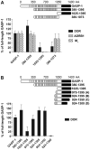
Figure 3. Two portions of GASP-1 are implicated in the interaction with GPCRs.
A, GST-pull down experiments with three truncated mutants of GASP-1 and DOR, ADRB1 and M1 C-tails. Deletion analysis revealed the critical role played by the central part of GASP-1 (380–1073) in the interaction with the DOR, ADRB1 and M1 C-tails and especially with ADRB1 and M1. Surprisingly, the conserved carboxyl-terminal part of GASP-1 (1025–1395) displayed no significant interaction with these receptor C-tails, except DOR for which a 25% interaction was observed. B, GST-pull down experiments with additional truncated mutants of GASP-1 and DOR C-tail. Detailed deletion analysis showed that the interaction with DOR C-tail required the entire carboxyl-terminal part as well as the central part of GASP-1. Results are represented as percent of the full-length GASP-1 interaction and correspond to the mean ± S.E.M of three independent experiments.
Figure 4
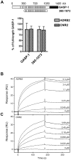
Figure 4. Purified full-length GPCRs dose-dependently bind to the central domain of GASP-1 in SPR experiments.
A, Interaction of the central domain of GASP-1 compared to the full-length protein with GST-fused ADRB2 and CNR2 C-tails by GST pull down experiments. The results show that both receptors interact in vitro with GASP-1 and that the central part of GASP-1 is strongly involved in the interaction with ADRB2 and CNR2. B, Binding of a range of concentrations of ADRB2 to the central domain of GASP-1. C, Binding of a range of concentrations of CNR2 to the central domain of GASP-1. Overall, we observed a dose-dependent binding of ADRB2 and CNR2 with the central domain of GASP-1. The receptor concentrations are indicated on the figures. All curves are double referenced and corrected for changes in capture density of the central domain of GASP-1. ADRB2, β2 adrenergic receptor; CNR2, cannabinoid receptor type 2.
Figure 5
Figure 5. The central domain of GASP-1 co-immunoprecipitates with GPCRs in cells.
The central domain of GASP-1 (amino-acids 380 to 1073 of GASP-1 in pcDNA3.1) was transiently transfected in HEK293 cells stably expressing GFP-tagged ADRB1, ADRB2, CALCR or M1 receptor. HEK293 cells stably expressing MyrPalm-mYFP and transiently transfected with the central domain of GASP-1 were used as a negative control. The central domain of GASP-1 co-immunoprecipitated with the four different GPCRs while no co-immunoprecipation was observed in cells expressing the central domain of GASP-1 alone or co-expressing this domain with myristoylated-palmitoylated mYFP (MyrPalm-mYFP).
Figure 6
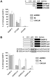
Figure 6. The GASP motif is critical for the interaction of GASP-2 with GPCRs.
A, GST-pull down experiments with two truncated mutants of GASP-2 and ADRB1, M1 and CALCR C-tails. Grey boxes represent the 15 AA GASP motifs. Deletion analysis revealed that the central domain of GASP-2, which contains the two GASP motifs, is critical for the interaction between GASP-2 and ADRB1, M1 and CALCR C-tails. B, GST-pull down experiments with full-length GASP-2 where one (GASP2-m1 and GASP2-m2) or both GASP motifs (GASP2-dm) were mutated. Grey boxes represent the wild-type motifs and X represent the mutant motifs. Consensus sequences are given for wild-type and mutant motifs. Mutated amino acids are underlined. Site directed mutagenesis analysis of these two repeated motifs showed that they played a crucial role in the interaction of GASP-2 with the three receptor C-tails tested here. Results are shown as percent of the wild-type GASP-2 interaction and correspond to the mean ± S.E.M of three independent experiments.
Figure 7
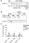
Figure 7. A small synthetic peptide derived from the GASP motif of GASP-2 blocks the interaction between GASPs and GPCR C-tails in GST-pull down experiments.
A, GASP peptide competes for the interaction between GASP-2 and GST-fused ADRB1 C-tail. The scrambled peptide displayed no significant effect on the interaction between GASP-2 and ADRB1. B, A fixed concentration of GASP peptide (150 µM) inhibits the interaction between GASP-1, -2 or -7 with ADRB1 C-tail, but not the scrambled peptide. C, Phosphor-imaging quantification of the competition experiments for the interaction between GASP-1, -2 and -7 and four different receptor C-tails with GASP peptide. A fixed concentration of GASP peptide (150 µM) strongly inhibited interactions of GASPs with ADRB1, M1, CALCR and TXA2 C-tails. Results are represented as percent of the interaction between the corresponding GASPs and GPCRs in absence of peptide (mean ± S.E.M of three independent experiments).
Figure 8
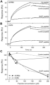
Figure 8. GASP peptide prevents receptor binding to the central domain of GASP-1 in SPR experiments.
A, Binding of 1.1 µM ADRB2 alone or preincubated with either 250 µM GASP peptide or 250 µM control peptide to captured central domain of GASP-1. B, Binding of 0.30 µM CNR2 alone or preincubated with either 250 µM GASP peptide or 250 µM control peptide to captured central domain of GASP-1. All curves are double referenced and corrected for changes in captured GASP density. C, Endpoint responses from competition binding curves for 1.1 µM ADRB2 preincubated with a range of concentrations of either GASP peptide (▪) or control peptide (•) and 0.30 µM CNR2 preincubated with either GASP peptide (□) or control peptide (○). The responses are normalized to the endpoint response from an injection with receptor only (0 µM peptide).

Similar articles

Cited by

References

    1. Ferguson SS (2001) Evolving concepts in G protein-coupled receptor endocytosis: the role in receptor desensitization and signaling. Pharmacol Rev 53: 1–24. - PubMed
    1. Cao TT, Deacon HW, Reczek D, Bretscher A, von Zastrow M (1999) A kinase-regulated PDZ-domain interaction controls endocytic sorting of the beta2-adrenergic receptor. Nature 401: 286–290. - PubMed
    1. Li JG, Chen C, Liu-Chen LY (2002) Ezrin-radixin-moesin-binding phosphoprotein-50/Na+/H+ exchanger regulatory factor (EBP50/NHERF) blocks U50,488H-induced down-regulation of the human kappa opioid receptor by enhancing its recycling rate. J Biol Chem 277: 27545–27552. - PubMed
    1. Cong M, Perry SJ, Hu LA, Hanson PI, Claing A, et al. (2001) Binding of the beta2 adrenergic receptor to N-ethylmaleimide-sensitive factor regulates receptor recycling. J Biol Chem 276: 45145–45152. - PubMed
    1. Gullapalli A, Wolfe BL, Griffin CT, Magnuson T, Trejo J (2006) An essential role for SNX1 in lysosomal sorting of protease-activated receptor-1: evidence for retromer-, Hrs-, and Tsg101-independent functions of sorting nexins. Mol Biol Cell 17: 1228–1238. - PMC - PubMed

Publication types

MeSH terms

Grants and funding

This work was supported by Ministère de l’Education Nationale de l’Enseignement Supérieur et de la Recherche (MENSER) fellowship, Association pour la Recherche sur le Cancer (N°3423), Fondation pour la Recherche Médicale, Centre National de la Recherche Scientifique, Université Louis Pasteur, the Lundbeck Foundation Center for Biomembranes and Nanomedicine, and the programs BioScaRT and UNIK Synthetic Biology. The funders had no role in study design, data collection and analysis, decision to publish, or preparation of the manuscript.