. 2013 Mar 4;200(5):619-33.
doi: 10.1083/jcb.201211069. Epub 2013 Feb 25.

Arp2/3 complex ATP hydrolysis promotes lamellipodial actin network disassembly but is dispensable for assembly

Affiliations

Arp2/3 complex ATP hydrolysis promotes lamellipodial actin network disassembly but is dispensable for assembly

Elena Ingerman et al. J Cell Biol. .

Abstract

We examined the role of ATP hydrolysis by the Arp2/3 complex in building the leading edge of a cell by studying the effects of hydrolysis defects on the behavior of the complex in the lamellipodial actin network of Drosophila S2 cells and in a reconstituted, in vitro, actin-based motility system. In S2 cells, nonhydrolyzing Arp2 and Arp3 subunits expanded and delayed disassembly of lamellipodial actin networks and the effect of mutant subunits was additive. Arp2 and Arp3 ATP hydrolysis mutants remained in lamellipodial networks longer and traveled greater distances from the plasma membrane, even in networks still containing wild-type Arp2/3 complex. In vitro, wild-type and ATP hydrolysis mutant Arp2/3 complexes each nucleated actin and built similar dendritic networks. However, networks constructed with Arp2/3 hydrolysis-defective mutants were more resistant to disassembly by cofilin. Our results indicate that ATP hydrolysis on both Arp2 and Arp3 contributes to dissociation of the complex from the actin network but is not strictly necessary for lamellipodial network disassembly.


Figures

Figure 1.
Figure 1.
Arp2/3 ATP hydrolysis mutants nucleate actin at levels near that of wild-type Arp2/3 complex. (A) Arp2Q137A does not hydrolyze ATP, in contrast to wild-type Arp2. We cross-linked azido-γ-[32P]ATP to WT and Arp2Q137A yeast Arp2/3. The VCA domain of the yeast NPF, Las17, was added to stimulate ATP hydrolysis by Arp2/3. Samples were removed from the reaction tubes at the indicated times, methanol precipitated, resuspended in sample buffer, and resolved by SDS-PAGE. Wild-type yeast Arp2/3 shows an appreciable loss of 32P signal over time, signifying hydrolysis and subsequent dissociation of the γ32-phosphate of cross-linked nucleotide from the ATP-binding pocket. (B) Fluorimetry of pyrene-labeled actin demonstrating Arp2/3-accelerated actin polymerization with WT and ATP hydrolysis mutant human Arp2/3 variants. Reactions were performed in 1× KMEI (50 mM KCl, 1 mM MgCl2, 1 mM EGTA, and 10 mM imidazole, pH 7.0) with 4 µM actin (5% pyrene labeled), 41 nM human Arp2/3, and 100 nM nWASP WWCA. Each pyrene reaction was performed two times. The data shown are from a single representative experiment.
Figure 2.
Figure 2.
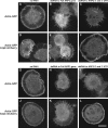
Double-stranded RNA (dsRNA) directed against the 5′ and 3′ untranslated regions (UTRs) of ARP2 or ARP3 depletes the targeted endogenous protein, producing the serrated phenotype. Arp2-GFP and Arp3-GFP each rescue cells depleted of endogenous Arp2 and Arp3, respectively. We created stable S2 cells lines that express actin-GFP, either alone or with Arp2-mCherry (or Arp3-mCherry). dsRNA targeting the coding sequence of ARP2 (or ARP3) depletes the corresponding protein, resulting in the serrated phenotype (B, E, H, and K). In contrast, cells transfected with dsRNA targeting the 5′ and 3′ UTRs of ARP2 or ARP3 are selectively depleted of endogenous Arp2 or Arp3 (C and I), and are rescued by exogenously expressed Arp2-mCherry (F) and Arp3-mCherry (L). Bar, 10 µm.
Figure 3.
Figure 3.
ATP hydrolysis mutant Arp2/3 complex produces dendritic actin network disassembly defects. This is demonstrated by the longer lifetimes and greater distances traveled of Arp2/3 speckles, as compared with WT Arp2/3. (A) We created the following Drosophila S2 stable cell lines: Arp2(WT)-GFP (3); Arp2Q137A-GFP (4); Arp3(WT)-GFP (6); and Arp3Q137A-GFP (7) (see Table 1 for complete list of cell lines). We imaged these stable cell lines and created kymographs from the movies (B–E, second column). We quantified the speckle distances traveled and speckle lifetimes. Endogenous Arp2 (B and C) and Arp3 (D and E) were depleted by dsRNA directed against the 5′ and 3′ UTRs, leaving Arp2-GFP (B and C) and Arp3-GFP (D and E) as the sole copy of Arp2 (B and C) and Arp3 (D and E) in the cell. Bar, 10 µm. (F) We replotted our speckle lifetime and distance-traveled measurements using a box-and-whisker plot. We used the nonparametric Kolmogorov-Smirnov test to compare ATP hydrolysis mutant data against WT data. We calculated p-values to be less than 0.001. The central mark in the box represents the median value; the edges of the box represent the 25th and 75th percentiles. Whiskers extend to the most extreme data points not considered outliers, and outliers are plotted individually, in red.
Figure 4.
Figure 4.
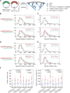
ATP hydrolysis is necessary for timely detachment of Arp2/3 from the lamellipodial actin network, as seen from the ATP hydrolysis mutant’s longer lifetime and longer distance traveled. Arp2/3 containing a single ATP hydrolysis mutant subunit behaves differently than wild-type Arp2/3 within the same cell. (A) We expressed wild-type mCherry-tagged Arp2 (or Arp3) along with ATP hydrolysis mutant GFP-tagged Arp2 (or Arp3) in the same cells. (B and C) Imaging of stable S2 cell lines (1-3): Arp2(WT)-GFP, Arp2(WT)-mCherry (30 cells, 907 GFP speckles, 244 mCherry speckles); and (1-4): Arp2Q137A-GFP, Arp2(WT)-mCherry (34 cells, 972 GFP speckles, 321 mCherry speckles). (D and E) Imaging of stable S2 cell lines (2-6): Arp3(WT)-GFP, Arp3(WT)-mCherry (41 cells, 915 GFP speckles, 692 mCherry speckles.); and (2-7): Arp3Q137A-GFP, Arp3(WT)-mCherry (45 cells, 1,673 GFP speckles, 952 mCherry speckles). We created kymographs showing speckle trajectories, which we used to determine speckle distances traveled and speckle lifetimes. Median values for speckle distances traveled (B and D) or speckle lifetimes (C and E) are noted. Each imaging experiment was repeated on three separate occasions and speckle data were combined. (F) As in Fig. 3, F and G, we replotted our data in a box-and-whisker plot and used the nonparametric Kolmogorov-Smirnov test to compare ATP hydrolysis mutant data against WT data. We calculated p-values to be less than 0.001.
Figure 5.
Figure 5.
Replacing both the Arp2 and the Arp3 subunits of the Arp2/3 complex with ATP hydrolysis mutant variants produces a more severe disassembly phenotype than replacing a single subunit. (A) We created stable cell lines that express mCherry-tagged Arp2 ATP hydrolysis mutant and GFP-tagged Arp3 ATP hydrolysis mutant. We also created stable cell lines in which we switched the fluorescent tags on Arp2 and Arp3; thus, cells express GFP-tagged Arp2 ATP hydrolysis mutant and mCherry-tagged Arp3 ATP hydrolysis mutant. In all cases, we combined speckle measurements from cells expressing Arp2-GFP and Arp3-mCherry with measurements from cells expressing Arp2-mCherry and Arp3-GFP. We plotted our speckle lifetimes (B) and speckle distances traveled (C) in a box-and-whisker plot and used the nonparametric Kolmogorov-Smirnov test to compare ATP hydrolysis mutant data against WT data. We calculated p-values to be less than 0.001.
Figure 6.
Figure 6.
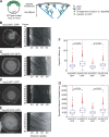
Wild-type and ATP hydrolysis mutant variants of Arp2/3 complex build actin networks in a reconstituted actin-based motility system. (A) The VCA domain of ActA, an Arp2/3-activating protein from the pathogen Listeria monocytogenes, is covalently attached to polystyrene beads. In the presence of 3% Alexa Fluor 488–labeled cytoplasmic actin, Arp2/3 complex, and capping protein, a dendritic actin network grows in a spherical array from the bead surface, creating a shell of actin. After the actin shell breaks symmetry, a sustained tail of actin continues to grow from the bead surface. At 50 nM, wild-type Arp2/3, Arp2Q137A Arp2/3, Arp3Q137A Arp2/3, and Arp2Q137A/Arp3Q137A Arp2/3 all build (B) similar actin networks (C) with comparable actin tail lengths, and similar actin intensities in (C) the tail and (D) shell regions. Bar, 10 µm.
Figure 7.
Figure 7.
Differences in actin disassembly between networks constructed with wild-type vs. ATP hydrolysis mutant Arp2/3 complex become apparent under recycling conditions. (A) The addition of cofilin and profilin to the in vitro actin-based motility system causes turnover and recycling of actin monomers by promoting disassembly of actin filaments. Cofilin severs the actin shell, detaching it from the tail. (B and E) Severing of shells from growing tails occurs 14 min earlier in networks built with wild-type Arp2/3 complex (top) than in networks built with double ATP hydrolysis mutant Arp2/3 complex (bottom). (C) Shells built with ATP hydrolysis mutant Arp2/3 grow to longer lengths than shells built with wild-type Arp2/3 complex. (D) Actin intensities of shells constructed with wild-type Arp2/3 are comparable or slightly lower than intensities of shells constructed with ATP hydrolysis mutant Arp2/3. (E) Quantification of the timing of tail severing for WT (13 tails), Arp2Q137A (9 tails), Arp3Q137A (9 tails), and double ATP hydrolysis mutant (8 tails). Arp2Q137A, Arp3Q137A, and double mutant tails result in shells severed later than wild-type tails by 3.5, 4.5, and 12 min, respectively. Bar, 10 µm.

Similar articles

Cited by

References

    1. Akin O., Mullins R.D. 2008. Capping protein increases the rate of actin-based motility by promoting filament nucleation by the Arp2/3 complex. Cell. 133:841–851 10.1016/j.cell.2008.04.011 - DOI - PMC - PubMed
    1. Blanchoin L., Pollard T.D. 1999. Mechanism of interaction of Acanthamoeba actophorin (ADF/Cofilin) with actin filaments. J. Biol. Chem. 274:15538–15546 10.1074/jbc.274.22.15538 - DOI - PubMed
    1. Blanchoin L., Pollard T.D. 2002. Hydrolysis of ATP by polymerized actin depends on the bound divalent cation but not profilin. Biochemistry. 41:597–602 10.1021/bi011214b - DOI - PubMed
    1. Blanchoin L., Amann K.J., Higgs H.N., Marchand J.B., Kaiser D.A., Pollard T.D. 2000. Direct observation of dendritic actin filament networks nucleated by Arp2/3 complex and WASP/Scar proteins. Nature. 404:1007–1011 10.1038/35010008 - DOI - PubMed
    1. Bork P., Sander C., Valencia A. 1992. An ATPase domain common to prokaryotic cell cycle proteins, sugar kinases, actin, and hsp70 heat shock proteins. Proc. Natl. Acad. Sci. USA. 89:7290–7294 10.1073/pnas.89.16.7290 - DOI - PMC - PubMed

Publication types

MeSH terms