. 2013 Mar 20;32(6):874-85.
doi: 10.1038/emboj.2013.32. Epub 2013 Feb 22.

NDR2-mediated Rabin8 phosphorylation is crucial for ciliogenesis by switching binding specificity from phosphatidylserine to Sec15

Affiliations

NDR2-mediated Rabin8 phosphorylation is crucial for ciliogenesis by switching binding specificity from phosphatidylserine to Sec15

Shuhei Chiba et al. EMBO J. .

Abstract

Primary cilia are antenna-like sensory organelles protruding from the plasma membrane. Defects in ciliogenesis cause diverse genetic disorders. NDR2 was identified as the causal gene for a canine ciliopathy, early retinal degeneration, but its role in ciliogenesis remains unknown. Ciliary membranes are generated by transport and fusion of Golgi-derived vesicles to the pericentrosome, a process requiring Rab11-mediated recruitment of Rabin8, a GDP-GTP exchange factor (GEF) for Rab8, and subsequent Rab8 activation and Rabin8 binding to Sec15, a component of the exocyst that mediates vesicle tethering. This study shows that NDR2 phosphorylates Rabin8 at Ser-272 and defects in this phosphorylation impair preciliary membrane assembly and ciliogenesis, resulting in accumulation of Rabin8-/Rab11-containing vesicles at the pericentrosome. Rabin8 binds to and colocalizes with GTP-bound Rab11 and phosphatidylserine (PS) on pericentrosomal vesicles. The phospho-mimetic S272E mutation of Rabin8 decreases affinity for PS but increases affinity for Sec15. These results suggest that NDR2-mediated Rabin8 phosphorylation is crucial for ciliogenesis by triggering the switch in binding specificity of Rabin8 from PS to Sec15, thereby promoting local activation of Rab8 and ciliary membrane formation.


Conflict of interest statement

The authors declare that they have no conflict of interest.

Figures
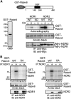
Figure 1
Figure 1
NDR kinases phosphorylate Rabin8 at Ser-272. (A) NDRs phosphorylate Rabin8. GST-Rabin8 was purified from E. coli (left panel). Myc-NDR1/2(WT) or KD mutants, which were expressed in COS-7 cells untreated or treated with OA, were immunoprecipitated and subjected to in vitro kinase assays using GST-Rabin8 as a substrate. Schematic structure of GST-Rabin8 is shown. Black box indicates the coiled–coil domain, which probably functions as a GEF domain. (B) NDRs phosphorylate Rabin8 at Ser-272. NDR1 (left panel) and NDR2 (right panel) were immunoprecipitated and subjected to in vitro kinase assays with GST-Rabin8(WT or S272A), as in (A). Source data for this figure is available on the online supplementary information page.
Figure 2
Figure 2
NDR2-mediated Rabin8 phosphorylation is crucial for ciliogenesis. (A) Effect of siRNAs on NDR1/2 expression. RPE1 cells were transfected with control, NDR1, or NDR2 siRNAs (25 nM), and cultured for 72 h. Cell lysates were analysed by immunoblotting with anti-NDR1 or anti-NDR2 antibody. (B) Effect of NDR1/2 or Rabin8 depletion on ciliogenesis. RPE1 cells transfected with siRNAs (25 nM) were cultured for 48 h and serum-starved for 48 h. Cells were stained for Ac-tubulin (red) and DAPI (blue). Scale bar, 10 μm; insets magnification, × 2.7. (C) Quantification of the number of ciliated cells. Data are means±s.e.m. (n=4; >100 cells per experiment, one-way ANOVA followed by Turkey test). ***P<0.001 compared with control siRNA. (D) Effect of NDR1/2 depletion on the cilium length (n=7 in control and NDR1 siRNA cells; n=6 in NDR2 siRNA cells; n=5 in NDR1/NDR2 double siRNA cells; >50 axonemes per experiment). Scatter plots show the median, 25th and 75th percentiles. Dots indicate individual data points. *P<0.05; **P<0.01; ***P<0.001, by paired t-test. (E) Kinase activity of NDR2 is required for ciliogenesis. RPE1 cells stably expressing control vector or sr mouse (m) NDR2(WT or KD) were transfected with NDR2 siRNA, cultured for 36 h and serum-starved for 36 h. Data are means±s.e.m. (n=3; >100 cells per experiment). ***P<0.001, by one-way ANOVA followed by Turkey test. (F) Localization of NDR1 and NDR2. RPE1 cells were transfected with control YFP, YFP-NDR1, or -NDR2, fixed and imaged by YFP fluorescence. Bar, 20 μm. (G) Quantification of the number of cells with punctate NDR localization. Data are means±s.e.m. (n=3; >40 cells per experiment). (H) Time-lapse fluorescent images of YFP-NDR1/2 and mCherry-centrin2 in serum-starved RPE1 cells. See also Supplementary Movie S1 and S2. Bar, 10 μm. (I) Expression levels of Rabin8 and YFP-sr-Rabin8(WT or S272A). RPE1 cells or those stably expressing YFP-sr-Rabin8(WT or S272A) were transfected with Rabin8 siRNA. Cell lysates were analysed by anti-Rabin8 immunoblotting. (J) Effect of S272A mutation on ciliogenesis. RPE1 cells or those stably expressing YFP-sr-Rabin8(WT or S272A) were transfected with Rabin8 siRNA and treated as in (C). Data are means±s.e.m. (n=3; >100 cells per experiment). *P<0.05, **P<0.01, by one-way ANOVA followed by Turkey test. Source data for this figure is available on the online supplementary information page.
Figure 3
Figure 3
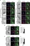
Non-phosphorylatable mutation of Rabin8 or depletion of NDR2 causes pericentrosomal accumulation of Rabin8. (A) Time course of Rabin8 localization after serum starvation. RPE1 cells stably expressing YFP-Rabin8(WT or S272A) were serum-starved. At the indicated times, cells were fixed and imaged by YFP fluorescence (inverted images or green) and pericentrin (red) and Ac-tubulin (blue) immunostaining. Arrowheads indicate the position of the centrosome. Bar, 10 μm. (B) S272A mutation causes pericentrosomal accumulation of Rabin8. RPE1 cells transfected with YFP-Rabin8(WT or S272A) were serum-starved for 24 h. Cells were imaged as in (A). Bar, 10 μm. (C) Quantification of the number of cells with pericentrosomal localization of YFP-Rabin8. (D) NDR2 depletion causes pericentrosomal accumulation of Rabin8. RPE1 cells transfected with YFP-Rabin8(WT) and control or NDR2 siRNA were serum-starved for 24 h. Cells were imaged as in (A). Bar, 10 μm. (E) Quantification of the number of cells with pericentrosomal localization of YFP-Rabin8. In (C) and (E), data are means±s.e.m. (n=3; >100 cells per experiment). **P<0.01, by paired t-test.
Figure 4
Figure 4
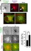
Colocalization of Rabin8 with Rab11. (A) Rabin8 and Rab11 colocalize on pericentrosomal vesicles in NDR2-depleted cells. RPE1 cells were transfected with YFP-Rabin8(WT) and NDR2 siRNA and serum-starved. Cells were imaged by YFP fluorescence (green) and Rab11 (red) and pericentrin (blue) staining. Bar, 10 μm. (B) Rabin8(S272A) colocalizes with Rab11(QL) on pericentrosomal vesicles. RPE1 cells were cotransfected with YFP-Rabin8(WT or S272A) (green) and CFP-Rab11(QL) (red). Bar, 20 μm (top panel) and 5 μm (bottom panel). Right panel shows the quantification of the number of cells with YFP-Rabin8 colocalization with CFP-Rab11(QL) on pericentrosomal vesicles. Data are means±s.e.m. (n=3; >50 cells per experiment). ***P<0.001, by paired t-test.
Figure 5
Figure 5
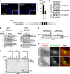
Rabin8 binds to and colocalizes with PS, and the phospho-mimic mutation of Rabin8 inhibits binding to PS. (A) Phospholipid blot assays. Rabin8 or the S272A and S272E mutants were purified from Sf-21 cells, incubated with phospholipid blots, and immunoblotted with anti-Rabin8 antibody. (B, C) Phospholipid-cosedimentation assays using multilamellar vesicles. In (B), purified Rabin8 was incubated with multilamellar vesicles composed of PC (100%) or PS/PC (15:85 molar ratio). In (C), purified Rabin8(S272A or S272E) mutants were incubated with multilamellar vesicles composed of PS/PC (15:85 molar ratio). After centrifugation, pellets were analysed by CBB staining. (D) Quantitative analysis of Rabin8 binding to PS vesicles. Purified Rabin8(S272A or S272E) was incubated with various amounts of multilamellar vesicles composed of PS/PC (15:85 molar ratio) and analysed, as in (C). The apparent dissociation constants (Kd) for the binding of Rabin8(S272A) and Rabin8(S272E) to total lipids of PS/PC vesicles were calculated. Data are means±s.e.m. (n=3). *P<0.05, by paired t-test. (E) Colocalization of Rab11 and PS. RPE1 cells transfected with mCherry-evt2(PH) were analysed by Rab11 staining (green) and mCherry fluorescence (red). Bar, 10 μm. (F) Colocalization of Rabin8(S272A) and PS on pericentrosomal vesicles. RPE1 cells transfected with YFP-Rabin8(S272A) and mCherry-evt2(PH) were serum-starved and imaged by YFP (green) and mCherry (red) fluorescence. Bar, 10 μm. (G) Mapping of the region of Rabin8 required for PS binding. Rabin8 deletion mutants purified from Sf-21 cells were subjected to phospholipid-cosedimentation assays, as in (C). Source data for this figure is available on the online supplementary information page.
Figure 6
Figure 6
Phospho-mimic mutation of Rabin8 promotes binding to Sec15 and characterization of Rabin8(I329S) mutant. (A) Effect of Sec15 depletion on ciliogenesis. RPE1 cells transfected with Sec15A and Sec15B siRNAs were cultured for 48 h in growth medium and serum-starved for 48 h. Cells were fixed and stained for Ac-tubulin (red) and DNA (blue). Scale bar, 20 μm; insets magnification, × 2.7. Right panel shows the quantification of the number of ciliated cells. Data are means±s.e.m. (n=3; >100 cells per experiment). **P<0.01, by paired t-test. (B) The Rabin8 S272E mutant has increased binding ability to Sec15. RPE1 cells stably expressing Rabin8 (WT, S272A, or S272E) were transfected with Flag-Sec15. Cell lysates were immunoprecipitated with anti-Flag antibody and then analysed by anti-GFP immunoblotting. (C) Alignment of the sequences of human Rabin8 and yeast Sec2p. Black and grey boxes indicate identical and homologous residues, respectively. (D) Rabin8(I329S) has a lower affinity for Rab11 and a higher affinity for Sec15. GFP-Rab11 or GFP-Sec15 expressed in 293T cells was subjected to GST pull-down assays using GST (−) or GST-Rabin8(WT or I329S). (E) Rabin8(I329S) has decreased affinity for PS. Rabin8(WT or I329S) purified from Sf-21 cells was incubated with multilamellar vesicles composed of different PS/PC ratios and analysed as in Figure 5B. (F) Rabin8(Δ300–305) has decreased affinity for PS. GST-Rabin8(WT or Δ300–305) purified from Sf-21 cells was incubated with multilamellar vesicles composed of PS/PC (15:85 molar ratio) and analysed as in Figure 5B. (G) Rabin8(I329S) does not accumulate on the pericentrosome. RPE1 cells cotransfected with Flag-Rabin8(WT or I329S) and GFP-Rab11(QL) were serum-starved for 3 h. Cells were analysed by anti-Flag staining and GFP fluorescence. Bar, 10 μm. Source data for this figure is available on the online supplementary information page.

Similar articles

Cited by

References

    1. Berta AI, Boesze-Battaglia K, Genini S, Goldstein O, O'Brien PJ, Szel A, Acland GM, Beltran WA, Aguirre GD (2011) Photoreceptor cell death, proliferation and formation of hybrid rod/S-cone photoreceptors in the degenerating STK38L mutant retina. PLoS One 6: e24074. - PMC - PubMed
    1. Bryant DM, Datta A, Rodriguez-Fraticelli AE, Peränen J, Martin-Belmonte F, Mostov KE (2010) A molecular network for de novo generation of the apical surface and lumen. Nat Cell Biol 12: 1035–1045 - PMC - PubMed
    1. Chiba S, Ikeda M, Katsunuma K, Ohashi K, Mizuno K (2009) MST2- and Furry-mediated activation of NDR1 kinase is critical for precise alignment of mitotic chromosomes. Curr Biol 19: 675–681 - PubMed
    1. Cornils H, Stegert MR, Hergovich A, Hynx D, Schmitz D, Dirnhofer S, Hemmings BA (2010) Ablation of the kinase NDR1 predisposes mice to the development of T cell lymphoma. Sci Signal 3: ra47. - PubMed
    1. Devroe E, Erdjument-Bromage H, Tempst P, Silver PA (2004) Human Mob proteins regulate the NDR1 and NDR2 serine-threonine kinases. J Biol Chem 279: 24444–24451 - PubMed

Publication types