. 2013;9(2):e1003252.
doi: 10.1371/journal.pgen.1003252. Epub 2013 Feb 7.

Antagonism versus cooperativity with TALE cofactors at the base of the functional diversification of Hox protein function

Affiliations

Antagonism versus cooperativity with TALE cofactors at the base of the functional diversification of Hox protein function

María Luisa Rivas et al. PLoS Genet. 2013.

Abstract

Extradenticle (Exd) and Homothorax (Hth) function as positive transcriptional cofactors of Hox proteins, helping them to bind specifically their direct targets. The posterior Hox protein Abdominal-B (Abd-B) does not require Exd/Hth to bind DNA; and, during embryogenesis, Abd-B represses hth and exd transcription. Here we show that this repression is necessary for Abd-B function, as maintained Exd/Hth expression results in transformations similar to those observed in loss-of-function Abd-B mutants. We characterize the cis regulatory module directly regulated by Abd-B in the empty spiracles gene and show that the Exd/Hth complex interferes with Abd-B binding to this enhancer. Our results suggest that this novel Exd/Hth function does not require the complex to bind DNA and may be mediated by direct Exd/Hth binding to the Abd-B homeodomain. Thus, in some instances, the main positive cofactor complex for anterior Hox proteins can act as a negative factor for the posterior Hox protein Abd-B. This antagonistic interaction uncovers an alternative way in which MEIS and PBC cofactors can modulate Abd-B like posterior Hox genes during development.


Conflict of interest statement

The authors have declared that no competing interests exist.

Figures

Figure 1
Figure 1. Hth and Exd downregulation by Abd-B in the posterior abdominal segments.
(A–B) Hth and Abd-B expression in wild type embryos at stage 12 (A) and stage13 (B). Hth protein disappears from the dorsal area where the spiracles are formed (A′ and A″ show a close up of A rotated 180° to keep dorsal up and anterior left). Note that in the spiracle region only a cluster of 8 cells retain high Hth levels. (C) Abd-B mutant showing elevated levels of Hth in the dorsal region. (D) Wild type st14 embryo stained with anti-Exd (D, green) and anti-AbdB (D′, red). Exd protein localizes to the nucleus in anterior segments (T2 inset) while is cytoplasmic in the posterior domain where Abd-B is expressed (A8 insets). (E) Dorsal view of a st11 embryo overexpressing Exd and Hth. Insets in E′ show that although the posterior A8 segment has high levels of nuclear Exd/Hth expression (only Hth shown in E) Abd-B expression is normal (E′). White arrows point to the A8 segment. Anterior left in all panels, and dorsal up in A–C.
Figure 2
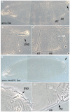
Figure 2. Downregulation of Exd/Hth expression is required for the normal development of the posterior abdominal segments.
(A) Ectopic expression of Exd results in viable larvae with normal posterior spiracles (psp, black arrow A′) and a normal denticle pattern with the A8 denticle belt immediately abutting the anal pads (ap, white arrow A″). (B) Simultaneous ectopic expression of Exd/Hth results in larvae that form aberrant posterior spiracles (B′), a reduced A8 denticle belt and an extra A9 belt anterior to the anal pad (B″).
Figure 3
Figure 3. Expression of early Abd-B downstream targets after Exd/Hth ectopic induction.
Expression of Cut protein (A–B), spalt RNA (C–D) and the ems posterior spiracle reporter gene (E–F) in wild type (A,C,E) or embryos expressing ectopically Hth and Exd with the arm-Gal4 line (B, D, F). Arrows point to the posterior spiracle site. Embryos in (A–D) have retracted the germ band while those in (E–F) are at extended germ band, and are thus folded with the A8 segment close to the head.
Figure 4
Figure 4. Effect of the ectopic expression of Abd-B isoforms on the development of embryos expressing different levels of Hth and Exd.
(A) Abd-Bm ectopic expression with the arm-Gal4 line induces posterior spiracles in ectopic positions (In this figure arrows point to normal A8 posterior spiracles and arrowheads to ectopic spiracles). (B) Ectopic expression of Abd-Bm can only form small remnants of posterior spiracles when co-expressed with the Hth and Exd proteins. (C) Abd-Br ectopic expression with the arm-Gal4 line is not capable of inducing ectopic posterior spiracles in a wild type background. (D) Abd-Br can induce ectopic posterior spiracles in hthP2 null embryos. (E) Abd-Br can induce small ectopic posterior spiracles in hthP2 heterozygous embryos. Note that the ectopic spiracles in the posterior segments are more complete. (F) In hypomorphic hth100-1 alleles lacking the homeodomain containing isoform but still expressing the homeodomainless protein Abd-Br is not capable of efficiently inducing ectopic posterior spiracles (compare to panel D). These embryos have tiny spiracles that can be explained by the reduction of total Hth protein caused by this allele, resulting in levels more similar to those present in heterozygous hthP2 embryos shown in panel E. (G) Abd-Br expression in zygotic exdYO12 mutant embryos induces ectopic spiracles. (H) Close up of (G) showing some ectopic spiracles. All experiments in this figure were performed at 25°C.
Figure 5
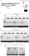
Figure 5. Characterization of the Abd-B binding sites in the ems posterior spiracle enhancer.
(A) Dissection of the ems1.2 posterior spiracle enhancer. Black bars represent fragments tested in transgenic lacZ constructs. Grey bars represent oligos tested in EMSA. Asterisks indicate the location of putative Abd-B binding sites in the ems0.35 fragment. (B) EMSA showing Abd-B binding to the ems0.35 enhancer (lane 2 arrow), this band is supershifted by anti-AbdB confirming that the complex contains Abd-B (lane 3 arrowhead). (C) Abd-B binding to the ems0.35 enhancer is competed by cold oligos containing Abd-B sites. Oligos 4 and 6 show higher affinity for Abd-B. Oligo 3 that does not contain putative Abd-B sites does not compete (lanes 11–14). (D) EMSA showing that wild type cold oligo 4 (lanes 3–6) and oligo 6 (lanes 19–22) can compete for Abd-B binding to the ems0.35 enhancer while cold oligo 4 with a mutation on both 4A and 4B sites (lanes 15–18) or oligo 6 with a mutation on its only site (lanes 23–26) cannot compete Abd-B binding. Note that separately mutating in oligo 4 site 4A (lanes 7–10) or site 4B (lanes 11–14) shows that site 4A has higher affinity for Abd-B. However, comparison of the independent mutations to the double 4A4B mutant suggests both sites are functional (lanes 15–18). Triangles in panels C and D represent increasing amounts of the indicated cold oligo competitor. (E) EMSA showing that Abd-B binds to wild type oligos 4 and 6 through the predicted putative Abd-B sites, as binding to the oligos decreases when these sites are mutated (compare WT lanes 3–4 and 18–19 with lanes labelled as mut). Site 4A and 6 bind to Abd-B with higher affinity than site 4B.
Figure 6
Figure 6. Expression of different ems0.35 enhancer variants in st11 embryos.
(A) ßGal expression of the ems0.35 wild type enhancer. (B) Expression of ems0.35 with a mutation on the second putative Abd-B site. (C–E) Reduced expression of ems0.35 constructs carrying a single mutation on either the 4A (C), 4B (D) or 6 (E) putative Abd-B sites. (F) Expression of ems0.35 double mutant in site 4A and 6. (G) ems0.35 constructs double mutant for site 4A and 4B show no spiracle expression. Arrows point to the posterior spiracle primordium.
Figure 7
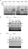
Figure 7. Exd/Hth interfere with Abd-B binding to the ems spiracle enhancer.
(A) EMSA showing that Exd/Hth does not bind to the ems0.35 oligos (lanes 5,10,15,20,25,30) in conditions where Abd-B binds to oligos 4 and 6 (lanes 18–19 and 28–29, asterisks). (B–C) EMSA showing that Abd-B binding to oligos 4 and 6 is partially competed by increasing amounts of Exd/Hth proteins (lanes 12–14). Separate expression of Hth or Exd has only a small effect on Abd-B binding to these oligos.
Figure 8
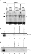
Figure 8. Direct Abd-B homeodomain binding to the Exd/Hth complex.
(A) EMSA showing that both Abd-Bm (lanes 3–5) and Abd-Br (lanes13–15) binding for oligo 4 is competed in the presence of Exd/Hth. Similar competition is observed over an Abd-B variant with a conserved W residue mutated (lanes 8–10). Note that in each lane several size bands appear (black arrows and grey arrowheads in panel A). These bands are specific as they are supershifted by anti-AbdB. We interpret them as due to Abd-B being translated in vitro from internal methionines as the smaller band in lane 1 coincides with the larger Abd-Br band in lane 11. (B–C) GST-Abd-B pull-down experiments with Exd and Hth. Beads binding GST or GST fused to the Abd-B C-terminal fragments were incubated with methionine-S35 labelled Exd (B) or Hth (C). (B) An Abd-B C-terminal fragment binds S35-Exd. This interaction is reduced when the homeodomain is deleted from the fragment, and the Abd-B homeodomain by itself can bind Exd. (C) Abd-B homeodomain only weakly binds S35-Hth (GST-AbdBHD, third lane), but the interaction is enhanced by the presence of unlabelled Exd protein (GST-AbdBHD+Exd, fifth lane) indicating the formation of a trimeric complex. For each of the S35-labelled proteins, 25% of the amount used in the binding reactions was directly loaded in the first lanes (Input).

Similar articles

Cited by

References

    1. Pearson JC, Lemons D, McGinnis W (2005) Modulating Hox gene functions during animal body patterning. Nat Rev Genet 6: 893–904. - PubMed
    1. Hueber SD, Weiller GF, Djordjevic MA, Frickey T (2010) Improving Hox protein classification across the major model organisms. PLoS ONE 5: e10820 doi:10.1371/journal.pone.0010820. - DOI - PMC - PubMed
    1. Merabet S, Hudry B, Saadaoui M, Graba Y (2009) Classification of sequence signatures: a guide to Hox protein function. Bioessays 31: 500–511. - PubMed
    1. Hombría JC-G, Rivas ML, Sotillos S (2009) Genetic control of morphogenesis- Hox induced organogenesis of the posterior spiracles. Int J Dev Biol 53: 1349–1358. - PubMed
    1. Hu N, Castelli-Gair J (1999) Study of the posterior spiracles of Drosophila as a model to understand the genetic and cellular mechanisms controlling morphogenesis. Developmental Biology 214: 197–210. - PubMed

Publication types

MeSH terms

Grants and funding

This work was supported by the Programa Consolider, MICINN, European Regional Development Fund (FEDER), and Junta de Andalucía to JC-GH and by an EMBO short-term fellowship to MLR. YG was supported by the CNRS, Université de la Méditerranée, and grants from CEFIPRA, ANR, FRM, and ARC, and by a fellowship from MRT CEFIPRA to NS. The funders had no role in study design, data collection and analysis, decision to publish, or preparation of the manuscript.