

Activation of WIP1 phosphatase by HTLV-1 Tax mitigates the cellular response to DNA damage
- PMID: 23405243
- PMCID: PMC3566092
- DOI: 10.1371/journal.pone.0055989
Activation of WIP1 phosphatase by HTLV-1 Tax mitigates the cellular response to DNA damage
Abstract
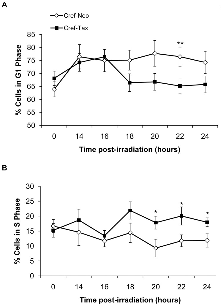
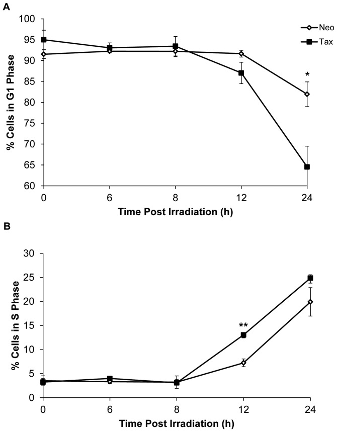
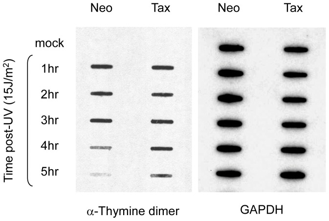
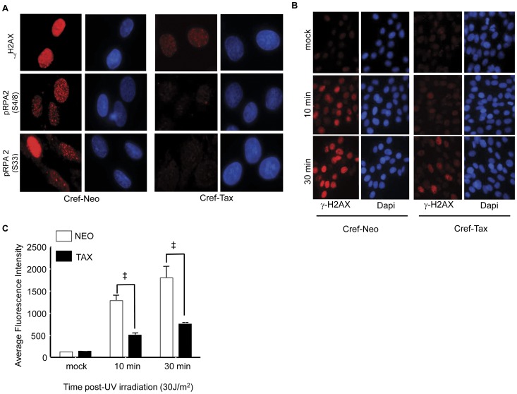
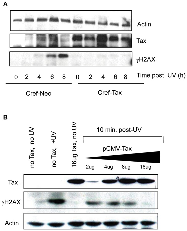
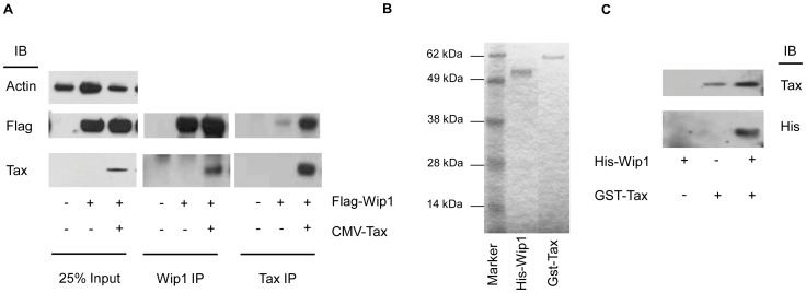
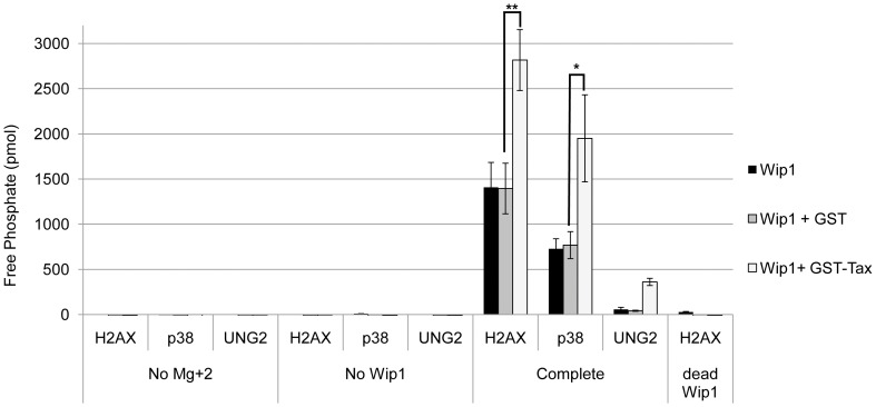
Genomic instability stemming from dysregulation of cell cycle checkpoints and DNA damage response (DDR) is a common feature of many cancers. The cancer adult T cell leukemia (ATL) can occur in individuals infected with human T cell leukemia virus type 1 (HTLV-1), and ATL cells contain extensive chromosomal abnormalities, suggesting that they have defects in the recognition or repair of DNA damage. Since Tax is the transforming protein encoded by HTLV-1, we asked whether Tax can affect cell cycle checkpoints and the DDR. Using a combination of flow cytometry and DNA repair assays we showed that Tax-expressing cells exit G(1) phase and initiate DNA replication prematurely following damage. Reduced phosphorylation of H2AX (γH2AX) and RPA2, phosphoproteins that are essential to properly initiate the DDR, was also observed in Tax-expressing cells. To determine the cause of decreased DDR protein phosphorylation in Tax-expressing cells, we examined the cellular phosphatase, WIP1, which is known to dephosphorylate γH2AX. We found that Tax can interact with Wip1 in vivo and in vitro, and that Tax-expressing cells display elevated levels of Wip1 mRNA. In vitro phosphatase assays showed that Tax can enhance Wip1 activity on a γH2AX peptide target by 2-fold. Thus, loss of γH2AX in vivo could be due, in part, to increased expression and activity of WIP1 in the presence of Tax. siRNA knockdown of WIP1 in Tax-expressing cells rescued γH2AX in response to damage, confirming the role of WIP1 in the DDR. These studies demonstrate that Tax can disengage the G(1)/S checkpoint by enhancing WIP1 activity, resulting in reduced DDR. Premature G(1) exit of Tax-expressing cells in the presence of DNA lesions creates an environment that tolerates incorporation of random mutations into the host genome.
Conflict of interest statement
Figures


Similar articles
-
Wip1 directly dephosphorylates gamma-H2AX and attenuates the DNA damage response.Cancer Res. 2010 May 15;70(10):4112-22. doi: 10.1158/0008-5472.CAN-09-4244. Epub 2010 May 11. Cancer Res. 2010. PMID: 20460517 Free PMC article.
-
Downregulation of Wip1 phosphatase modulates the cellular threshold of DNA damage signaling in mitosis.Cell Cycle. 2013 Jan 15;12(2):251-62. doi: 10.4161/cc.23057. Epub 2012 Jan 15. Cell Cycle. 2013. PMID: 23255129 Free PMC article.
-
Wild-type p53-induced phosphatase 1 (Wip1) forestalls cellular premature senescence at physiological oxygen levels by regulating DNA damage response signaling during DNA replication.Cell Cycle. 2014;13(6):1015-29. doi: 10.4161/cc.27920. Epub 2014 Jan 31. Cell Cycle. 2014. PMID: 24552809 Free PMC article.
-
HTLV-1 Tax: Linking transformation, DNA damage and apoptotic T-cell death.Chem Biol Interact. 2010 Nov 5;188(2):359-65. doi: 10.1016/j.cbi.2010.06.005. Epub 2010 Jun 15. Chem Biol Interact. 2010. PMID: 20558150 Review.
-
Impact of HTLV-I Tax on cell cycle progression and the cellular DNA damage repair response.Oncogene. 2005 Sep 5;24(39):5986-95. doi: 10.1038/sj.onc.1208976. Oncogene. 2005. PMID: 16155605 Review.
Cited by
-
Modulation of DNA damage and repair pathways by human tumour viruses.Viruses. 2015 May 22;7(5):2542-91. doi: 10.3390/v7052542. Viruses. 2015. PMID: 26008701 Free PMC article. Review.
-
Highlights on distinctive structural and functional properties of HTLV Tax proteins.Front Microbiol. 2013 Sep 9;4:271. doi: 10.3389/fmicb.2013.00271. Front Microbiol. 2013. PMID: 24058363 Free PMC article. Review.
-
The human T-cell leukemia virus type-1 tax oncoprotein dissociates NF-κB p65RelA-Stathmin complexes and causes catastrophic mitotic spindle damage and genomic instability.Virology. 2019 Sep;535:83-101. doi: 10.1016/j.virol.2019.07.003. Epub 2019 Jul 3. Virology. 2019. PMID: 31299491 Free PMC article.
-
Mechanisms of Oncogenesis by HTLV-1 Tax.Pathogens. 2020 Jul 7;9(7):543. doi: 10.3390/pathogens9070543. Pathogens. 2020. PMID: 32645846 Free PMC article. Review.
-
Inducible nitric oxide synthase mediates DNA double strand breaks in Human T-Cell Leukemia Virus Type 1-induced leukemia/lymphoma.Retrovirology. 2015 Aug 12;12:71. doi: 10.1186/s12977-015-0196-y. Retrovirology. 2015. PMID: 26265053 Free PMC article.
References
-
- McDonald ER, El Deiry WS (2001) Checkpoint genes in cancer. Ann Med JID - 8906388 33: 113–122. - PubMed
-
- Jallepalli PV, Lengauer C (2001) Chromosome segregation and cancer: cutting through the mystery. Nature Rev Cancer JID - 101124168 1: 109–117. - PubMed
-
- Bartek J, Bartkova J, Lukas J (2007) DNA damage signalling guards against activated oncogenes and tumour progression. Oncogene 26: 7773–7779. - PubMed
-
- Bartkova J, Horejsi Z, Koed K, Kramer A, Tort F, et al. (2005) DNA damage response as a candidate anti-cancer barrier in early human tumorigenesis. Nature 434: 864–870. - PubMed
Publication types
MeSH terms
Substances
Grants and funding
LinkOut - more resources
Full Text Sources
Other Literature Sources
