. 2013 Mar 21;38(3):581-95.
doi: 10.1016/j.immuni.2013.01.009. Epub 2013 Feb 7.

Luminal bacteria recruit CD103+ dendritic cells into the intestinal epithelium to sample bacterial antigens for presentation

Affiliations

Luminal bacteria recruit CD103+ dendritic cells into the intestinal epithelium to sample bacterial antigens for presentation

Julia Farache et al. Immunity. .

Abstract

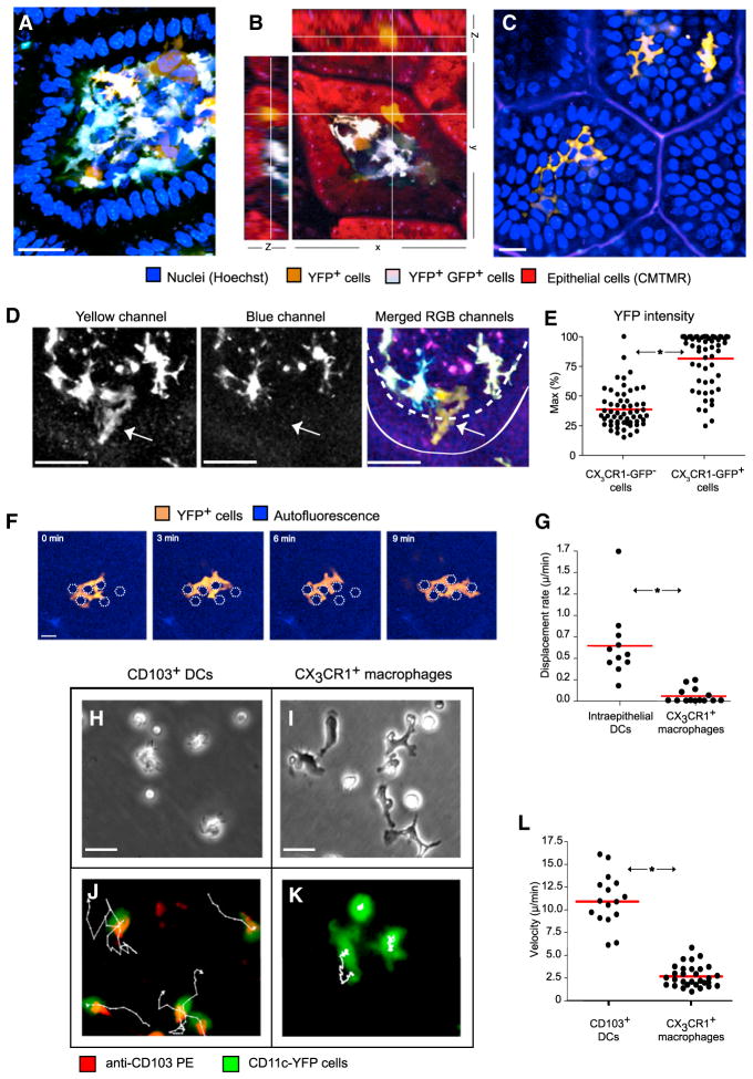
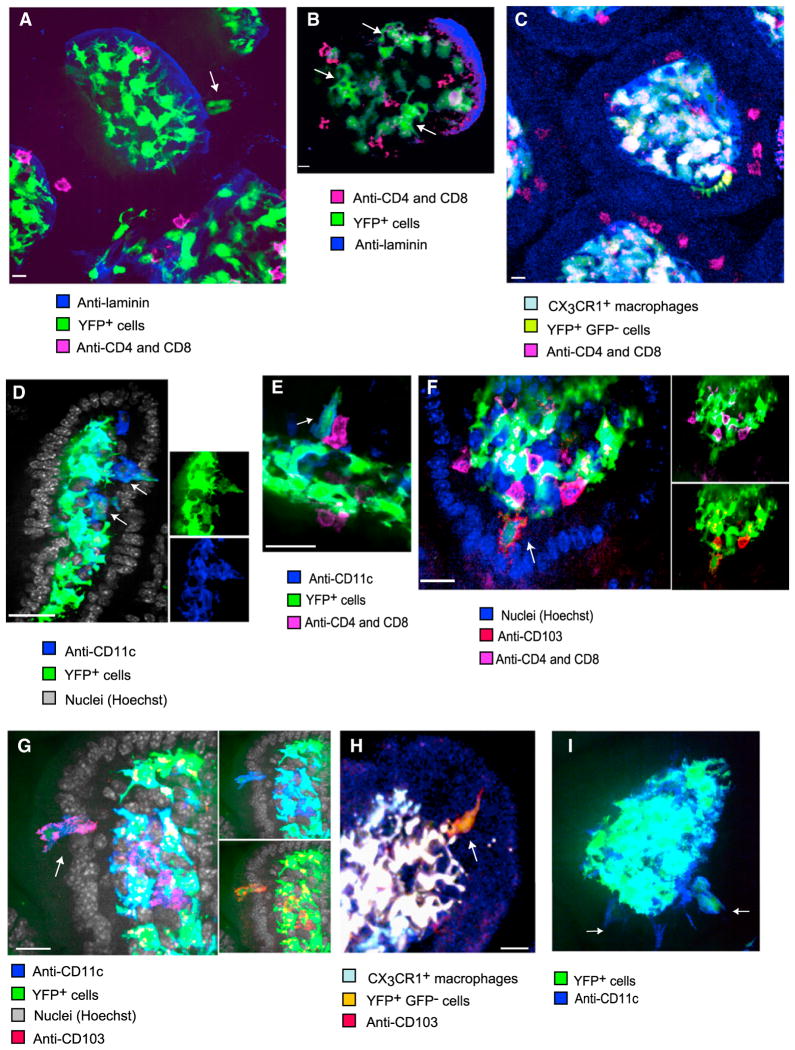
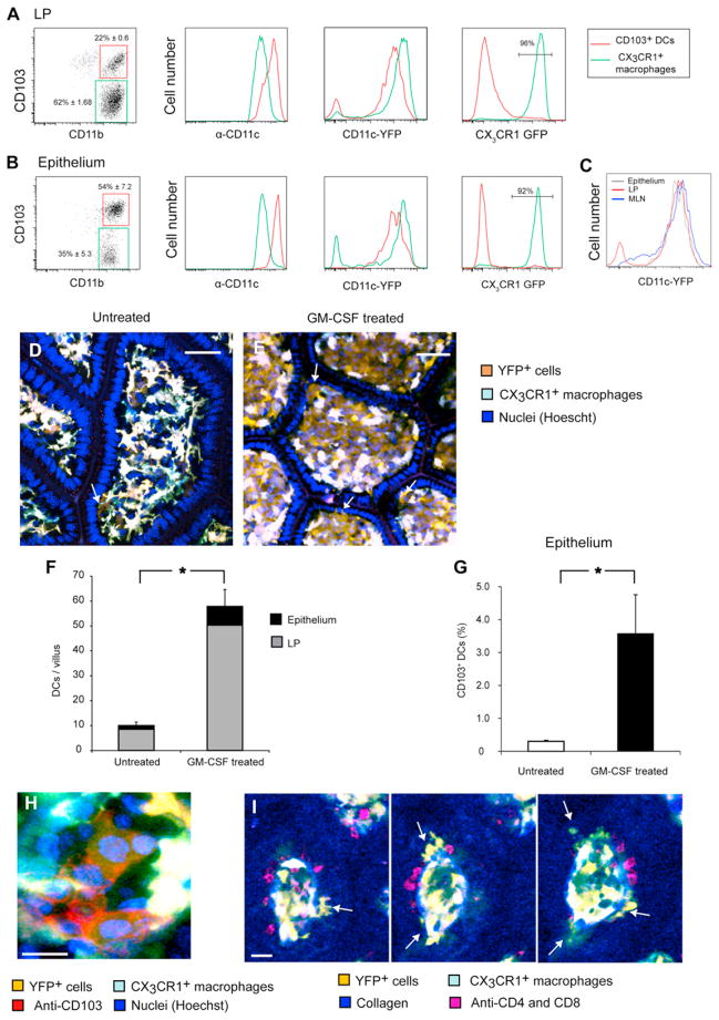
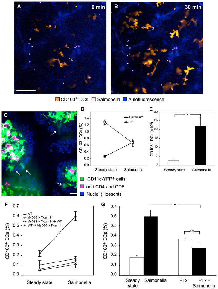
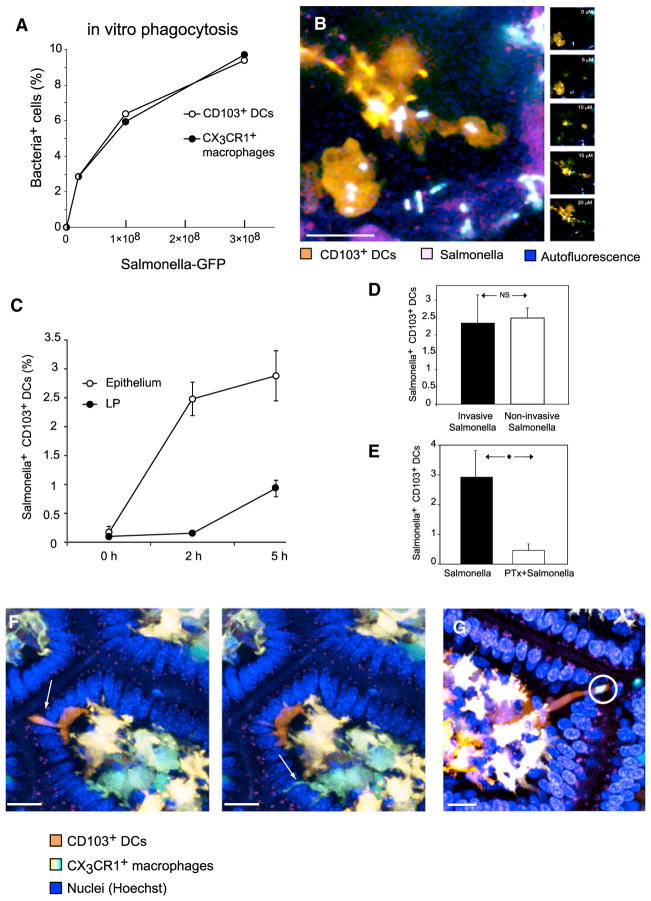
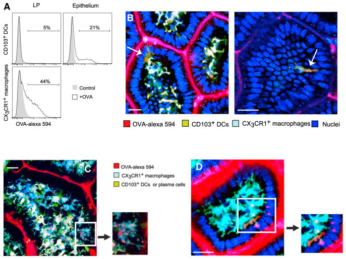
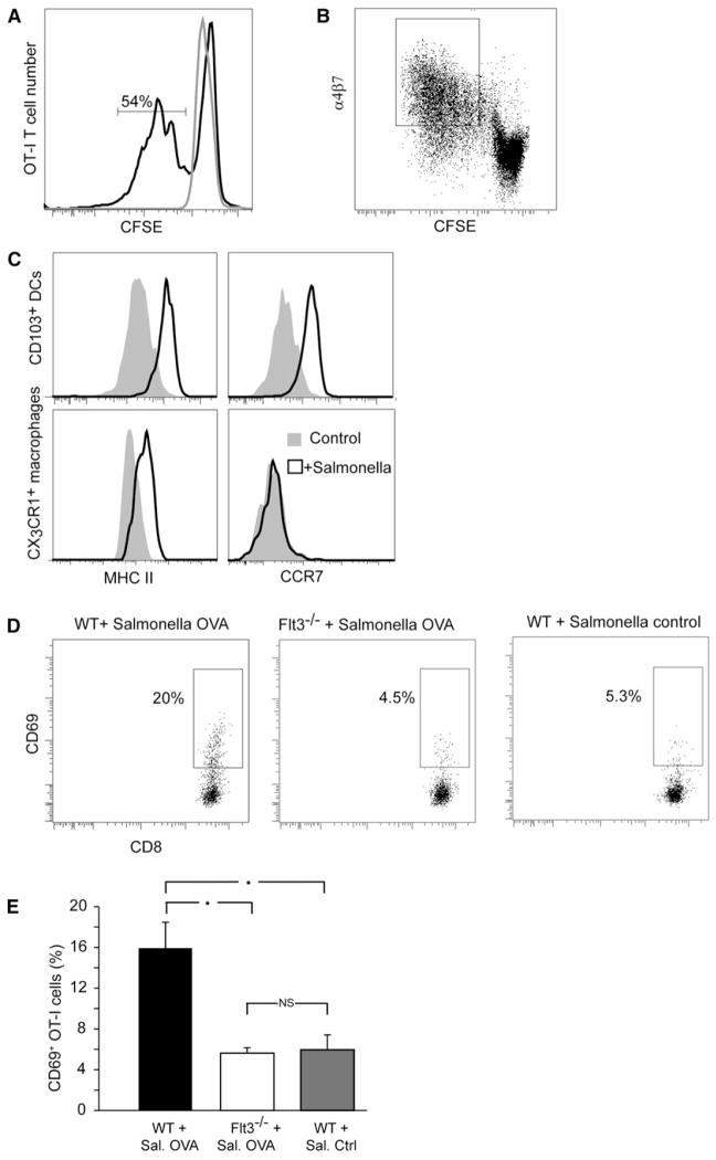
CD103+ dendritic cells (DCs) carry bacteria from the small intestine and can present antigens to T cells. Yet they have not been recorded sampling luminal bacteria or presenting bacterial antigens in mesentery lymph nodes. We used 2-photon microscopy in live Cx3cr1(+/gfp) ×Cd11c-YFP mice to study these processes. At steady state, sparse CD103+ DCs occupied the epithelium. They patrolled among enterocytes while extending dendrites toward the lumen, likely using tight-junction proteins to penetrate the epithelium. Challenge with Salmonella triggered chemokine- and toll-like receptor (TLR)-dependent recruitment of additional DCs from the lamina propria (LP). The DCs efficiently phagocytosed the bacteria using intraepithelial dendrites. Noninvasive bacteria were similarly sampled. In contrast, CD103+ DCs sampled soluble luminal antigen inefficiently. In mice harboring CD103+ DCs, antigen-specific CD8 T cells were subsequently activated in MLNs. Intestinal CD103+ DCs are therefore equipped with unique mechanisms to independently complete the processes of uptake, transportation, and presentation of bacterial antigens.


Figures

Figure 1
Figure 1. A Population of Motile CX3CR1-GFPCD11c-YFPint Cells Occupies the Intestinal Epithelium
Two-photon microscopy in the ileum of a live Cx3cr1+/gfp × Cd11c-YFP mice. (A) A cross-section through a villus; the LP is populated by sparse GFPYFPint cells and abundant GFP+YFPhi cells, whose exact hue depends on the ratio of GFP to YFP (scale bar represents 25 μM). (B) Examining reconstructed data from three axes highlights a GFPYFPint cell lodged within the epithelial layer. (C) A cross-section through the apical epithelium of several adjacent villi shows 3 GFPYFPint cells occupying the space between epithelial cells (scale bar represents 25 μM). (D) A GFPYFPintcell (arrow) fluoresces in the yellow channel but not the blue channel. Solid and dashed lines outline the luminal surface and basement membrane, respectively. (A–D, data are representative of at least 20 independent experiments). (E) In 2-photon microscopy, large motile amoeboid CX3CR1-GFP cells showed on average half the YFP intensity as CX3CR1GFP+ cells (horizontal bars denote averages, p < 0.0001, n = 65, five independent experiments). (F) A YFPint cell was followed in the epithelium for 9 min. The cell meandered between enterocytes, whose inferred positions are indicated by white polygons, without displacing them (scale bar represents 30 μM). (G) Based on intravital 2-photon microscopy, the displacement rate of CD103+ DCs was significantly (p < 0.0001) higher than of X3CR1+ macrophages imaged in the same villi (n = 27, three independent experiments). In vitro live-cell microscopy of CD103+ DCs (H and J) and CX3CR1+ macrophages (I and K) sorted using flow cytometry from the LP revealed their intrinsic motility patterns (scale bars represent 30 μM). (H) CD103+ DCs displayed amoeboid morphology. (I) CX3CR1+ macrophages adhered to the plate showing stellate morphology. (J) CD103+ DCs crawled vigorously, using lamellipodia at the leading edge and trailing a distinct CD103rich uropod; tracks follow 3 cells for 30 min. (K) Similarly tracked CX3CR1+ macrophages displayed little lateral displacement. (L) Image analysis demonstrate that, in vitro, CD103+ DCs crawled four times faster than CX3CR1+ macrophages (n = 50, p < 0.000.1, horizontal bars denote averages, representative of three independent experiments).
Figure 2
Figure 2. Immunohistology of the Epithelium Identifies CD11c-YFPint Cells as CD103+ DCs
Samples of the small intestine from untreated mice were rapidly excised and fixed for immunohistological analysis of whole-mounted tissues. Single Z-planes are shown (scale bars represent 10 μM). (A) In Cd11c-YFP mice, not only CD4+CD8+ IELs but also some CD11c-YFP+ cells (arrow) were located directly above the laminin+ basement membrane among enterocytes. Such cells expressed less YFP than the highly-branched cells confined to the LP. (B) Within the epithelial layer, amoeboid YFP+ cells (arrows) could be seen alongside IELs. (C) A CX3CR1-GFPCD11c-YFP+ cell (in yellow) in the epithelium exhibited an elaborate shape and no T cell markers (D) YFPint cells (arrows), located beneath and among enterocytes expressed membranal CD11c. (E) A YFPint cell (arrow) expresses CD11c, but not CD4 or CD8, whereas adjacent IELs do. (F) CD103 is expressed by the large YFP+ cells (arrow), which do not express CD4 or CD8. (G) CD103 and CD11c are coexpressed by a large YFP+ cell (arrow) in the epithelium. (H) CX3CR1GFPCD11c-YFPint cells, marked with membranal CD103+, can be located in the epithelium. (I) YFP+ CD11c+ cells were also observed in the epithelium of Rag1−/− mice (arrows). All micrographs are representative of at least six independent samples.
Figure 3
Figure 3. CX3CR1-GFPCD11c-YFPint Cells Are CD103+ DCs Responsive to GM-CSF
(A and B) Following separation of the ileal tissue to the LP (A) and intraepithelial compartment (B), live MHC-II+ CD11c+ cells were analyzed by flow cytometry as detailed in Figure S1. Data are representative of at least five independent experiments. (A) In the LP, CX3CR1+ macrophages outnumbered CD103+ DCs 3- to 4-fold (left). Even though CD103+ DCs expressed more membranal CD11c (center left), they showed less YFP fluorescence (center right). (B) In the epithelium, CD103+ DCs dominated, although contaminant CX3CR1+ cells from the LP were also detectable. A similar relation between YFP and CD11c was observed. (C) MHC-II+CD11b+CD11c+CD103+ DCs from the epithelium showed the same YFP intensity as did CD103+ DCs in the LP and MLN. (D–I) GM-CSF specifically expanded GFPYFPint cells. Using 2-photon microscopy, we compared untreated Cx3cr1+/gfp × Cd11c-YFP mice (D) with mice inoculated with a GM-CSF-secreting tumor (E). Arrows point to DCs in the epithelium (scale bar represents 50 μM). (F) GM-CSF expanded GFPYFPint cells 6-fold (p < 0.0001) in both the LP and epithelium, while GFP+YFPhi cells were not affected noticeably (16 villi from two experiments, error bars indicate SEM for total numbers). (G) Flow cytometry analysis confirmed the proliferation of CD103+ DCs in the epithelium. Columns depict the percentages of CD103+ DCs out of hematopoietic cells based on data pooled from three experiments. (H) Whole-mount immunohistology confirmed that GFPYFPint cells in GM-CSF-treated mice expressed membranal CD103 (scale bars represent 10 μM). (I) Serial Z sections through a villus in a GM-CSF-treated mouse show multiple CX3CR1-GFP CD11c-YFPint cells (arrows), which did not express CD4 or CD8 markers (scale bars represent 30 μM). Images are representative of two experiments.
Figure 4
Figure 4. Luminal Salmonella Recruit CD103+ DCs to the Epithelium
(A and B) Two-photon microscopy of the epithelial layer in several adjacent villi exposed to Salmonella. (A) Some intraepithelial YFPint CD103+ DCs were already visible at time 0, but within 30 min (B) they were joined by several other DCs that had migrated up into the epithelium (scale bar represents 50 μM). (C) Immunohistology following Salmonella challenge captured several CD103+ DCs (arrows) within the epithelium. Image is representative of three experiments. (D and E) By flow cytometry, Salmonella challenge increased the percentage of CD103+ DCs in the epithelium (p < 0.0001) and concomitantly decreased it in the LP (p = 0.003, data pooled from four experiments; percentages of DCs were calculated out of total live cells). (E) This translated to a marked increase in the total number of CD103+ DCs in the epithelium (p < 0.0001). (F) We examined the ileal epithelium of WT mice, mice deficient in MyD88 and Ticam1 (TRIF) and chimeric mice whose hematopoietic cells are either MyD88/ × Ticam1/ or WT. Myd88/ × Ticam1/ mice had significantly lower percentages of intraepithelial CD103+ DCs (p = 0.012) and only in WT mice did Salmonella challenge significantly recruit CD103+ DCs, as indicated by ANOVA (p < 0.001, n = 27, data were pooled from two experiments and percentages calculated out of total live cells). (G) Overnight treatment of mice with PTx significantly reduced the recruitment of CD103+ DCs to the epithelium (significant interaction at p < 0.001, n = 18, data pooled from two experiments). Error bars indicate SEM. Columns depict the percentages of CD103+ DCs out of total live cells.
Figure 5
Figure 5. Salmonella Uptake by CD103+ DCs Is Active and Chemokine-Dependent
(A) We incubated 2 × 10⁶ cells containing all subsets of mononuclear phagocytes from the LP with increasing numbers of GFP+ Salmonella. CD103+ DCs were as efficient as CX3CR1+ macrophages at ingesting Salmonella (representative of two experiments). (B) After applying Salmonella to the ileal lumen, 2-photon microscopy could visualize GFP+ bacteria accumulating, within 30 min, inside intraepithelial CD103+ DCs (maximum intensity projection, left). Individual Z-planes (depths indicated) show that bacteria were internalized. (C–E) Ileal loops were injected with GFP+ Salmonella. In vivo bacterial sampling by intestinal CD103+ DCs was assessed by flow cytometry. Error bars indicate SEM (C) CD103+ DCs in the epithelium were the first to capture Salmonella: by 2 hr, 2.5% the DCs in this compartment became GFP+, whereas no ingestion was detected in the LP. At 5 hr, bacteria further accumulated in intra-epithelial DCs but were also detected in DCs in the LP. (D) Intestines were injected with the invasive Salmonella strain χ4550 or with the noninvasive strain SB161. Both strains were sampled as efficiently by CD103+ DCs isolated from the epithelium at 2 hr (n = 13, data pooled from two experiments). (E) Overnight treatment of mice with PTx almost ablated Salmonella uptake at 2 hr (p < 0.0001, n = 18, two combined experiments). (F) As CD103+ DCs crawl above the basement membrane, they extend dendrites through the epithelium (arrow on left). These extensions are thicker than the TEDs extended by CX3CR1+ macrophages (arrow on right). Data are representative of at least ten independent experiments. (G) In several instances, the dendrites of CD103+ DCs could be seen engulfing Salmonella (two bacteria circled), and retracting them toward their soma (scale bar represents 20 μM).
Figure 6
Figure 6. CD103+ DCs in the Epithelium Sample Soluble Ag
(A) The ligated ileum was injected with Alexa-549-OVA and harvested at 2 hr to assess Ag uptake by MHCII+CD11c+ mononuclear phagocytes in the LP and epithelium using flow cytometry. Although CD103+ DCs in the LP failed to ingest OVA (top left), significantly more (p < 0.002) DCs in the epithelium could ingest it (top right). CX3CR1+ macrophages in the LP (bottom) were significantly more efficient at ingesting OVA. Data are representative of three independent experiments. (B and C) Two-photon microscopy of a Cx3cr1-GFP ×Cd11c-YFP mouse 20 min after Alexa-549-OVA was applied to the mucosa (scale bar represents 20 μM). (B) CD103+ DCs in the epithelium extend dendrites and accumulate Ag inclusions (arrows). (C) Most CX3CR1+ macrophages accumulate OVA in vacuoles. Ag uptake was not confined to macrophages contacting the epithelium. Ovoid CD11c-YFPhi plasma cells are also visible. In the insets, OVA+ vesicles propagate along macrophages TEDs. (D) OVA+ vacuoles are engulfed by macrophage membranes. The scale bar represents 30 μM; images are representative of at least five independent experiments.
Figure 7
Figure 7. CD103+ DCs from the Epithelium Are Capable APCs, and Their Absence Impairs T Cell Activation
(A and B) CD103+ DCs, either pulsed with OVA or used as controls were cocultured with CSFE-labeled OT-I CD8+ T cells. At 3 days, 54% of T cells have proliferated (A) and upregulated the gut-tropic α4β7 integrin (B). (A) and (B) are representative of three independent experiments. (C) Following in vitro exposure to Salmonella for 5 hr, both CD103+ DCs and CX3CR1+ macrophages upregulated MHC-II but only CD103+ DCs also upregulated CCR7 (is representative of two independent experiments). (D) WT and Flt3/ mice were transferred i.v. with OT-I cells and inoculated with WT or OVA+ Salmonella in the gut. Analysis of CD69 expression in MLNs 40 hr later showed Ag-specific activation of OT-I T cells in WT (p < 0.001) but not Flt3/ (p = 0.84, n = 8) mice compared to the response to WT Salmonella, as quantified in (E). Error bars represent SEM.

Similar articles

Cited by

References

    1. Abreu MT. Toll-like receptor signalling in the intestinal epithelium: how bacterial recognition shapes intestinal function. Nat Rev Immunol. 2010;10:131–144. - PubMed
    1. Adachi O, Kawai T, Takeda K, Matsumoto M, Tsutsui H, Sakagami M, Nakanishi K, Akira S. Targeted disruption of the MyD88 gene results in loss of IL-1- and IL-18-mediated function. Immunity. 1998;9:143–150. - PubMed
    1. Anjuère F, Luci C, Lebens M, Rousseau D, Hervouet C, Milon G, Holmgren J, Ardavin C, Czerkinsky C. In vivo adjuvant-induced mobilization and maturation of gut dendritic cells after oral administration of cholera toxin. J Immunol. 2004;173:5103–5111. - PubMed
    1. Annacker O, Coombes JL, Malmstrom V, Uhlig HH, Bourne T, Johansson-Lindbom B, Agace WW, Parker CM, Powrie F. Essential role for CD103 in the T cell-mediated regulation of experimental colitis. J Exp Med. 2005;202:1051–1061. - PMC - PubMed
    1. Arques JL, Hautefort I, Ivory K, Bertelli E, Regoli M, Clare S, Hinton JC, Nicoletti C. Salmonella induces flagellin- and MyD88-dependent migration of bacteria-capturing dendritic cells into the gut lumen. Gastroenterology. 2009;137:579–587. e1–2. - PubMed

Publication types

MeSH terms