Concurrent chemotherapy inhibits herpes simplex virus-1 replication and oncolysis
- PMID: 23348635
- PMCID: PMC3865714
- DOI: 10.1038/cgt.2012.97
Concurrent chemotherapy inhibits herpes simplex virus-1 replication and oncolysis
Abstract
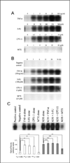
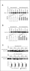
Herpes simplex virus-1 (HSV-1) replication in cancer cells leads to their destruction (viral oncolysis) and has been under investigation as an experimental cancer therapy in clinical trials as single agents, and as combinations with chemotherapy. Cellular responses to chemotherapy modulate viral replication, but these interactions are poorly understood. To investigate the effect of chemotherapy on HSV-1 oncolysis, viral replication in cells exposed to 5-fluorouracil (5-FU), irinotecan (CPT-11), methotrexate (MTX) or a cytokine (tumor necrosis factor-α (TNF-α)) was examined. Exposure of colon and pancreatic cancer cells to 5-FU, CPT-11 or MTX in vitro significantly antagonizes both HSV-1 replication and lytic oncolysis. Nuclear factor-κB (NF-κB) activation is required for efficient viral replication, and experimental inhibition of this response with an IκBα dominant-negative repressor significantly antagonizes HSV-1 replication. Nonetheless, cells exposed to 5-FU, CPT-11, TNF-α or HSV-1 activate NF-κB. Cells exposed to MTX do not activate NF-κB, suggesting a possible role for NF-κB inhibition in the decreased viral replication observed following exposure to MTX. The role of eukaryotic initiation factor 2α (eIF-2α) dephosphorylation was examined; HSV-1-mediated eIF-2α dephosphorylation proceeds normally in HT29 cells exposed to 5-FU, CPT-11 or MTX. This report demonstrates that cellular responses to chemotherapeutic agents provide an unfavorable environment for HSV-1-mediated oncolysis, and these observations are relevant to the design of both preclinical and clinical studies of HSV-1 oncolysis.
Conflict of interest statement
We declare that we have no conflict of interest.
Figures


Similar articles
-
Positron emission tomography of herpes simplex virus 1 oncolysis.Cancer Res. 2007 Apr 1;67(7):3295-300. doi: 10.1158/0008-5472.CAN-06-4062. Cancer Res. 2007. PMID: 17409438
-
Resveratrol suppresses nuclear factor-kappaB in herpes simplex virus infected cells.Antiviral Res. 2006 Dec;72(3):242-51. doi: 10.1016/j.antiviral.2006.06.011. Epub 2006 Jul 14. Antiviral Res. 2006. PMID: 16876885
-
Oncolysis by viral replication and inhibition of angiogenesis by a replication-conditional herpes simplex virus that expresses mouse endostatin.Cancer. 2004 Aug 15;101(4):869-77. doi: 10.1002/cncr.20434. Cancer. 2004. PMID: 15305421
-
HSV-1 viral oncolysis and molecular imaging with PET.Curr Cancer Drug Targets. 2007 Mar;7(2):175-80. doi: 10.2174/156800907780058871. Curr Cancer Drug Targets. 2007. PMID: 17346109 Review.
-
Replication and Spread of Oncolytic Herpes Simplex Virus in Solid Tumors.Viruses. 2022 Jan 10;14(1):118. doi: 10.3390/v14010118. Viruses. 2022. PMID: 35062322 Free PMC article. Review.
Cited by
-
An Extensive Review on Preclinical and Clinical Trials of Oncolytic Viruses Therapy for Pancreatic Cancer.Front Oncol. 2022 May 24;12:875188. doi: 10.3389/fonc.2022.875188. eCollection 2022. Front Oncol. 2022. PMID: 35686109 Free PMC article. Review.
-
NF-κB Signaling in Targeting Tumor Cells by Oncolytic Viruses-Therapeutic Perspectives.Cancers (Basel). 2018 Nov 8;10(11):426. doi: 10.3390/cancers10110426. Cancers (Basel). 2018. PMID: 30413032 Free PMC article. Review.
-
Immunogenicity of oncolytic vaccinia viruses JX-GFP and TG6002 in a human melanoma in vitro model: studying immunogenic cell death, dendritic cell maturation and interaction with cytotoxic T lymphocytes.Onco Targets Ther. 2017 May 2;10:2389-2401. doi: 10.2147/OTT.S126320. eCollection 2017. Onco Targets Ther. 2017. PMID: 28496337 Free PMC article.
-
Promising oncolytic agents for metastatic breast cancer treatment.Oncolytic Virother. 2015 Jun 3;4:63-73. doi: 10.2147/OV.S63045. eCollection 2015. Oncolytic Virother. 2015. PMID: 27512671 Free PMC article. Review.
-
Chemotherapy and Oncolytic Virotherapy: Advanced Tactics in the War against Cancer.Front Oncol. 2014 Jun 11;4:145. doi: 10.3389/fonc.2014.00145. eCollection 2014. Front Oncol. 2014. PMID: 24967214 Free PMC article. Review.
References
-
- Pawlik TM, Nakamura H, Yoon SS, Mullen JT, Chandrasekhar S, Chiocca EA, et al. Oncolysis of diffuse hepatocellular carcinoma by intravascular administration of a replication-competent, genetically engineered herpesvirus. Cancer Res. 2000;60(11):2790–2795. - PubMed
-
- Yoon SS, Nakamura H, Carroll NM, Bode BP, Chiocca EA, Tanabe KK. An oncolytic herpes simplex virus type 1 selectively destroys diffuse liver metastases from colon carcinoma. FASEB J. 2000;14(2):301–311. - PubMed
-
- Mullen JT, Donahue JM, Chandrasekhar S, Yoon SS, Liu W, Ellis LM, et al. Oncolysis by viral replication and inhibition of angiogenesis by a replication-conditional herpes simplex virus that expresses mouse endostatin. Cancer. 2004;101(4):869–877. - PubMed
-
- Nakamura H, Mullen JT, Chandrasekhar S, Pawlik TM, Yoon SS, Tanabe KK. Multimodality therapy with a replication-conditional herpes simplex virus 1 mutant that expresses yeast cytosine deaminase for intratumoral conversion of 5-fluorocytosine to 5-fluorouracil. Cancer Res. 2001;61(14):5447–5452. - PubMed
Publication types
MeSH terms
Substances
Grants and funding
LinkOut - more resources
Full Text Sources
Other Literature Sources
Medical

