

Cigarette smoke increases BLT2 receptor functions in bronchial epithelial cells: in vitro and ex vivo evidence
- PMID: 23347335
- PMCID: PMC3647190
- DOI: 10.1111/imm.12077
Cigarette smoke increases BLT2 receptor functions in bronchial epithelial cells: in vitro and ex vivo evidence
Abstract
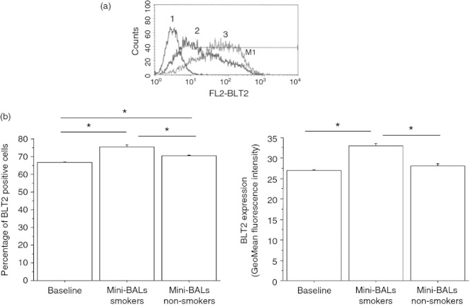
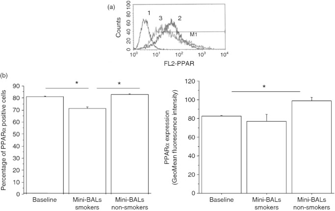
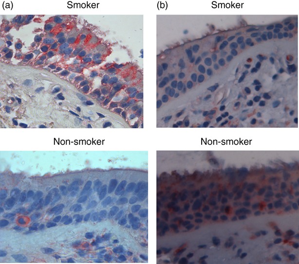
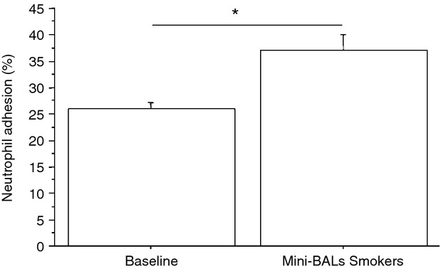
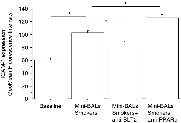
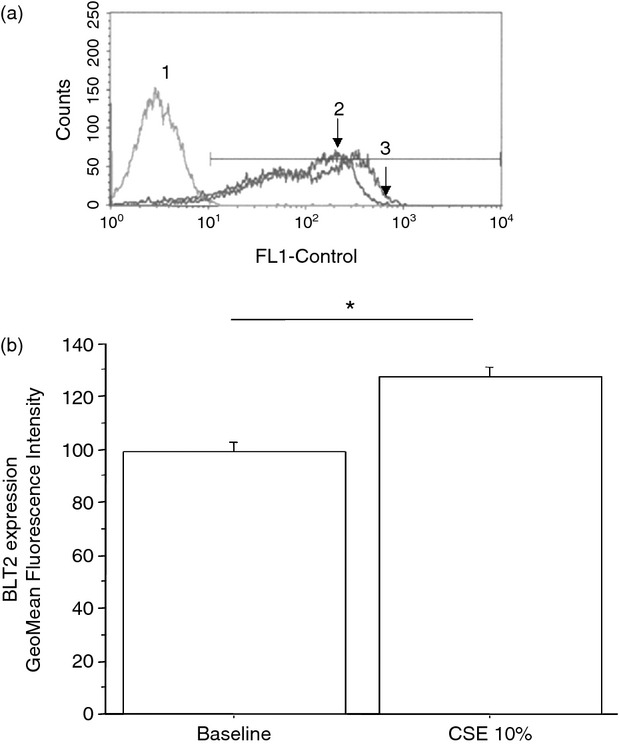
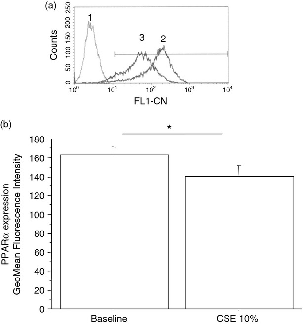
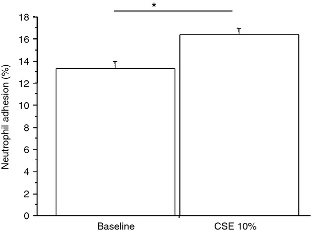
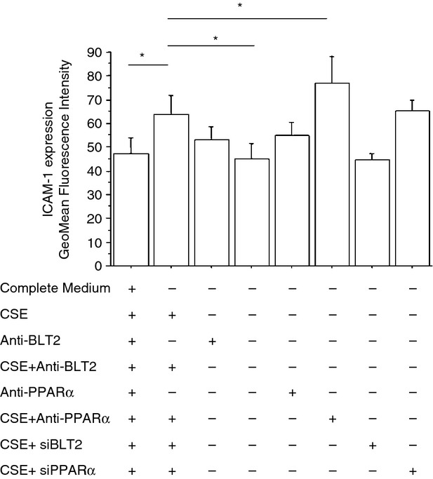
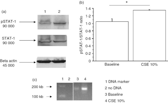
Leukotriene B(4) (LTB(4)) is a neutrophil chemotactic molecule with important involvement in the inflammatory responses of chronic obstructive pulmonary disease (COPD). Airway epithelium is emerging as a regulator of innate immune responses to a variety of insults including cigarette smoke, the major risk factor for COPD. In this study we have explored whether cigarette smoke extracts (CSE) or soluble mediators present in distal lung fluid samples (mini-bronchoalveolar lavages) from smokers alter the expression of the LTB(4) receptor 2 (BLT2) and peroxisome proliferator-activated receptor-α (PPAR-α) in bronchial epithelial cells. We also evaluated the effects of CSE on the expression of intercellular adhesion molecule 1 (ICAM-1) and on the binding of signal transducer and activator of transcription 1 (STAT-1) to ICAM-1 promoter as well as the adhesiveness of neutrophils to bronchial epithelial cells. CSE and mini-bronchoalveolar lavages from smokers increased BLT2 and ICAM-1 expression as well as the adhesiveness of neutrophils to bronchial epithelial cells and decreased PPAR-α expression. CSE induced the activation of STAT-1 and its binding to ICAM-1 promoter. These findings suggest that, in bronchial epithelial cells, CSE promote a prevalent induction of pro-inflammatory BLT2 receptors and activate mechanisms leading to increased neutrophil adhesion, a mechanism that contributes to airway neutrophilia and to tissue damage.
© 2013 Blackwell Publishing Ltd.
Figures
Similar articles
-
Carbocysteine regulates innate immune responses and senescence processes in cigarette smoke stimulated bronchial epithelial cells.Toxicol Lett. 2013 Nov 25;223(2):198-204. doi: 10.1016/j.toxlet.2013.09.013. Epub 2013 Sep 25. Toxicol Lett. 2013. PMID: 24076166
-
Aryl hydrocarbon receptor (AhR) attenuation of subchronic cigarette smoke-induced pulmonary neutrophilia is associated with retention of nuclear RelB and suppression of intercellular adhesion molecule-1 (ICAM-1).Toxicol Sci. 2014 Jul;140(1):204-23. doi: 10.1093/toxsci/kfu068. Epub 2014 Apr 20. Toxicol Sci. 2014. PMID: 24752502
-
Pleural mesothelial cells express both BLT2 and PPARalpha and mount an integrated response to pleural leukotriene B4.J Immunol. 2008 Nov 15;181(10):7292-9. doi: 10.4049/jimmunol.181.10.7292. J Immunol. 2008. PMID: 18981151
-
Cysteinyl leukotriene-1 receptor activation in a human bronchial epithelial cell line leads to signal transducer and activator of transcription 1-mediated eosinophil adhesion.J Pharmacol Exp Ther. 2008 Jun;325(3):1024-30. doi: 10.1124/jpet.107.131649. Epub 2008 Feb 27. J Pharmacol Exp Ther. 2008. PMID: 18305014
-
Bacterial-induced release of inflammatory mediators by bronchial epithelial cells.Eur Respir J. 1996 Sep;9(9):1913-22. doi: 10.1183/09031936.96.09091913. Eur Respir J. 1996. PMID: 8880112 Review.
Cited by
-
Lactobacillus rhamnosus Restores Antiviral Signaling and Attenuates Cytokines Secretion from Human Bronchial Epithelial Cells Exposed to Cigarette Smoke and Infected with SARS-CoV-2.Probiotics Antimicrob Proteins. 2023 Dec;15(6):1513-1528. doi: 10.1007/s12602-022-09998-2. Epub 2022 Nov 8. Probiotics Antimicrob Proteins. 2023. PMID: 36346611 Free PMC article.
-
Leukotriene B4 Receptors Are Necessary for the Stimulation of NLRP3 Inflammasome and IL-1β Synthesis in Neutrophil-Dominant Asthmatic Airway Inflammation.Biomedicines. 2021 May 11;9(5):535. doi: 10.3390/biomedicines9050535. Biomedicines. 2021. PMID: 34064821 Free PMC article.
-
Leukotriene B4 Receptor 2 Mediates the Production of G-CSF That Plays a Critical Role in Steroid-Resistant Neutrophilic Airway Inflammation.Biomedicines. 2022 Nov 19;10(11):2979. doi: 10.3390/biomedicines10112979. Biomedicines. 2022. PMID: 36428547 Free PMC article.
-
Leukotriene B4 receptor-2 contributes to KRAS-driven lung tumor formation by promoting interleukin-6-mediated inflammation.Exp Mol Med. 2021 Oct;53(10):1559-1568. doi: 10.1038/s12276-021-00682-z. Epub 2021 Oct 11. Exp Mol Med. 2021. PMID: 34635780 Free PMC article.
-
Importance of the leukotriene B4-BLT1 and LTB4-BLT2 pathways in asthma.Semin Immunol. 2017 Oct;33:44-51. doi: 10.1016/j.smim.2017.08.005. Semin Immunol. 2017. PMID: 29042028 Free PMC article. Review.
References
-
- Proud D, Leigh R. Epithelial cells and airway diseases. Immunol Rev. 2011;242:186–204. - PubMed
-
- Crooks SW, Bayley DL, Hill SL, Stockley RA. Bronchial inflammation in acute bacterial exacerbations of chronic bronchitis: the role of leukotriene B4. Eur Respir J. 2000;15:274–80. - PubMed
-
- Hill AT, Bayley D, Stockley RA. The interrelationship of sputum inflammatory markers in patients with chronic bronchitis. Am J Respir Crit Care Med. 1999;160:893–8. - PubMed
-
- Gompertz S, O'Brien C, Bayley DL, Hill SL, Stockley RA. Changes in bronchial inflammation during acute exacerbations of chronic bronchitis. Eur Respir J. 2001;17:1112–9. - PubMed
Publication types
MeSH terms
Substances
LinkOut - more resources
Full Text Sources
Other Literature Sources
Research Materials
Miscellaneous
