

Magnetic nanoparticles for tumor imaging and therapy: a so-called theranostic system
- PMID: 23344909
- PMCID: PMC4171953
- DOI: 10.1007/s11095-013-0982-y
Magnetic nanoparticles for tumor imaging and therapy: a so-called theranostic system
Abstract
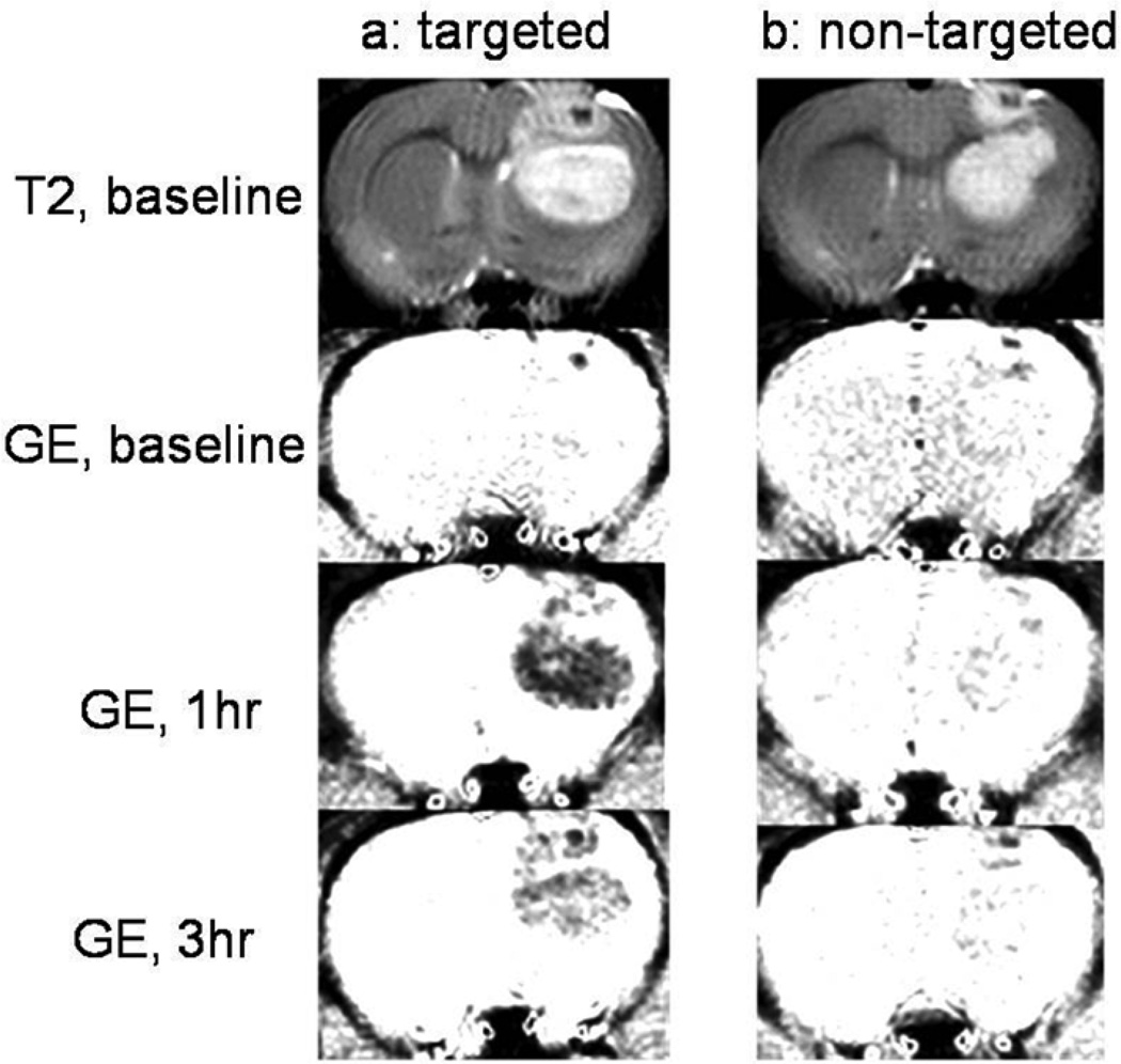
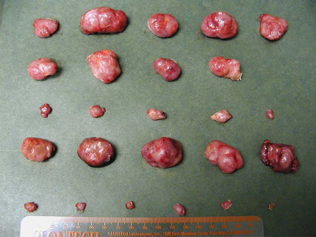
In this review, we discussed the establishment of a so-called "theranostic" system by instituting the basic principles including the use of: [1] magnetic iron oxide nanoparticles (MION)-based drug carrier; [2] intra-arterial (I.A.) magnetic targeting; [3] macromolecular drugs with unmatched therapeutic potency and a repetitive reaction mechanism; [4] cell-penetrating peptide-mediated cellular drug uptake; and [5] heparin/protamine-regulated prodrug protection and tumor-specific drug re-activation into one single drug delivery system to overcome all possible obstacles, thereby achieving a potentially non-invasive, magnetic resonance imaging-guided, clinically enabled yet minimally toxic brain tumor drug therapy. By applying a topography-optimized I.A. magnetic targeting to dodge rapid organ clearance of the carrier during its first passage into the circulation, tumor capture of MION was enriched by >350 folds over that by conventional passive enhanced permeability and retention targeting. By adopting the prodrug strategy, we observed by far the first experimental success in a rat model of delivering micro-gram quantity of the large β-galactosidase model protein selectively into a brain tumor but not to the ipsi- or contra-lateral normal brain regions. With the therapeutic regimens of most toxin/siRNA drugs to fully (>99.9%) eradicate a tumor being in the nano-molar range, the prospects of reaching this threshold become practically accomplishable.
Figures


Similar articles
-
Brain tumor targeting of magnetic nanoparticles for potential drug delivery: effect of administration route and magnetic field topography.J Control Release. 2011 Nov 7;155(3):393-9. doi: 10.1016/j.jconrel.2011.06.033. Epub 2011 Jul 7. J Control Release. 2011. PMID: 21763736 Free PMC article.
-
Magnetically-enabled and MR-monitored selective brain tumor protein delivery in rats via magnetic nanocarriers.Biomaterials. 2011 Sep;32(26):6245-53. doi: 10.1016/j.biomaterials.2011.05.004. Epub 2011 May 31. Biomaterials. 2011. PMID: 21632104 Free PMC article.
-
Polymeric magnetic nanoparticles: a multitargeting approach for brain tumour therapy and imaging.Drug Deliv Transl Res. 2022 Jul;12(7):1588-1604. doi: 10.1007/s13346-021-01063-9. Epub 2021 Sep 19. Drug Deliv Transl Res. 2022. PMID: 34537930 Review.
-
Development of a magnetic nano-graphene oxide carrier for improved glioma-targeted drug delivery and imaging: In vitro and in vivo evaluations.Chem Biol Interact. 2018 Nov 1;295:97-108. doi: 10.1016/j.cbi.2018.08.027. Epub 2018 Aug 28. Chem Biol Interact. 2018. PMID: 30170108
-
Magnetic iron oxide nanoparticles as drug carriers: clinical relevance.Nanomedicine (Lond). 2018 Apr;13(8):953-971. doi: 10.2217/nnm-2017-0336. Epub 2018 Jan 29. Nanomedicine (Lond). 2018. PMID: 29376469 Review.
Cited by
-
Targeted iron-oxide nanoparticle for photodynamic therapy and imaging of head and neck cancer.ACS Nano. 2014 Jul 22;8(7):6620-32. doi: 10.1021/nn501652j. ACS Nano. 2014. PMID: 24923902 Free PMC article.
-
Nanocarriers Call the Last Shot in the Treatment of Brain Cancers.Technol Cancer Res Treat. 2022 Jan-Dec;21:15330338221080974. doi: 10.1177/15330338221080974. Technol Cancer Res Treat. 2022. PMID: 35253549 Free PMC article. Review.
-
Superparamagnetic iron oxide nanoparticles mediated (131)I-hVEGF siRNA inhibits hepatocellular carcinoma tumor growth in nude mice.BMC Cancer. 2014 Feb 21;14:114. doi: 10.1186/1471-2407-14-114. BMC Cancer. 2014. PMID: 24555445 Free PMC article.
-
Status Quo and Trends of Intra-Arterial Therapy for Brain Tumors: A Bibliometric and Clinical Trials Analysis.Pharmaceutics. 2021 Nov 6;13(11):1885. doi: 10.3390/pharmaceutics13111885. Pharmaceutics. 2021. PMID: 34834300 Free PMC article. Review.
-
Lipopeptide-Coated Iron Oxide Nanoparticles as Potential Glycoconjugate-Based Synthetic Anticancer Vaccines.ACS Appl Mater Interfaces. 2015 Aug 12;7(31):17535-44. doi: 10.1021/acsami.5b05497. Epub 2015 Aug 3. ACS Appl Mater Interfaces. 2015. PMID: 26200668 Free PMC article.
References
-
- National cancer institute at the u.S. National institute of health webpage. Available from: http://www.cancer.gov.
-
- The children’s brain tumor foundation webpage. Available from: http://www.cbtf.org/medical.b.html.
-
- The brain tumor society webpage. Available from: http://www.tbts.org.
-
- Grieg NH, Ries LG, Yancik R, Rapoport SI. Increasing annual incidence of primary malignant brain tumors in the elderly. J Natl Cancer I. 1990;82(20):1621–1624. - PubMed
-
- Black PM. Brain tumors. New Engl J Med. 1991;324(21):1471–1476. - PubMed
Publication types
MeSH terms
Substances
Grants and funding
LinkOut - more resources
Full Text Sources
Other Literature Sources
Medical
