. 2013 Mar 15;288(11):7781-7790.
doi: 10.1074/jbc.M112.441907. Epub 2013 Jan 22.

Ubiquitinated proteins activate the proteasomal ATPases by binding to Usp14 or Uch37 homologs

Affiliations

Ubiquitinated proteins activate the proteasomal ATPases by binding to Usp14 or Uch37 homologs

Andreas Peth et al. J Biol Chem. .

Abstract

Degradation of ubiquitinated proteins by 26 S proteasomes requires ATP hydrolysis, but it is unclear how the proteasomal ATPases are regulated and how proteolysis, substrate deubiquitination, degradation, and ATP hydrolysis are coordinated. Polyubiquitinated proteins were shown to stimulate ATP hydrolysis by purified proteasomes, but only if the proteins contain a loosely folded domain. If they were not ubiquitinated, such proteins did not increase ATPase activity. However, they did so upon addition of ubiquitin aldehyde, which mimics the ubiquitin chain and binds to 26 S-associated deubiquitinating enzymes (DUBs): in yeast to Ubp6, which is essential for the ATPase activation, and in mammalian 26 S to the Ubp6 homolog, Usp14, and Uch37. Occupancy of either DUB by a ubiquitin conjugate leads to ATPase stimulation, thereby coupling deubiquitination and ATP hydrolysis. Thus, ubiquitinated loosely folded proteins, after becoming bound to the 26 S, interact with Ubp6/Usp14 or Uch37 to activate ATP hydrolysis and enhance their own destruction.


Figures

FIGURE 1.
FIGURE 1.
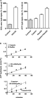
Ub conjugates stimulate ATPase activity of 26 S proteasomes. A, ATP hydrolysis by rabbit muscle 26 S proteasomes was measured in the presence of E6AP or ubiquitinated E6AP. Production of free phosphate (Pi) was detected using the malachite green assay. B, the rabbit muscle 26 S ATPase activity and peptide hydrolysis were measured simultaneously in the presence of E6AP or ubiquitinated E6AP, which were added at the outset (time 0), and the increase in activity was observed with time. Proteasomal ATPase activity at the outset was taken as 100% and was measured using the coupled pyruvate kinase/lactate dehydrogenase enzymatic assay (upper panel), and peptidase activity using suc-GGL-amc (lower panel). All values are the means of at least three experiments ± S.E.
FIGURE 2.
FIGURE 2.
The stimulation of ATPase activity requires binding of the substrate to two sites on the 26 S proteasome. A, peptide hydrolysis by MEF 26 S proteasomes was measured in the presence or absence of hexaubiquitin (HexUb; left panel). Peptidase activity in the absence of hexaubiquitin was taken at 100%. Rates of ATP hydrolysis by 26 S proteasomes were measured in the presence of casein (1 μm), linear hexaubiquitin (1 μm), or a combination of the two, by measuring the accumulation of free phosphate over time (right panel). Rate of ATP hydrolysis in the absence of casein or hexaubiquitin was taken as 100%. B, ATPase activity of rabbit 26 S proteasomes was measured by the release of free phosphate over time in the presence of casein (top panel). ATPase activity was measured in the presence of Ub or Ub aldehyde (middle panel). ATPase activity was measured in the presence of casein plus Ub or casein plus Ub aldehyde (bottom panel). The variations in the basal rate of ATP hydrolysis between the top/middle panel and the bottom panel are caused by using different 26 S preparations. All values are the means of at least three experiments ± S.E.
FIGURE 3.
FIGURE 3.
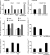
The stimulation of ATPase activity by Ub conjugates in yeast proteasomes requires Ubp6. A, ATP hydrolysis by ΔUbp6 yeast 26 S proteasomes was measured in the presence of E6AP or ubiquitinated E6AP or casein plus Ub or casein plus Ub aldehyde. The stimulation of ATP hydrolysis was restored by addition of purified Ubp6p (gray bars) in the presence of ubiquitinated E6AP or casein plus Ub aldehyde. ATP hydrolysis in the presence of E6AP was taken as 100%. B, ATP hydrolysis by ΔUbp6 yeast 26 S proteasomes was measured in the presence of Ub, casein, or casein plus Ub. The stimulation of ATP hydrolysis was restored by addition of purified Ubp6C118Ap (gray bars), which lacks DUB activity in the presence of casein plus Ub. ATP hydrolysis in the presence of Ub was taken as 100%. C, peptide hydrolysis of GGL-amc in wt and Δrpn10 yeast 26 S proteasomes was measured in the presence of E6AP, ubiquitinated E6AP, Ub, and Ub aldehyde. D, total ubiquitin-amc hydrolysis by wt 26 S particles (1 nm) was measured. IU1 (50 μm) shows only a slight inhibition, whereas ubiquitin aldehyde (500 nm) completely blocks DUB activity. E, 26 S ATPase activity (10 nm) was measured in the presence of E6AP (500 nm), ubiquitinated E6AP, or ubiquitinated E6AP after the proteasomes where pretreated with 50 μm IU1. F, 26 S ATPase activity (10 nm) was measured in the presence of casein (1 μm), casein plus IU1 (50 μm), or casein plus ubiquitin aldehyde (500 nm). All values are the means of at least three experiments ± S.E.
FIGURE 4.
FIGURE 4.
Uch37 also can promote the stimulation of ATP hydrolysis 26 S proteasomes by Ub conjugates. A, 26 S associated DUBs in proteasomes from wt and Usp14−/− MEF cells were labeled with HA-Ub vinylsulfone and detected by Western blotting with anti-HA, anti-Usp14, and anti-Uch37 antibodies. B, stimulation of gate opening (i.e. enhanced peptide hydrolysis) by ubiquitinated E6AP was measured in wt and ΔUsp14 proteasomes. C, stimulation of 26 S ATP hydrolysis was measured by the addition of ubiquitinated E6AP in wt and ΔUsp14 proteasomes. D, stimulation of gate opening (i.e. enhanced peptide hydrolysis) by ubiquitinated Sic1 was measured in ΔUsp14 proteasomes with and without blocking of Uch37 active site with Ub vinylsulfone (Ub-Vs). All values are the means of at least three experiments ± S.E. AU, arbitrary units.
FIGURE 5.
FIGURE 5.
Loosely folded (but not tightly folded) substrates stimulate ATP hydrolysis. A, ATP hydrolysis by rabbit muscle 26 S proteasomes was measured as described in Fig. 1 in the presence of Ub5-DHFR with and without methotrexate (MTA). 26 S ATPase activity in the presence of MTA was used as a control. B, ATP hydrolysis was measured in the presence of DHFR with and without Ub aldehyde or methotrexate (MTA). ATPase activity in the presence of DHFR was taken as 100%. C, ATP hydrolysis by 26 S particles purified from yeast ubp6C118A mutant strain was measured in the presence of Ub and native or molten globular lysozyme, which was pretreated with 800 mm guanidine hydrochloride. ATPase activity in the presence of Ub was taken as 100%. D, ATP hydrolysis by rabbit muscle 26 S proteasomes was measured in the presence of non-ubiquitinated E6AP (black bars) or Nedd4 (gray bars) or after both Ub ligases were allowed to autoubiquitinate with Lys-48 or Lys-63 linked tetra-Ub. ATPase activity in the presence of E6AP was taken as 100%. E, ATP hydrolysis was measured in the presence of non-ubiquitinated E6AP or Nedd4 together with Ub (black bars) or Ub aldehyde (gray bars). ATPase activity in the presence of the unmodified E3s was taken as 100%. All values are the means of at least three experiments ± S.E.
FIGURE 6.
FIGURE 6.
Ubiquitin conjugates activate their own degradation by the 26 S proteasome. Ub conjugates initially bind reversibly to Rpn10 and Rpn13. The Ub chain interacts then with the 19 S-associated DUBs Usp14 and Uch37 trimming the chain from its distal end leading to gate opening substrates with a loosely folded domain suitable for the initiation of degradation also stimulate ATP hydrolysis when binding directly to the ATPase subunits. This transient increase in ATP hydrolysis and gate opening allows for maximum rates of degradation while the polypeptide is hydrolyzed.

Similar articles

Cited by

References

    1. Groll M., Bajorek M., Köhler A., Moroder L., Rubin D. M., Huber R., Glickman M. H., Finley D. (2000) A gated channel into the proteasome core particle. Nat. Struct. Biol. 7, 1062–1067 - PubMed
    1. Elsasser S., Chandler-Militello D., Müller B., Hanna J., Finley D. (2004) Rad23 and Rpn10 serve as alternative ubiquitin receptors for the proteasome. J. Biol. Chem. 279, 26817–26822 - PubMed
    1. Husnjak K., Elsasser S., Zhang N., Chen X., Randles L., Shi Y., Hofmann K., Walters K. J., Finley D., Dikic I. (2008) Proteasome subunit Rpn13 is a novel ubiquitin receptor. Nature 453, 481–488 - PMC - PubMed
    1. Liu C. W., Li X., Thompson D., Wooding K., Chang T. L., Tang Z., Yu H., Thomas P. J., DeMartino G. N. (2006) ATP binding and ATP hydrolysis play distinct roles in the function of 26 S proteasome. Mol. Cell 24, 39–50 - PMC - PubMed
    1. Peth A., Uchiki T., Goldberg A. L. (2010) ATP-dependent steps in the binding of ubiquitin conjugates to the 26S proteasome that commit to degradation. Mol. Cell 40, 671–681 - PMC - PubMed

Publication types