. 2013 Jul;70(14):2555-68.
doi: 10.1007/s00018-012-1242-8. Epub 2013 Jan 19.

Immobile survival of motoneuron (SMN) protein stored in Cajal bodies can be mobilized by protein interactions

Affiliations

Immobile survival of motoneuron (SMN) protein stored in Cajal bodies can be mobilized by protein interactions

Benjamin Förthmann et al. Cell Mol Life Sci. 2013 Jul.

Abstract

Reduced levels of survival of motoneuron (SMN) protein lead to spinal muscular atrophy, but it is still unknown how SMN protects motoneurons in the spinal cord against degeneration. In the nucleus, SMN is associated with two types of nuclear bodies denoted as gems and Cajal bodies (CBs). The 23 kDa isoform of fibroblast growth factor-2 (FGF-2(23)) is a nuclear protein that binds to SMN and destabilizes the SMN-Gemin2 complex. In the present study, we show that FGF-2(23) depletes SMN from CBs without affecting their general structure. FRAP analysis of SMN-EGFP in CBs demonstrated that the majority of SMN in CBs remained mobile and allowed quantification of fast, slow and immobile nuclear SMN populations. The potential for SMN release was confirmed by in vivo photoconversion of SMN-Dendra2, indicating that CBs concentrate immobile SMN that could have a specialized function in CBs. FGF-2(23) accelerated SMN release from CBs, accompanied by a conversion of immobile SMN into a mobile population. Furthermore, FGF-2(23) caused snRNP accumulation in CBs. We propose a model in which Cajal bodies store immobile SMN that can be mobilized by its nuclear interaction partner FGF-2(23), leading to U4 snRNP accumulation in CBs, indicating a role for immobile SMN in tri-snRNP assembly.


Figures

Fig. 1
Fig. 1
Different size of Cajal bodies and gems in HEK cells. HEK293T cells were cotransfected with pSMN-EGFP and pFGF-223-DsRed2 or, as a control, pFGF-218-DsRed2 24 h before image acquisition. Subsequently, the diameters of coilin-positive (FGF-218: n = 52; FGF-223: n = 24) and coilin-negative (FGF-218: n = 19, FGF-223: n = 16) nuclear bodies were determined. The diameters of SMN- and coilin-positive nuclear bodies (CBs) were significantly larger than nuclear bodies that were only SMN positive (gems). The overexpression of FGF-223 showed no influence on the size of nuclear bodies compared to FGF-218 overexpressing cells (means ± SEM; ***p < 0.001; n.s. non-significant; Mann-Whitney test)
Fig. 2
Fig. 2
FRAP reveals mobilization of SMN from Cajal bodies by FGF-223. For live cell imaging, single CBs of pSMN-EGFP and pFGF-223-DsRed2 or pFGF-218-DsRed2 or pDsRed2 coexpressing HEK293T cells were bleached for 1 s (405 nm). Fluorescence recovery was measured 10 s before and 10 s after photobleaching and then every minute up to 20 min unless the focused nuclear body left the volume as defined by z-sections. a Example of a single FRAP measurement. Fitting of a one-exponential curve is represented by the black line; fitting of a two-exponential curve is shown by the green line. Data display best fit to a two-exponential model. b Example of a SMN-EGFP and FGF-223-DsRed2 coexpressing cell before and after photobleaching. Scale bar 10 μm. c Summary kinetics of averaged FRAP measurements (n = 10). FGF-223-DsRed2 increased the fluorescence recovery of SMN-EGFP to CBs. d The immobile populations of SMN-EGFP was significantly decreased after overexpression of FGF-223-DsRed2. The fast population of SMN-EGFP was significantly increased compared to the DsRed2 control. The slow populations were not significantly altered. The recovery halftimes (t 1/2) of the slow and fast populations of SMN-EGFP were non-significantly changed after overexpression of FGF-223-DsRed2 (means ± SEM; *p < 0.05; n.s. non-significant; unpaired, two-tailed t test)
Fig. 3
Fig. 3
Photoconversion of Dendra2-tagged SMN confirms mobilization of SMN by FGF-223. Live cell photoconversion experiments were performed using HEK293T cells coexpressing pSMN-Dendra2 and pFGF-223-ECFP or pFGF-218-ECFP or pECFP. SMN-Dendra2 in CBs was photoconverted for 1 s (405 nm). Fluorescence loss was measured 10 s before, 10 s after conversion and then every minute up to 20 min unless the nuclear body left the defined volume. a Example of a SMN-Dendra2 and FGF-223-ECFP (not shown) coexpressing cell before and after photoconversion. Scale bar 10 μm. b Averaged photoconversion measurements were fitted to a one-exponential curve and statistically analyzed (n = 10). Coexpression of FGF-223-ECFP increased the loss of red fluorescence of SMN-Dendra2 from large nuclear bodies after photoconversion. c The immobile population was significantly decreased after overexpression of FGF-223-ECFP; the mobile population of SMN-EGFP was significantly increased compared to the ECFP control. The slow populations were not significantly altered. The recovery halftimes (t 1/2) of the mobile population of SMN-EGFP were non-significantly changed after overexpression of FGF-223-ECFP (means ± SEM; *p < 0.05; ***p < 0.001; n.s. non-significant; unpaired two tailed t test)
Fig. 4
Fig. 4
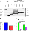
FGF-223 reduces SMN binding to coilin. a Endogenous coilin was purified from lysates of HEK293T cells using recombinantly expressed FGF-223, FGF-218 and GST fusion of SMN coupled to glutathione beads (SMN-GST) and analyzed by Western blot. GST was used as a control (GST control). FGF-223 decreased binding of coilin to SMN-GST. Because of the high amounts of FGF-218 used, a slight unspecific binding of FGF-218 to SMN-GST was found. After SMN-GST pulldown, also fragments of coilin (anti-coilin) and fragments of SMN-GST (anti-GST) were detected. b The level of endogenous, SMN-bound coilin was decreased in lysates of cells with FGF-223 (densitometrical analysis of the highest band of coilin). c To determine changes of coilin-SMN association in CBs in vivo, HEK293T cells were transfected with pFGF-223-DsRed2 (dd’) or pFGF-218-DsRed2 (as a control, ee”). Cells were fixed and immunostained with anti-coilin (d3e3”) and anti-SMN (d2e2”). Nuclei were analyzed by counting SMN-positive or -negative CBs (FGF-218: n = 119, FGF-223: n = 132) as demonstrated in the merged pictures (d4e4”). The distribution of SMN-positive (yellow arrows) and -negative (red arrows) CBs was significantly changed after overexpression of FGF-223-DsRed2 (c, ***p < 0.001, χ 2 test)
Fig. 4
Fig. 4
FGF-223 reduces SMN binding to coilin. a Endogenous coilin was purified from lysates of HEK293T cells using recombinantly expressed FGF-223, FGF-218 and GST fusion of SMN coupled to glutathione beads (SMN-GST) and analyzed by Western blot. GST was used as a control (GST control). FGF-223 decreased binding of coilin to SMN-GST. Because of the high amounts of FGF-218 used, a slight unspecific binding of FGF-218 to SMN-GST was found. After SMN-GST pulldown, also fragments of coilin (anti-coilin) and fragments of SMN-GST (anti-GST) were detected. b The level of endogenous, SMN-bound coilin was decreased in lysates of cells with FGF-223 (densitometrical analysis of the highest band of coilin). c To determine changes of coilin-SMN association in CBs in vivo, HEK293T cells were transfected with pFGF-223-DsRed2 (dd’) or pFGF-218-DsRed2 (as a control, ee”). Cells were fixed and immunostained with anti-coilin (d3e3”) and anti-SMN (d2e2”). Nuclei were analyzed by counting SMN-positive or -negative CBs (FGF-218: n = 119, FGF-223: n = 132) as demonstrated in the merged pictures (d4e4”). The distribution of SMN-positive (yellow arrows) and -negative (red arrows) CBs was significantly changed after overexpression of FGF-223-DsRed2 (c, ***p < 0.001, χ 2 test)
Fig. 5
Fig. 5
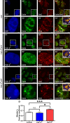
SnRNPs accumulate at CBs after reduction of immobile SMN by FGF-223. HEK293T cells were transfected with pFGF-223-ECFP (c1, c1’) or as controls pFGF-218-ECFP (b1, b1’) or pECFP (a1, a1’) 24 h before fixation. Cells were fixed and immunostained with anti-coilin (a2c2’) and anti-SmE (a3c3’). Coilin and SmE showed colocalization in the nucleus as demonstrated in the merged images (a4c4’) primarily in CBs. Arrows point to CBs that were enlarged. Intensity correlation analysis demonstrated in PDM (product of differences from the mean) images (a4’, b4’, c4’; LUT: orange, positive PDM; blue, negative PDM) showed a significant increase in colocalization of coilin and SmE in CBs after overexpression of pFGF-223-ECFP (M means ± SEM; *p < 0.05, ***p < 0.001; Mann-Whitney test; CFP: n = 45, FGF-218: n = 45, FGF-223: n = 45, pooled from 3 independent experiments). Scale bar 10 μm (a, b, c), 2 μm (a’, b’, c’), 0.2 μm (PDM images)
Fig. 6
Fig. 6
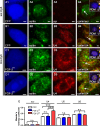
Reduction of immobile SMN by FGF-223 leads to accumulation of U4 snRNPs at CBs. Fluorescence in situ hybridization (FISH, a3d3) with probes for U4, U5 and U6 snRNAs and immunostainings with anti-coilin (a2d2) were performed in HEK293T cells transfected with pFGF-223-ECFP (d1) or, as controls, pFGF-218-ECFP (c1) or pECFP (a1, b1) 24 h before fixation. Arrows point to the CBs in the merged images (a4d4) that were enlarged in the inserted PDM images. Here, intensity correlation analyses are demonstrated (LUT: orange, positive PDM; blue, negative PDM). Colocalized pixels (both channels vary synchronically from the mean pixel intensity) are represented by positive PDM (yellow). A significant increase in colocalization of coilin and U4 snRNP in CBs after overexpression of pFGF-223-ECFP was detected (E means ± SEM; ***p < 0.001; Mann-Whitney test; CFP: n = 45, FGF-218: n = 45, FGF-223: n = 45, pooled from 3 independent experiments). Scale bar, 2 μm (a1d4), 0.2 μm (insets). Stainings of U5 and U6 snRNPs and additional stainings of U4 snRNPs are shown in Supplemental Fig. 2
Fig. 7
Fig. 7
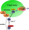
Molecular model for functional effects of FGF-223/SMN complex formation. Complex formation with FGF-223 results in a release of SMN stored and immobilized in CBs. Different populations of mobile and immobile SMN indicate equilibrium with several other interacting proteins. Additional interaction partners like FGF-223 change the balance of this network, leading to a mobilization of SMN from the CBs. Immobile SMN at CBs appears to have a function in the assembly of tri-snRNPs. Therefore, decreasing the amount of immobile SMN likely results in an accumulation of U4 snRNPs and indicates a dysfunction in tri-snRNP assembly

Similar articles

Cited by

References

    1. Liu Q, Dreyfuss G. A novel nuclear structure containing the survival of motor neurons protein. EMBO J. 1996;15:3555–3565. - PMC - PubMed
    1. Matera AG, Frey MR. Coiled bodies and gems: janus or gemini? Am J Hum Genet. 1998;63:317–321. doi: 10.1086/301992. - DOI - PMC - PubMed
    1. Meister G, Eggert C, Fischer U. SMN-mediated assembly of RNPs: a complex story. Trends Cell Biol. 2002;12:472–478. doi: 10.1016/S0962-8924(02)02371-1. - DOI - PubMed
    1. Paushkin S, Gubitz AK, Massenet S, Dreyfuss G. The SMN complex, an assemblyosome of ribonucleoproteins. Curr Opin Cell Biol. 2002;14:305–312. doi: 10.1016/S0955-0674(02)00332-0. - DOI - PubMed
    1. Lefebvre S, Burglen L, Reboullet S, Clermont O, Burlet P, Viollet L, Benichou B, Cruaud C, Millasseau P, Zeviani M, et al. Identification and characterization of a spinal muscular atrophy-determining gene. Cell. 1995;80:155–165. doi: 10.1016/0092-8674(95)90460-3. - DOI - PubMed

Publication types

MeSH terms

LinkOut - more resources