. 2013 Mar;24(6):704-14.
doi: 10.1091/mbc.E12-06-0471. Epub 2013 Jan 16.

p120-catenin and β-catenin differentially regulate cadherin adhesive function

Affiliations

p120-catenin and β-catenin differentially regulate cadherin adhesive function

Rebecca G Oas et al. Mol Biol Cell. 2013 Mar.

Abstract

Vascular endothelial (VE)-cadherin, the major adherens junction adhesion molecule in endothelial cells, interacts with p120-catenin and β-catenin through its cytoplasmic tail. However, the specific functional contributions of the catenins to the establishment of strong adhesion are not fully understood. Here we use bioengineering approaches to identify the roles of cadherin-catenin interactions in promoting strong cellular adhesion and the ability of the cells to spread on an adhesive surface. Our results demonstrate that the domain of VE-cadherin that binds to β-catenin is required for the establishment of strong steady-state adhesion strength. Surprisingly, p120 binding to the cadherin tail had no effect on the strength of adhesion when the available adhesive area was limited. Instead, the binding of VE-cadherin to p120 regulates adhesive contact area in a Rac1-dependent manner. These findings reveal that p120 and β-catenin have distinct but complementary roles in strengthening cadherin-mediated adhesion.


Figures
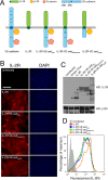
FIGURE 1:
FIGURE 1:
Chimeric receptors are expressed at comparable levels at the plasma membrane. (A) The four chimeric constructs used in this work as adhesive receptors are depicted, along with wild-type VE-cadherin. The extracellular domain of VE-cadherin is replaced with IL-2R to generate IL-2R–VE-cadcyto. A triple-alanine mutation in the p120-binding site that uncouples the cadherin tail from p120 is introduced to generate IL-2R–VE-cadEMD→AAA. The catenin-binding domain is deleted to generate IL-2R–VE-cadΔCBD. The four constructs are introduced into cells by way of adenoviral vectors. (B–D) Human microvascular endothelial cells were transduced with chimeric constructs containing the IL-2R extracellular domain fused to the cytoplasmic domain of VE-cadherin. (B) Expression of the constructs was verified by immunofluorescence microscopy. Cells were stained for IL-2R to detect the receptors and DAPI to show the cell nuclei and enable evaluation of infection rates. IL-2R staining was absent in uninfected cells, and for all four constructs we observed infection rates of 80% or higher. Scale bar, 100 μm. (C) Expression levels were also assessed by Western blot in which whole-cell lysates were probed for IL-2R and with p120 as a loading control. Arrowheads indicate main bands. Higher–molecular weight bands result from IL-2R glycosylation. (D) The levels of expression of the chimeric receptors at the cell surface were assessed using flow cytometry. Unpermeabilized cells were fixed and stained for IL-2R and tested for their peak fluorescence values as compared with those of uninfected control cells. Peak values for each construct occurred within a similar range, indicating that their surface expression was comparable.
FIGURE 2:
FIGURE 2:
Cytoplasmic domains of chimeric receptors recruit p120 and β-catenin to the site of adhesion. (A) Immunoprecipitations were performed to verify that the chimeric receptors were able to complex with the appropriate catenins. Magnetic beads conjugated to antibodies against IL-2R were used to pull down the chimeric receptors, which were subsequently probed for p120 and β-catenin. p120 coprecipitated with IL-2R–VE-cadcyto and IL- 2R–VE-cadΔCBD, and β-catenin was pulled down by IL-2R–VE-cadcyto and IL-2R–VE-cadEMD→AAA. (B, C) To ensure that catenins were recruited to the sites of adhesion mediated by the chimeric constructs, cells expressing the constructs were seeded on micropatterned coverslips (see Figure 3) using IL-2R antibodies as an adhesive ligand. The cells were then extracted using Triton X-100 in a cytoskeleton stabilization buffer and stained for p120, β-catenin, and IL-2R. p120 colocalized with IL-2R–VE-cadcyto and IL-2R–VE-cadΔCBD (B), whereas β-catenin colocalized with IL-2R–VE-cadcyto and IL-2R–VE-cadEMD→AAA (C). Bars, 20 μm. (D, E) Colocalization of IL-2R chimeras with p120 (D) and β-catenin (E) was quantified as Pearson's r. Thick line, median (n = 5–6 cells per group); box, interquartile range; whiskers, full range.
FIGURE 3:
FIGURE 3:
Micropatterned coverslips feature adhesive islands that allow for the generation of regular arrays of evenly spaced cells with well-defined morphologies. Micropatterning occurs through a stepwise process. (A) Glass coverslips are coated with titanium and gold using an electron beam evaporator, and then a PDMS stamp is used to print islands of the self-assembling monolayer across the surface of the coverslip (A, 1). This generates adhesive islands that can passively adsorb ligands that will interact specifically with adhesive receptors on the surface of cells (A, 2). Spaces between the adhesive islands are backfilled with polyethylene glycol to create nonadhesive surfaces around sites of cell adhesion (A, 3). Cells are seeded on the micropatterned coverslips and adhere individually to adhesive islands (A, 4). (B) Cells expressing IL-2R and chimeric constructs adhere to adhesive islands. Micropatterned coverslips are treated with immunoglobulin G directed against the IL-2 receptor, which can be detected with fluorescently labeled secondary antibodies. Cells expressing IL-2R seeded on the micropatterned surfaces form a regular array of single cells attached to adhesive islands.
FIGURE 4:
FIGURE 4:
Linkage between cadherins and the actin cytoskeleton is necessary to strengthen steady-state adhesion. (A) A hydrodynamic spinning-disk device was used to measure the adhesive strength of populations of cells. Cells were seeded onto micropatterned coverslips and allowed to adhere for 16 h. A coverslip with adherent cells was mounted onto the spinning-disk apparatus, and a vacuum pump was used to hold the sample in place. The sample was then submerged into the spin chamber filled with PBS+ with 2 mM dextrose. The chamber was equipped with baffles at the edges, which prevented the spinning motion of the sample from creating a vortex. The sample was then spun for 5 min at a controlled speed (ω), resulting in a gradient of shear force (τ) proportional to the distance from the coverslip center (r). Samples were then fixed, permeabilized, and stained for microscopy and quantification of adherent cells remaining on the coverslip. (B) The cells remaining attached to the coverslip were counted at various positions across the coverslip, and the values were plotted and sigmoid curves were fitted to the combined count totals. (C) Comparisons of adhesion strength values (τ50) among the chimeric constructs. IL-2R was significantly less adhesive than IL-2R–VE-cadcyto and IL-2R–VE-cadEMD→AAA (Tukey test; p < 0.050) but not significantly different from IL-2R–VE-cadΔCBD.
FIGURE 5:
FIGURE 5:
The interaction between p120 and the cadherin juxtamembrane domain is required to promote cell spreading. (A, B) Cells expressing the chimeric constructs were seeded sparsely on unpatterned coverslips coated with antibodies against IL-2R. The cells were allowed to adhere for 30 min before fixation. (A) Representative images of adherent cells expressing the IL-2R constructs. Uninfected cells and those infected with an empty adenoviral vector were not able to attach to the IL-2R–antibody-coated coverslips. Bar, 20 μm. (B) The spread areas of 100 cells per condition, chosen at random, were measured and plotted by quantifying the number of cells per condition whose spread areas were larger than a given area in microns. On these inverse cumulative distribution plots, each data point indicates the percentage of cells (y-axis) that have spread areas greater than a given value (x-axis). Thus a population of cells exhibiting comparatively larger spread areas will generate data points that fall further to the right on the graph than the other populations being compared. The median values of the different groups were found to be statistically different (Kruskal–Wallis test; p < 0.001). IL-2R and IL-2R–VE-cadEMD→AAA were not statistically different from each other, and IL-2R–VE-cadcyto and IL-2R–VE-cadΔCBD were not statistically different from each other. However, both members of the former pair were statistically different from both members of the latter pair (Tukey test; p < 0.05). (C, D) IL-2R, IL-2R–VE-cadcyto, and IL-2R–VE-cadEMD→AAA were expressed in mouse endothelial cells that were either control or p120 null, and their spreading ability was measured as in the previous experiment. (C) In control cells, IL-2R–VE-cadcyto exhibited significantly increased spreading over IL-2R and IL-2R–VE-cadEMD→AAA (Kruskal–Wallis test; p < 0.001; Tukey test; p < 0.05). (D) In p120-null cells, the increased spreading observed in IL-2R–VE-cadcyto was lost (Kruskal–Wallis test; p = 0.230). (D, inset) Surface expression levels of the various chimeras were similar, as measured by immunofluorescence. Thick line, median (n = 28–38 cells per group); box, interquartile range; whiskers, 90% range.
FIGURE 6:
FIGURE 6:
Rac1 inhibition phenocopies the p120-dependent spreading defect. (A) The adhesion strength of primary microvascular endothelial cells expressing IL-2R and IL-2R–VE-cadcyto with and without Rac1 inhibitor NSC23766 treatment was assayed using the hydrodynamic spinning-disk device. The addition of NSC23766 did not significantly affect adhesion strength mediated by the VE-cadherin cytoplasmic tail (Tukey test; p < 0.050). (B–D) Cell spreading was measured in untreated endothelial cells (B), NSC23766-treated endothelial cells (C), and endothelial cells expressing a constitutively active Rac1 mutant (D). Rac1 inhibition prevented cell spreading and eliminated differences between chimeras, whereas constitutively active Rac1 increased spreading.
FIGURE 7:
FIGURE 7:
Interaction between p120 and cadherins strengthens cell adhesion by promoting cell spreading. A proposed model demonstrating the distinct contributions of p120 and β-catenin to strengthening adhesion.

Similar articles

Cited by

References

    1. Ades EW, Candal FJ, Swerlick RA, George VG, Summers S, Bosse DC, Lawley TJ. HMEC-1: establishment of an immortalized human microvascular endothelial cell line. J Invest Dermatol. 1992;99:683–690. - PubMed
    1. Anastasiadis PZ. p120-ctn: a nexus for contextual signaling via Rho GTPases. Biochim Biophys Acta. 2007;1773:34–46. - PubMed
    1. Anastasiadis PZ, Moon SY, Thoreson MA, Mariner DJ, Crawford HC, Zheng Y, Reynolds AB. Inhibition of RhoA by p120 catenin. Nat Cell Biol. 2000;2:637–644. - PubMed
    1. Beckers CM, van Hinsbergh VW, van Nieuw Amerongen GP. Driving Rho GTPase activity in endothelial cells regulates barrier integrity. Thromb Haemost. 2010;103:40–55. - PubMed
    1. Bodeau AL, Berrier AL, Mastrangelo AM, Martinez R, LaFlamme SE. A functional comparison of mutations in integrin beta cytoplasmic domains: effects on the regulation of tyrosine phosphorylation, cell spreading, cell attachment and beta1 integrin conformation. J Cell Sci. 2001;114:2795–2807. - PubMed

Publication types

LinkOut - more resources