. 2013 Mar;87(6):3447-60.
doi: 10.1128/JVI.02324-12. Epub 2013 Jan 9.

Daxx upregulation within the cytoplasm of reovirus-infected cells is mediated by interferon and contributes to apoptosis

Affiliations

Daxx upregulation within the cytoplasm of reovirus-infected cells is mediated by interferon and contributes to apoptosis

Kalen R Dionne et al. J Virol. 2013 Mar.

Abstract

Reovirus infection is a well-characterized experimental system for the study of viral pathogenesis and antiviral immunity within the central nervous system (CNS). We have previously shown that c-Jun N-terminal kinase (JNK) and the Fas death receptor each play a role in neuronal apoptosis occurring in reovirus-infected brains. Death-associated protein 6 (Daxx) is a cellular protein that mechanistically links Fas signaling to JNK signaling in several models of apoptosis. In the present study, we demonstrate that Daxx is upregulated in reovirus-infected brain tissue through a type I interferon-mediated mechanism. Daxx upregulation is limited to brain regions that undergo reovirus-induced apoptosis and occurs in the cytoplasm and nucleus of neurons. Cytoplasmic Daxx is present in Fas-expressing cells during reovirus encephalitis, suggesting a role for Daxx in Fas-mediated apoptosis following reovirus infection. Further, in vitro expression of a dominant negative form of Daxx (DN-Daxx), which binds to Fas but which does not transmit downstream signaling, inhibits apoptosis of reovirus-infected cells. In contrast, in vitro depletion of Daxx results in increased expression of caspase 3 and apoptosis, suggesting that Daxx plays an antiapoptotic role in the nucleus. Overall, these data imply a regulatory role for Daxx in reovirus-induced apoptosis, depending on its location in the nucleus or cytoplasm.


Figures
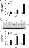
Fig 1
Fig 1
Daxx is upregulated in the reovirus-infected brain. Two- to 3-day-old Swiss Webster mice were inoculated i.c. with 1,000 PFU T3A, T3D, or an equal volume of PBS (mock). Brains were harvested for total RNA purification or protein lysate preparation at 4, 6, or 8 dpi. (a) RT-qPCR quantification of Daxx transcript relative to actin transcript for individual brains (n = 3) revealed progressive Daxx mRNA upregulation during T3A infection. (b) Representative Western blot demonstrating reovirus-induced Daxx protein upregulation. (c) Western blot densitometry results with T3A-infected brains (n = 3), showing progressive Daxx upregulation during reovirus infection.
Fig 2
Fig 2
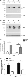
Reovirus-induced Daxx expression is type I interferon dependent. (a and b) L929 fibroblasts (a) or Neuro-2a cells (b) were treated with 1,000 U/ml IFN-β or PBS control, and whole-cell lysates were prepared at 12, 24, and 36 h for Western blotting detection of Daxx. (c) BSCs, prepared from 2- to 3-day-old Swiss Webster mice, were cultured for 5 days prior to exposure to cycloheximide and 1,000 U/ml universal type I IFN. Slices were harvested, and total RNA was purified at 1 and 2 days after treatment. RT-qPCR quantification of Daxx transcript relative to actin transcript for individual cultures (n = 5) revealed type I interferon-induced Daxx expression. (d) BSCs, prepared from 2- to 3-day-old type I interferon knockout mice (IFNAR−/−) and congenic wild-type mice (B6wt) were infected with T3A immediately upon plating. RT-qPCR quantification of Daxx transcript relative to actin transcript for individual cultures (n = 3) showed reovirus-induced Daxx mRNA upregulation infection, which was blocked in the absence of type I interferon signaling.
Fig 3
Fig 3
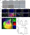
Daxx is upregulated in reovirus-infected brain regions and cells. Two- to 3-day-old Swiss Webster mice were inoculated i.c. with 1,000 PFU T3A, T3D, or an equal volume of PBS (mock). Brains were harvested at 8 dpi for histological processing. (a) Immunohistochemical staining of Daxx protein specifically in brain regions that were characteristically infected by reovirus (cingulate, hippocampus, and thalamus). The parietal cortex served as an internal negative control, as it is not infected by reovirus to any significant degree. (b) Following T3A infection, fluorescently labeled Daxx (red) was found largely within cells that were positively labeled for reovirus antigen σ3 (green). Magnification, ×630. Bar, 10 μm. (c) An isolated cell in the thalamus (inset) is enlarged by an additional 10× for demonstration of Daxx and reovirus in the same cell. (d) The percentage of cells that expressed high levels of Daxx within populations of either reovirus antigen-negative (-) or positive (+) cells. (e) The percentage of cells that expressed reovirus antigen within populations of cells expressing either low (-) or high (+) levels of Daxx. For both panels d and e, cells were quantified from 2 different fields obtained from the hippocampi of 2 individual reovirus-infected mice.
Fig 4
Fig 4
Upregulated Daxx protein is localized to the cytoplasm of reovirus-infected neurons and L929 cells. (a) Primary hippocampal neurons were prepared from Swiss Webster mouse pups at the age of 0 to 1 day. Neurons were infected with T3A at 5 days in vitro and processed for immunocytochemistry at 7 dpi. Labeled Daxx (red) was found in neurons that were reovirus antigen σ3 positive (Reo; green). Magnification, ×400. Bar, 10 μm. (b) Two- to 3-day-old Swiss Webster mice were inoculated i.c. with 1,000 PFU T3A or an equal volume of PBS (mock). Brains were harvested at 4 to 8 dpi for immunohistochemical labeling of Daxx (red) and NeuN (green), a neuron-specific marker. Magnification, ×1,000. Bar, 10 μm. (c) An isolated cell in the thalamus (inset) is enlarged an additional 10× for demonstration of Daxx in NeuN-positive neurons. (d) Cytoplasmic and nuclear fractions prepared from brains extracted at 8 dpi from Swiss Webster mice that had been intracerebrally inoculated with 100 PFU T3A or an equal volume of PBS (mock). The cytoplasmic and nuclear extracts were analyzed by Western blotting for detection of Daxx and lamin B nuclear marker. (e) L929 fibroblasts were treated with T3A reovirus at an MOI 20 or PBS (mock), and cells were harvested at 2 dpi for preparation of cytoplasmic (Cyto) and nuclear (Nuc) fractions. Western blotting was performed for detection of Daxx and the lamin B nuclear marker.
Fig 5
Fig 5
Daxx colocalizes with Fas in reovirus-infected brains. Two-to 3-day-old Swiss Webster mice were intracerebrally inoculated with 1,000 PFU T3A. Brains were harvested at 8 dpi for immunohistochemical labeling of Daxx (red) and Fas (green). (a) The images show Daxx in the cytoplasm of cells expressing Fas (white arrows). Magnification, ×630. (b) The percentage of cells that expressed Fas within populations of cells expressing either low (-) or high (+) levels of cytoplasmic Daxx. Percentages were derived from 2 different fields obtained from the hippocampi of 3 individual reovirus-infected animals. (c) Subcellular colocalization of Daxx and Fas, demonstrated by digitally deconvoluted z-stacked imaging. Magnification, ×1,000. Bar, 10 μm.
Fig 6
Fig 6
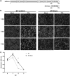
In vitro DN-Daxx expression reduces reovirus-induced cell death. (a) Schematic of the DN-Daxx construct stably expressed in L929 fibroblasts. DN-Daxx is composed solely of the C-terminal end of Daxx, corresponding to the Fas-binding region. Functionally, this mutant binds Fas but fails to recruit downstream effectors that initiate apoptosis. (b) L929 fibroblasts were T3A infected at an MOI of 20 or PBS (mock) treated. Photomicrographs were taken at 1, 2, and 3 dpi to demonstrate resistance to reovirus-induced cell death in DN-Daxx-expressing cells. Magnification, ×100. (c) DN-Daxx and EV L929 fibroblasts were T3A infected at an MOI 20. MTT was added to cell medium (n = 3) at specified time points, and the relative cell number was quantified by using a colorimetric plate reader.
Fig 7
Fig 7
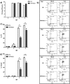
In vitro DN-Daxx expression reduces reovirus-induced apoptosis. L929 fibroblasts, stably expressing either EV or DN-Daxx, were T3A infected at an MOI of 20. (a) Cells were harvested, and total RNA was isolated at different time points as indicated. cDNA was synthesized from each experimental sample, and T3A cDNA standards (with known virus titers, as determined by a traditional plaque assay) were subjected to RT-qPCR amplification of the reovirus L1 gene. PFUe/ml indicates the equivalent number of PFU/ml as determined by placement of each data point on the standard curve. (b) Representative scatter plots of flow cytometry-based quantification of caspase 3/7 FLICA and PI staining at specified time points postinoculation with T3A. (c) Graphic representation of flow cytometry data (n = 4), showing the percentage of cells that positively stained with PI in T3A samples. (d) Graphic representation of flow cytometry data (n = 4), showing the percentage of cells that were positively stained with caspase 3/7 FLICA in T3A samples. (e) DN-Daxx or EV L929 fibroblasts were treated with 1 μM staurosporine. At specified time points, total cells were analyzed with flow cytometry for quantification of caspase 3/7 FLICA and PI staining. The data are representative of three experiments with similar results.
Fig 8
Fig 8
In vitro DN-Daxx expression does not affect staurosporine-induced apoptosis. L929 fibroblasts, stably expressing either EV or DN-Daxx, were treated with staurosporine. At specified time points, total cells were analyzed with flow cytometry for quantification of caspase 3/7 FLICA and PI staining. The data are representative of three experiments with similar results.
Fig 9
Fig 9
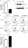
In vitro Daxx knockdown potentiates reovirus-induced apoptosis. (a) L929 fibroblasts were transfected with 30 nM siControl (siCon) or siDaxx. Total RNA was isolated 48 h after transfection. The uninfected L929 (no-si) was used as an additional control. RT-qPCR quantification of Daxx transcripts relative to actin transcripts for individual cultures (n = 3) revealed siDaxx knockdown of Daxx mRNA. (b) Total cell lysates were prepared 72 h after siRNA transfection and analyzed by SDS-PAGE and Western blotting for quantitation of Daxx and tubulin. The data are representative of three experiments with similar results. (c) Two days after siRNA transfection, cells were infected with T3A reovirus at an MOI of 20 or 200. At 3 dpi, floating cells and trypsin-treated cells were harvested and stained with caspase 3/7 FLICA and PI. The quantifications of FLICA- and PI-positive cells were achieved by flow cytometry. The data are representative of three independent experiments with similar results. (d) Total RNA was isolated 48 h after siRNA transfection. RT-qPCR quantification of caspase 3 transcript relative to actin transcript for individual cultures (n = 3) revealed that siDaxx upregulated caspase 3 expression.

Similar articles

Cited by

References

    1. Tan K, Patel S, Gandhi N, Chow F, Rumbaugh J, Nath A. 2008. Burden of neuroinfectious diseases on the neurology service in a tertiary care center. Neurology 71:1160–1166 - PubMed
    1. Tyler K. L. 2004. Herpes simplex virus infections of the central nervous system: encephalitis and meningitis, including Mollaret's. Herpes 11:57A–64A - PubMed
    1. Raschilas F, Wolff M, Delatour F, Chaffaut C, De Broucker T, Chevret S, Lebon P, Canton P, Rozenberg F. 2002. Outcome of and prognostic factors for herpes simplex encephalitis in adult patients: results of a multicenter study. Clin. Infect. Dis. 35:254–260 - PubMed
    1. Richardson-Burns SM, Tyler KL. 2004. Regional differences in viral growth and central nervous system injury correlate with apoptosis. J. Virol. 78:5466–5475 - PMC - PubMed
    1. Clarke P, Debiasi RL, Goody R, Hoyt CC, Richardson-Burns SM, Tyler KL. 2005. Mechanisms of reovirus-induced cell death and tissue injury: role of apoptosis and virus-induced perturbation of host-cell signaling and transcription factor activation. Viral Immunol. 18:89–115 - PMC - PubMed

Publication types

LinkOut - more resources