. 2012;7(12):e52519.
doi: 10.1371/journal.pone.0052519. Epub 2012 Dec 27.

Discovery of platyhelminth-specific α/β-integrin families and evidence for their role in reproduction in Schistosoma mansoni

Affiliations

Discovery of platyhelminth-specific α/β-integrin families and evidence for their role in reproduction in Schistosoma mansoni

Svenja Beckmann et al. PLoS One. 2012.

Abstract

In all metazoa, the response of cells to molecular stimuli from their environment represents a fundamental principle of regulatory processes controlling cell growth and differentiation. Among the membrane-linked receptors mediating extracellular communication processes are integrin receptors. Besides managing adhesion to the extracellular matrix or to other cells, they arrange information flow into the cells by activating intracellular signaling pathways often acting synergistically through cooperation with growth factor receptors. Although a wealth of information exists on integrins in different model organisms, there is a big gap of knowledge for platyhelminths. Here we report on the in silico detection and reconstruction of α and β integrins from free-living and parasitic platyhelminths, which according to structural and phylogenetic analyses form specific clades separate from each other and from further metazoan integrins. As representative orthologs of parasitic platyhelminths we have cloned one beta-integrin (Smβ-Int1) and four alpha-integrins (Smα-Int1 - Smα-Int4) from Schistosoma mansoni; they were characterized by molecular and biochemical analyses. Evidence is provided that Smβ-Int1 interacts and co-localizes in the reproductive organs with known schistosome cellular tyrosine kinases (CTKs), of which the Syk kinase SmTK4 appeared to be the strongest interaction partner as shown by yeast two-hybrid analyses and coimmunoprecipitation experiments. By a novel RNAi approach with adult schistosomes in vitro we demonstrate for the first time multinucleated oocytes in treated females, indicating a decisive role Smβ-Int1 during oogenesis as phenotypically analyzed by confocal laser scanning microscopy (CLSM). Our findings provide a first comprehensive overview about platyhelminth integrins, of which the parasite group exhibits unique features allowing a clear distinction from the free-living groups. Furthermore, we shed first lights on the functions of integrins in a trematode model parasite, revealing the complexity of molecular processes involved in its reproductive biology, which may be representative for other platyhelminths.


Conflict of interest statement

Competing Interests: The authors have declared that no competing interests exist.

Figures

Figure 1
Figure 1. Domain structures of S. mansoni integrin receptors.
Schematic structure of the schistosome α- and β-integrin receptors. Amino acid positions of the predicted conserved domains: Smα-Int1: 1273 aa; Int α 255–334, 359–418, 449–496; TM 1212–1234; Smα-Int2: 1492 aa; Int α 404–472, 476–546, 552–603; SP 1–30, 1296–1318; Smα-Int3: 1091 aa; Int α 257–316, 345–399; TM 980–1008; Smα-Int4: 1411 aa; SP 1–36; Int α 419–477; TM 1292–1314; Smβ-Int1: 865 aa; INB 36–470; EGF 567–604; TM 794–816;. (EGF: epidermal growth factor-like domain, Int α: integrin α domain (beta-propeller repeats), INB: integrin β domain, SP: signal peptide, TM: transmembrane domain).
Figure 2
Figure 2. Phylogenetic analyses showing the unique status of plathyhelminth α-integrins.
Phylogram of the analysis of the full-length sequences of the S. mansoni α-integrin receptors Smα-Int1, Smα-Int2, Smα-Int3, and other α-integrin receptors using CLUSTAL X (www.clustal.org) and TreeViewX. The phylogenetic relationship was deduced using the Bootstrap Neighbour-Joining (N–J) method and the bootstrap values were generated based on 1000 bootstrap trails with a random number generator seed of 100. Sequences were obtained from the National Centre for Biotechnology Information using the WWW Entrez Browser (www.ncbi.nlm.nih.gov), Swiss-Prot (www.uniprot.org), GeneDB (www.genedb.org), and the Schmidtea mediterranea Genome Database (http://smedgd.neuro.utah.edu/). The corresponding protein numbers are: Sha a1 (α-integrin 1 receptor, S. haematobium; Sha_102401), Sm a1 (α-integrin 1 receptor, S. mansoni; FR749887), Sjp a1 (α-integrin 1 receptor, S. japonicum; Sjp_0037690), Cs a5 (α-integrin 5 receptor, Clonorchis sinesis; GAA56616.1), Em a1 (α-integrin 1 receptor, Echinococcus multilocularis; EmuJ_000215000 ), Sm a4 (α-integrin 4 receptor, S. mansoni; Smp_1735401, Smp_181010), Sha a4 (α-integrin 4 receptor, S. haematobium; Sha_104436, Sha_106831), Sjp a4 (α-integrin 4 receptor, S. japonicum; Sjp_0046780, Sjp_0046790), Cs a4 (α-integrin 4 receptor, Clonorchis sinesis; GAA28731), Em a4 (α-integrin 4 receptor, Echinococcus multilocularis; EmuJ_000573500), Sha a2 (α-integrin 2 receptor, S. haematobium; Sha_106921), Sm a2 (α-integrin 2 receptor, S. mansoni; FR749888), Sjp a2 (α-integrin 2 receptor, S. japonicum; Sjp_0069490), Cs a-ps (α-integrin-ps receptor, Clonorchis sinesis; GAA54095, GAA49531, GAA49530), Em a2 (α-integrin 2 receptor, Echinococcus multilocularis; EmuJ_000192500 ), Smed a3 (α-integrin 3 receptor, Schmidtea mediterranea; lcl|mk4.000046.14.01), Smed a1 (α-integrin 1 receptor, Schmidtea mediterranea; lcl|mk4.001411.00.01), Smed a2 (α-integrin 2 receptor, Schmidtea mediterranea; lcl|mk4.003797. 00.01), Sha a3 (α-integrin 3 receptor, S. haematobium; Sha_102914), Sm a3 (α-integrin 3 receptor, S. mansoni; FR749889, Smp_156610, Smp_156620), Sjp a3 (α-integrin 3 receptor, S. japonicum; Sjp_0063430, Sjp_0063420), Cs a7 (α-integrin 7 receptor, Clonorchis sinesis; GAA52225.1), Em a3 (α-integrin 3 receptor, Echinococcus multilocularis; EmuJ_000782500), Sp aP (α-integrin P receptor, Strongylocentrotus purpuratus, AF177914), Dm aPS2 (α-integrin PS2 receptor, Drosophila melanogaster, Q24247), Mm a2b (α-integrin 2b receptor, Mus musculus; EDL34136.1), Hs a2b (α-integrin 2b receptor, Homo sapiens; EAW51595.1), Xl a2b (α-integrin 2b receptor, Xenopus laevis; NP_001088223.1), Mm a5 (α-integrin 5 receptor, Mus musculus; CAA55638.1), Rn a5 (α-integrin 5 receptor, Rattus norvegicus; NP_001101588.1), Hs a5 (α-integrin 5 receptor, Homo sapiens; NP_002196.2), Xl a5 (α-integrin 5 receptor, Xenopus laevis; NP_001081072.1), Hs aV (α-integrin V receptor, Homo sapiens; P06756), Hs a8 (α-integrin 8 receptor, Homo sapiens; P53708), Ce a-pat2 (α-integrin pat-2, Ceanorhabditis elegans; P34446), Gc a (α-integrin receptor, Geodia cydonium; X97283), Hs a1 (α-integrin 1 receptor, Homo sapiens; P56199), Hs a2 (α-integrin 2 receptor, Homo sapiens; P17301), Hs a10 (α-integrin 10 receptor, Homo sapiens; O75578), Hs a11 (α-integrin 11 receptor, Homo sapiens; Q9UKX5), Hs aD (α-integrin D receptor, Homo sapiens; Q13349), Hs aX (α-integrin X receptor, Homo sapiens; P20702), Hs aM (α-integrin M receptor, Homo sapiens; P11215), Hs aL (α-integrin L receptor, Homo sapiens; P20701), Hs aE (α-integrin E receptor, Homo sapiens; P38579), Hs a4 (α-integrin 4 receptor, Homo sapiens; P13612), Hs a9 (α-integrin 9 receptor, Homo sapiens; Q13797), Mm a7 (α-integrin 7 receptor, Mus musculus; AAA16600.1), Rn a7 (α-integrin 7 receptor, Rattus norvegicus; NP_110469.1), Hs a7 (α-integrin 7 receptor, Homo sapiens; EAW96822.1), Hs a6 (α-integrin 6 receptor, Homo sapiens; P23229), Hs a3 (α-integrin 3 receptor, Homo sapiens; P26006), Dm aPSI (α-integrin PSI receptor, Drosophila melanogaster, Q24247), and Ce a-ina1 (α-integrin ina1, Ceanorhabditis elegans; Q03600).
Figure 3
Figure 3. Phylogenetic analyses showing the unique status of plathyhelminth β-integrins.
Phylogram of the analysis of the full-length sequences of the S. mansoni β-integrin receptor Smβ-Int1 and other β-integrin receptors using CLUSTAL X (www.clustal.org) and TreeViewX. The phylogenetic relationship was deduced using the Bootstrap Neighbour-Joining (N–J) method and the bootstrap values were generated based on 1000 bootstrap trails with a random number generator seed of 100. Sequences were obtained from the National Centre for Biotechnology Information using the WWW Entrez Browser (www.ncbi.nlm.nih.gov), Swiss-Prot (www.uniprot.org), GeneDB (www.genedb.org), and the Schmidtea mediterranea Genome Database (http://smedgd.neuro.utah.edu/). The corresponding protein accession numbers are: Dm bPS (β-integrin PS, Drosophila melanogaster; P11584), Bg b (β-integrin, Biomphalaria glabrata; AF060203), Ce b-pat3 (β-integrin pat-3, Ceanorhabditis elegans; Q27874), Sp bL (β-integrin L subunit, Strongylocentrotus purpuratus; NP_999731), Sp bG (β-integrin G subunit, Strongylocentrotus purpuratus; NP_999732), Sp bC (β-integrin C subunit, Strongylocentrotus purpuratus; AF0559607), Mm b1 (β-integrin 1 receptor, Mus musculus; NP_034708.1), Rn b1 (β-integrin 1 receptor, Rattus norvegicus; NP_058718.2), Hs b1 (β-integrin 1 receptor, Homo sapiens; P05556), Gg b1 (β-integrin 1 receptor, Gallus gallus; NP_001034343.2), Xl b2 (β-integrin 2 receptor, Xenopus laevis; NP_001080017.1), Hs b2 (β-integrin 2 receptor, Homo sapiens; NP_000202.2), Hs b7 (β-integrin 7 receptor, Homo sapiens; NP_000880.1), HS b3 (β-integrin 3 receptor, Homo sapiens; P05106), Hs b5 (β-integrin 5 receptor, Homo sapiens; P18084), Hs b6 (β-integrin 6 receptor, Homo sapiens; P18564), Hs b 8 (β-integrin 8 receptor, Homo sapiens; P26012), Gc b (β-integrin receptor, Geodia cydonium; O97189), Am b (β-integrin, Acropora millepora; AF005356), Hs b4 (β-integrin 4 receptor, Homo sapiens; P16144), Sha b1 (β-integrin 1 receptor, S. haematobium; Sha_105750), Sm b1 (β-integrin 1 receptor, S. mansoni; FR749886), Sjp b1 (β-integrin 1 receptor, S. japonicum; Sjp_0081260), Cs b1 (β-integrin 1 receptor, Clonorchis sinesis; GAA31131.2), Em b1 (β-integrin 1 receptor, Echinococcus multilocularis; EmuJ_000528400), and Smed b1 (β-integrin 1 receptor, Schmidtea mediterranea; lcl|mk4.001280.01.01).
Figure 4
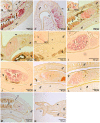
Figure 4. In situ-hybridization localized transcripts of Smβ-Int1, Smα-Int1- Smα-Int4 in the gonads and gonad-associated tissues of adult S. mansoni.
Representative sections (5 µm) of adult schistosome couples are shown (males and females are indicated), which were hybridized with DIG-labeled antisense-RNA probes of Smβ-Int1 (A–C), Smα-Int1 (D–F), Smα-Int2 (G, H), Smα-Int3 (I), Smα-Int4 (K) and for control with a DIG-labeled sense-RNA probe of Smα-Int3 (J), Smα-Int4 (L), or Smβ-Int1 (M, N). mRNA transcripts of Smβ-Int1 were detected in the ovary (o), the ootype-surrounding area (ot) anterior the ovary, the vitellarium (v), the subtegument (st), the testes (te), and the parenchyma (p) of both genders. Transcripts of Smα-Int1 were also detected in the ovary (o), the ootype-surrounding area (ot), and the vitellarium (v) of the female, the testes of the male, and the parenchyma (p) of both genders. Transcripts of Smα-Int2 were exclusively detected in the ootype-surrounding area (ot) anterior the ovary (o). Antisense and sense transcripts of Smα-Int3 and Smα-Int4 were only detected in the ovary (o). No signals were detected using sense transcripts of Smβ-Int1 (K, L), Smα-Int1, and Smα-Int2 (unpublished). dp: dorsal part; vp: ventral part; vs: ventral sucker; scale bars as indicated.
Figure 5
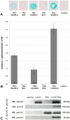
Figure 5. Interaction studies confirmed binding of Smβ-Int1 to the schistosome cellular kinases SmTK3, SmTK6, and SmTK4. A:
For binding studies in the YTH system, yeast cells (strain AH109) were co-transformed with the prey plasmid Smβ-Int1-C-term pACT2 together with the baits SmTK3-SH4SH3 pBridge, SmTK3-SH3 pBridge, SmTK6-SH4SH3 pBridge, SmTK6-SH3 pBridge, and SmTK4-SH2SH2 pBridge. Yeast clones were selected on Trp/Leu/His media (T/L/H) for interactions between bait and prey proteins, and β-galactosidase colony lift filter assays were performed for detection of LacZ expression. B: Comparative β-galactosidase liquid assays were performed with the yeast clones from A to determine the relative binding strengths. As control, untransformed yeast cells (AH109) were used (control). The statistical evaluation of seven independent measurements of β-Gal activity (n = 7) is shown (error bars are indicated). C: Co-Immunoprecipitation of HA-Smβ-Int1 and V5-SmTK4 expressed in Xenopus oocytes. Anti-HA antibodies immunoprecipitated Smβ-Int1 together with SmTK4 upon co-expression in oocytes. Inversely, anti-V5 antibodies immunoprecipitated SmTK4 with Smβ-Int1, when they were expressed together in oocytes.
Figure 6
Figure 6. RNAi knocking-down Smβ-Int1 and SmDia led to morphological changes in the ovary of S. mansoni females. A:
Using a combination of four siRNAs specific for Smβ-Int1 (β-Int1 siRNAs 1–4), a Smβ-Int1-transcript reduction down to 58% was determined by semi-quantitative RT-PCRs (n = 2) compared to control worms (no siRNAs), or worms electroporated with Smα-Int1-specific siRNAs (α-Int1 siRNA 1–4). B: The combination of two dsRNAs specific for SmDia led to a SmDia-transcript reduction down to 43% compared to control worms (no dsRNAs), which was determined by semi-quantitative RT-PCRs (n = 3). C: Confocal scanning laser microscope images of carmine red-stained whole-mount preparations of S. mansoni couples treated with siRNA specific for Smβ-Int1 (A, B), Smα-Int1 (C), without si/dsRNA (D), or with SmDia-specific dsRNAs (E, F). io: immature oocytes, mo: mature oocytes, arrows: poly-nucleated oocytes; scale bars 50 µm.
Figure 7
Figure 7. Model for integrin receptor and RTK-induced signaling pathways in S. mansoni.
The Syk kinase SmTK4, but also the Src kinase SmTK3, and the Src/Abl kinase SmTK6 are able to bind to the intracellular part of Smβ-Int1. Results of previous studies had already indicated that these three kinases interacted with each other and with SmVKR1, and all co-localized in the ovary of females , , . Furthermore, SmTK3 was found to interact with the diaphanous homolog SmDia , which is a binding partner of the Rho-GTPase SmRho1. Both SmDia and SmRho1 were suggested to organize the actin cytoskeleton within the gonads of schistosomes . As downstream partners of SmTK4, MAPK-activating protein (PM20/21) and mapmodulin were found, which may be involved in cytoskeleton reorganization and mitosis . SmDLG as a binding partner of SmTK6 may become activated upon complex formation and may subsequently interact with SmLGL and Scribble to control processes of cell growth and/or cell polarity (Buro et al., unpublished).

Similar articles

Cited by

References

    1. Fu G, Wang W, Luo BH (2012) Overview: structural biology of integrins. Methods Mol Biol 757: 81–99. - PubMed
    1. Anthis NJ, Campbell ID (2011) The tail of integrin activation. Trends Biochem Sci 36: 191–198. - PMC - PubMed
    1. Kim C, Ye F, Ginsberg MH (2011) Regulation of integrin activation. Annu Rev Cell Dev Biol 27: 321–345. - PubMed
    1. Campbell ID, Humphries MJ (2011) Integrin structure, activation, and interactions. Cold Spring Harb Perspect Biol 3: pii, a004994. - PMC - PubMed
    1. Guo W, Giancotti FG (2004) Integrin signalling during tumour progression. Nat Rev Mol Cell Biol 5: 816–826. - PubMed

Publication types

MeSH terms