. 2013 Feb 28;121(9):1584-94.
doi: 10.1182/blood-2012-08-450775. Epub 2013 Jan 7.

Robust T-cell stimulation by Epstein-Barr virus-transformed B cells after antigen targeting to DEC-205

Affiliations

Robust T-cell stimulation by Epstein-Barr virus-transformed B cells after antigen targeting to DEC-205

Carol S Leung et al. Blood. .

Abstract

DEC-205 is a type I transmembrane multilectin receptor that is predominantly expressed on dendritic cells (DCs). Therefore, previous studies primarily focused on processing of DEC-205–targeted antigens by this potent antigen presenting cell type. Here we show that Epstein-Barr virus (EBV) transformed lymphoblastoid B-cell lines (LCLs) not only express DEC-205 at similar levels to DCs, but also efficiently present targeted EBV nuclear antigen 1 (EBNA1) and EBV-latent membrane protein 1 (LMP1) to EBNA1- and LMP1-specific CD4+ and CD8+ T-cell clones in vitro. Targeting of antigens to DEC-205 on B cells led to more efficient MHC class II than I loading, and stimulated T cells more efficiently than targeting to DEC-205 on DCs. Although LCLs internalized DEC-205–targeted antigens less efficiently than DCs, they retained them for longer time periods and delivered them to endosomal compartments that receive also B-cell receptor targeted proteins. This could facilitate prolonged T-cell stimulation and efficient MHC class II loading, and, indeed, CD4+ T-cell expansion by DEC-205–targeted vaccination was significantly compromised in B-cell deficient mice. These studies suggest that B cells, activated by virus transformation or other means, can contribute to T-cell stimulation after DEC-205 targeting of antigens during vaccination.


Figures
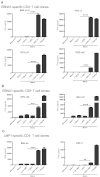
Figure 1
Figure 1
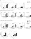
LCLs present DEC-205–targeted EBV antigens efficiently to T-cell clones of different peptide specificities and HLA restrictions. (A) HLA-matched (Match) LCLs for EBNA1-specific CD4+ T-cell clones of 4 different epitopes (SNP, VYG, KTS, and PQC) were incubated with medium, 1 μg/mL control Ig-EBNA1 (+ TIB-E1), αDEC-205 without EBNA1 fusion (+ αDEC), αDEC-205-EBNA1 (+ αDEC-E1) for 24 hours, or for 1 hour with epitope-specific peptide (+ Peptide). An HLA mismatched target LCL was also included into the analysis (Mismatch). T cells were incubated with these targets at an E/T ratio of 1:1. T-cell activity was determined after 18 hours by measuring IFNγ released into the supernatant. (B) As in panel A, T-cell responses of EBNA1-specific CD8+ T-cell clones, HPVc35 and HPVc41, were tested against LCLs incubated without or with αDEC-EBNA1. (C) HLA-matched (Match) LCLs for 2 LMP1-specific CD4+ T-cell clones were incubated with medium, 1 μg/mL control Ig-LMP1 (+ Ctr-LMP1), αDEC-205 without LMP1 fusion (+ αDEC), αDEC-205-LMP1 (+ αDEC-LMP1) for 24 hours, or for 1 hour with epitope-specific peptide (+ Peptide). T-cell activity was determined as in panel A. One representative experiment of 4 per T-cell clone is shown. Statistical analysis of all available data from 4 independent experiments was performed by Mann-Whitney test and P values are represented as **P < .01, ***P < .005, and ****P < .0001.
Figure 2
Figure 2
DEC-205 targeting of EBV antigens to LCLs stimulates better antigen-specific CD4+ T-cell responses than targeting to DCs. (A) HLA-matched LCLs, mDCs, iDCs, or B cells from the same donor were treated with the indicated concentration of control antibody, or αDEC-205 with EBNA1 or LMP1 fusion, or peptide, then incubated with the corresponding matched EBNA1- or LMP1-specific T-cell clones. T-cell responses were measured as IFNγ release after 18 hours of coculture at an E/T ratio of 1:1. (B) HLA-matched CD19+ B cells were incubated with 1 μg/mL control antibody with EBNA1 or LMP1 fusion, αDEC-205-EBNA1 or LMP1 fusion, αDEC-205-EBNA1 or LMP1 fusion with or without 2.5 μg/mL CpG for 24 hours, or for 1 hour with epitope-specific peptide, then incubated with EBNA1- or LMP1-specific CD4+ T cells. T-cell activity was determined as in panel A. One of 2 experiments per T-cell clone is shown. P values were calculated from the data of 2 independent experiments with Mann-Whitney test and are represented as **P < .01.
Figure 3
Figure 3
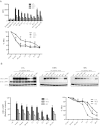
LCLs retain DEC-205–targeted antigen longer than DCs. (A) LCLs, mDCs, or iDCs were incubated with 1 μg/mL control Ig-EBNA1 or αDEC-205-EBNA1 on ice for 30 minutes. Cells were then washed and incubated at 37°C for the indicated time periods. As a control for internalization, cells were left on ice or incubated with 10μM cytochalasin B (cyto B) in addition to αDEC-205-EBNA1 pulsing. Cells were then stained with PE-conjugated anti-mouse antibody. Data are mean values plus SD from 3 independent experiments and relative internalization is shown as percent maximum. (B) LCLs, mDCs, or iDCs were incubated without (−ve) or with 4 μg/mL αDEC-205-LMP1 on ice for 30 minutes. Cells were then washed and incubated at 37°C or left on ice as a control. Cell samples were lysed at the indicated time points and frozen. Protein lysates were separated by SDS/PAGE, transferred to PVDF membranes for Western blotting, and probed with a LMP1-specific mAb. Blots were also probed for actin as a loading control. One representative blot of 3 experiments is shown; mean values plus SD of relative protein content and percentage maximum from 3 independent experiments are also shown.
Figure 4
Figure 4
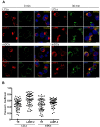
Intracellular localization of αDEC-205 in LCLs and mDCs. (A) LCLs and mDCs were incubated with αDEC-205, washed and incubated for 0 or 30 minutes at 37°C. Cells were then permeabilized, fixed, and stained with antibodies to transferrin receptor (TR) or lysosomal-associated membrane protein 2 (LAMP-2), αDEC-205 (DEC), and DAPI. Scale bars represent 5 μm. Representative cells from 1 experiment of 3 are shown. (B) Quantitative analysis for colocalization of αDEC-205 with TR and LAMP-2 in LCLs and mDCs loaded with αDEC-205 and incubated for 30 minutes at 37°C. Fifteen to 20 cells were analyzed from 1 independent experiment, data represent means from 50 to 60 cells from 3 experiments. Error bars indicate SD.
Figure 5
Figure 5
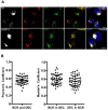
DEC-205 and B-cell receptor deliver antigens in LCLs with partial colocalization. LCLs were incubated with αDEC-205 and biotinylated F(ab′)2 anti-IgM Ab for 2 hours, washed, permeabilized, fixed, and stained with antibodies to transferrin receptor (TR) or lysosomal-associated membrane protein 2 (LAMP-2), αDEC-205 (DEC), αIgM (BCR), and DAPI. Scale bars represent 5 μm. Representative cells from 1 experiment of 3 are shown. (B) Quantitative analysis for colocalization of αDEC-205 (DEC) with αIgM (BCR) in LCLs incubated with αDEC-205 and αIgM for 30 minutes at 37°C. Fifteen to 20 cells were analyzed from 1 independent experiment, data represent means from 50 to 60 cells from 3 experiments. Error bars indicate SD.
Figure 6
Figure 6
B cells play a role in amplifying antigen-specific CD4+ T-cell responses toward DEC-205–targeted antigen in vivo. (A) C57BL/6 mice or the B-cell deficient JHT mice were immunized with 5 μg of isotype control fusion mAb with 50 μg polyIC (IgG-p24 + polyIC), or αDEC-205 mAb conjugated with HIV gag p24 (αDEC-p24) without or with 50 μg polyIC (αDEC-p24 + polyIC) as adjuvant, and boosted 1 month later. The mice were killed a week after boost, bulk splenocytes were harvested, stimulated with either gag p24 peptides or gag p17 peptide mix, IFNγ production was evaluated by intracellular cytokine staining in the CD3+CD4+ gated cells. (B) As in panel A, mean ± SD from 2 independent experiments with 3 mice per group is shown. P value was calculated with 2-tailed Student t test.

Similar articles

Cited by

References

    1. Steinman RM. Dendritic cells: understanding immunogenicity. Eur J Immunol. 2007;37(Suppl 1):S53–S60. - PubMed
    1. Münz C, Steinman RM, Fujii S. Dendritic cell maturation by innate lymphocytes: coordinated stimulation of innate and adaptive immunity. J Exp Med. 2005;202(2):203–207. - PMC - PubMed
    1. Steinman RM. The control of immunity and tolerance by dendritic cell. Pathol Biol. 2003;51(2):59–60. - PubMed
    1. Palucka K, Ueno H, Roberts L, Fay J, Banchereau J. Dendritic cells: are they clinically relevant? Cancer J. 2010;16(4):318–324. - PMC - PubMed
    1. Bonifaz LC, Bonnyay DP, Charalambous A, et al. In vivo targeting of antigens to maturing dendritic cells via the DEC-205 receptor improves T-cell vaccination. J Exp Med. 2004;199(6):815–824. - PMC - PubMed

Publication types

MeSH terms