

doi: 10.1038/nchembio.1159.
Epub 2013 Jan 6.
A conserved asparagine has a structural role in ubiquitin-conjugating enzymes
Affiliations
- PMID: 23292652
- PMCID: PMC3578109
- DOI: 10.1038/nchembio.1159
Item in Clipboard
A conserved asparagine has a structural role in ubiquitin-conjugating enzymes
Nat Chem Biol.
2013 Mar.
Abstract
It is widely accepted that ubiquitin-conjugating enzymes contain an active site asparagine that serves as an oxyanion hole, thereby stabilizing a negatively charged transition state intermediate and promoting ubiquitin transfer. Using structural and biochemical approaches to study the role of the conserved asparagine to ubiquitin conjugation by Ubc13-Mms2, we conclude that the importance of this residue stems primarily from its structural role in stabilizing an active site loop.
Conflict of interest statement
The Authors declare no competing financial interests.
Figures
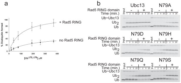
(a) Substrate partitioning experiments showing active fraction of Ubc13~Ub thioester as a function of acceptor ubiquitin concentration. Plot of percentage diubiquitin formed versus concentration of ubiquitin (Ub)Δ 75,Δ76 for Ubc13N79Q-Mms2 (filled circles) or Ubc13N79Q-Mms2 with the Rad5 RING domain (open circles). Points represent the average of 3 to 4 separate measurements with the standard deviation shown by the error bars. (b) Single discharge assays of diubiquitin formation by Ubc13-Mms2 containing wild-type Ubc13 or mutants with substitutions at N79, performed in the presence and absence of the Rad5 RING fragment. Full gels in Supplementary Figure 4.
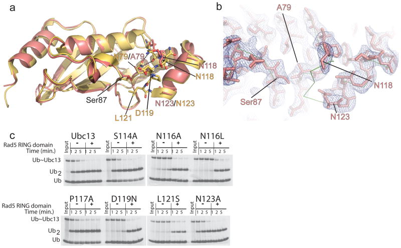
(a) Alignment of the structure of Ubc13N79A (salmon) with wild-type Ubc13 from 2GMI (yellow). Residue names are colored to match the coloring of the N79A or wild-type structures. (b) Electron density 2F0-Fc map contoured at 1.0σ showing the density for the active site loop in Ubc13N79A. The backbone Cα trace of wild-type Ubc13 from 2GMI is shown in green. (c) Single discharge assay for the Ubc13 active site loop mutants using ubiquitinΔ75,Δ76 (Ub) as the ubiquitin acceptor.
Similar articles
-
Ubc13: the Lys63 ubiquitin chain building machine.Oncotarget. 2016 Sep 27;7(39):64471-64504. doi: 10.18632/oncotarget.10948. Oncotarget. 2016. PMID: 27486774 Free PMC article. Review.
-
Architecture of the catalytic HPN motif is conserved in all E2 conjugating enzymes.Biochem J. 2012 Jul 15;445(2):167-74. doi: 10.1042/BJ20120504. Biochem J. 2012. PMID: 22563859
-
Molecular dynamics simulations reveal a new role for a conserved active site asparagine in a ubiquitin-conjugating enzyme.J Mol Graph Model. 2017 Sep;76:403-411. doi: 10.1016/j.jmgm.2017.07.006. Epub 2017 Jul 10. J Mol Graph Model. 2017. PMID: 28772203
-
A conserved asparagine in a ubiquitin-conjugating enzyme positions the substrate for nucleophilic attack.J Comput Chem. 2019 Aug 15;40(22):1969-1977. doi: 10.1002/jcc.25852. Epub 2019 May 9. J Comput Chem. 2019. PMID: 31070815
-
Ubiquitin-conjugating enzyme E2C: a potential cancer biomarker.Int J Biochem Cell Biol. 2014 Feb;47:113-7. doi: 10.1016/j.biocel.2013.11.023. Epub 2013 Dec 17. Int J Biochem Cell Biol. 2014. PMID: 24361302 Review.
Cited by
-
Distinct activation of an E2 ubiquitin-conjugating enzyme by its cognate E3 ligases.Proc Natl Acad Sci U S A. 2015 Feb 17;112(7):E625-32. doi: 10.1073/pnas.1415621112. Epub 2015 Feb 2. Proc Natl Acad Sci U S A. 2015. PMID: 25646477 Free PMC article.
-
E2~Ub conjugates regulate the kinase activity of Shigella effector OspG during pathogenesis.EMBO J. 2014 Mar 3;33(5):437-49. doi: 10.1002/embj.201386386. Epub 2014 Jan 20. EMBO J. 2014. PMID: 24446487 Free PMC article.
-
A comprehensive in silico analysis of sortase superfamily.J Microbiol. 2019 Jun;57(6):431-443. doi: 10.1007/s12275-019-8545-5. Epub 2019 May 27. J Microbiol. 2019. PMID: 30900148
-
Ubc13: the Lys63 ubiquitin chain building machine.Oncotarget. 2016 Sep 27;7(39):64471-64504. doi: 10.18632/oncotarget.10948. Oncotarget. 2016. PMID: 27486774 Free PMC article. Review.
-
Crystal structures of an E1-E2-ubiquitin thioester mimetic reveal molecular mechanisms of transthioesterification.Nat Commun. 2021 Apr 22;12(1):2370. doi: 10.1038/s41467-021-22598-y. Nat Commun. 2021. PMID: 33888705 Free PMC article.
References
-
- Pickart CM, Eddins MJ. Ubiquitin: structures, functions, mechanisms. Biochim Biophys Acta. 2004;1695:55–72. - PubMed
-
- Kerscher O, Felberbaum R, Hochstrasser M. Modification of proteins by ubiquitin and ubiquitin-like proteins. Annu Rev Cell Dev Biol. 2006;22:159–80. - PubMed
-
- Yunus AA, Lima CD. Lysine activation and functional analysis of E2-mediated conjugation in the SUMO pathway. Nat Struct Mol Biol. 2006;13:491–9. - PubMed
Publication types
MeSH terms
Substances
Associated data
- Actions
Grants and funding
LinkOut - more resources
Full Text Sources
Other Literature Sources
Molecular Biology Databases
