. 2013 Mar;33(3):501-9.
doi: 10.1161/ATVBAHA.112.300929. Epub 2013 Jan 3.

Pericytes from infantile hemangioma display proangiogenic properties and dysregulated angiopoietin-1

Affiliations

Pericytes from infantile hemangioma display proangiogenic properties and dysregulated angiopoietin-1

Elisa Boscolo et al. Arterioscler Thromb Vasc Biol. 2013 Mar.

Abstract

Objective: Infantile hemangioma (IH) is a rapidly growing vascular tumor affecting newborns. It is composed of immature endothelial cells and pericytes that proliferate into a disorganized mass of blood vessels. We isolated pericytes from IH (Hem-pericytes) to test our hypothesis that Hem-pericytes are unable to stabilize blood vessels.

Methods and results: We injected pericytes in vivo, in combination with endothelial cells, and found that Hem-pericytes formed more microvessels compared with control retinal pericytes. We, thereby, analyzed proangiogenic properties of the Hem-pericytes. They grew fast in vitro, and were unable to stabilize endothelial cell growth and migration, and expressed high levels of vascular endothelial growth factor-A compared with retinal pericytes. Hem-pericytes from proliferating phase IH showed lower contractility in vitro, compared with Hem-pericytes from the involuting phase and retinal pericytes. Consistent with a diminished ability to stabilize endothelium, angiopoietin 1 was reduced in Hem-pericytes compared with retinal pericytes. Normal retinal pericytes in which angiopoietin 1 was silenced produced conditioned medium that stimulated endothelial cell proliferation and migration.

Conclusions: We report the first successful isolation of patient-derived pericytes from IH tissue. Hem-pericytes exhibited proangiogenic properties and low levels of angiopoietin 1, consistent with a diminished ability to stabilize blood vessels in IH.


Figures
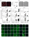
Figure 1
Figure 1. Hemangioma pericyte (Hem-pericyte) characterization
A- Proliferating and nvoluting IH tissue stained for Ki67 (green) and αSMA (red). Arrows indicate Ki67+ endothelial cells, arrowheads Ki67+ pericytes. Quantification of Ki67+ and Ki67+/αSMA+ cells in proliferating and involuting IH tissue. Data expressed as mean± SDM. B- In vitro morphology of proliferating and involuting Hem-pericytes. C- Real-time PCR mRNA expression levels for PDGFRβ, NG2, Desmin, Calponin, sm22α, αSMA, smMHC, CD90 and NOTCH3, in Hem-pericytes isolated from 4 different proliferating phase IH (146, 147, 154, 156) and 2 involuting phase IH (I-69, I-79). Human placental and retinal pericytes served as positive controls (n= triplicate for each cell type). Data expressed as mean± SDM. D- Pericytes from proliferating, involuting IH, human placenta and retina, stained for PDGFRβ, NG2, Desmin, Calponin, sm22α, αSMA, smooth muscle Myosin Heavy Chain (smMHC), CD90 and endothelial marker CD31. Scale bar 100μM.
Figure 2
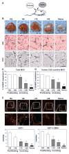
Figure 2. Hem-pericytes combined with ECFCs form blood vessels in Matrigel
A- Schematic of in vivo subcutaneous injection. B- Matrigel explants at day 7: ECFC combined with proliferating (154, 146), involuting (I-79, I-69) Hem-pericytes and retinal pericytes (top row). Representative histological sections from Matrigel explants stained with hematoxylin and eosin (middle row) and anti-human specific CD31 (bottom row). Scale bar 100μM. C- Blood vessels containing erythrocytes, indicating microvessels, were counted in hematoxylin and eosin sections. Vessels/mm2 in Hem-pericyte 154 explants set to 100%. (n=8 mice per group, 2 independent experiments, analysis carried out on 10 fields for each Matrigel explant). Data expressed as mean ± SEM. *p<0.02 compared to proliferating Hem-pericytes 154 and 146, ** p<0.05 compared to proliferating and involuting Hem-pericytes 146, 154, I-69 and I-79. D- Human CD31 stained vessels in sections were counted (n=8 mice per group, 2 independent experiments, analysis carried out on 10 fields for each Matrigel explant). Data expressed as mean ± SEM. *p<0.02 compared to proliferating Hem-pericytes 154 and 146. E- Representative images of explant sections stained for dividing cell marker Ki67 (green), αSMA (red), and DAPI (blue). Scale bar 100μM. Quantification of Ki67+ and Ki67+/αSMA+ cells in proliferating and involuting IH tissue. Data expressed as mean ± SEM. *p<0.05 compared to proliferating Hem-pericytes 154 and 146, ** p<0.05 compared to proliferating and involuting Hem-pericytes 146, 154, I-69 and I-79.
Figure 3
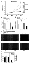
Figure 3. Proangiogenic properties of Hem-pericytes in vitro
A- Cell proliferation evaluated at 24, 48, 72 and 96 hours for proliferating (154, 146, 147, 156), involuting (I-69, I-79, I-81, I-82) Hem-pericytes and retinal pericytes. Cell count at 24 hours after seeding was set to 100% to normalize for differences in initial adherence to the well. Data expressed as mean ± SDM. B- Endothelial Colony Forming Cells (ECFC) proliferation analyzed after 48 hours of indirect contact with pericytes or medium alone (DMEM/10%FBS=no cells): schematic of experiment (top), quantification of ECFC proliferation (bottom). Data expressed as mean ± SEM. *p<0.05 compared to retinal pericytes, **p<0.05 compared to retinal pericytes and to control, no cells. C- ECFC migration towards pericyte-conditioned medium or medium alone (bottom chamber) was analyzed after 4 hours: schematic of experiment (top), quantification of ECFC migration (bottom). Data expressed as mean ± SEM. *p<0.05 compared to retinal pericytes. D- Scratch migration assay with ECFC migrating across the scraped region after 6-hour exposure to pericyte-conditioned medium. Cells fixed and nuclei stained with DAPI. E- VEGF-A protein levels in conditioned medium of Hem-pericytes and retinal pericytes analyzed by enzyme-linked immunoadsorbant assay (ELISA) (gray bars), and VEGF-A mRNA levels in cell lysates analyzed by real-Time PCR (black bars). *p<0.05 compared to retinal pericytes.
Figure 4
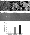
Figure 4. Hem-pericytes exhibit low contractility
A- Representative images of proliferating, involuting Hem- and retinal pericytes stained with phalloidin to evaluate F-actin filaments. Scale bare =100μM. B- Representative images of Hem-pericytes and retinal pericytes in silicone -based contractility assay. Arrow points to an example of a cell that has contracted and produced wrinkles in silicone substratum. C- Quantification of wrinkle-forming cells out of total (n=12 wells per each cell type, 5 fields per well). *p<0.05 compared to involuting Hem-pericytes and retinal pericytes. Data expressed as mean ± SDM.
Figure 5
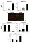
Figure 5. ANGPT1 expression is downregulated in Hem-pericytes
A- Real Time PCR analysis of ANGPT1 expression in proliferating and involuting Hem-pericytes and retinal pericytes. B- ELISA performed on the conditioned medium (48 hours) from proliferating and involuting Hem-pericytes and retinal pericytes. Data expressed as mean ± SDM. **p<0.05 compared to involuting Hem-pericytes and retinal pericytes, *p<0.05 compared to retinal pericytes. C Proliferating and involuting IH tissue stained for ANGPT1 activated- phosphoTIE2 (green), αSMA (red) and DAPI (blue). Scale bar 100μM. D- Real-time PCR mRNA expression levels for ANGPT1 in retinal pericytes, untreated (control), or treated with short interference (si)-RNA with a non targeting (si Non Target) sequence pool or ANGPT1 targeting sequence pool (si ANGPT1). Data expressed as mean ± SDM. E- ECFC proliferation analyzed after 48 hours of indirect contact with retinal pericytes (control, si Non Target, si ANGPT1). Values were normalized for retinal pericyte counts. Data expressed as mean ± SEM. *p<0.05. F- ECFC migration analyzed after 4 hours of contact with retinal pericyte (control, si Non Target, si ANGPT1)-conditioned medium. Values were normalized for retinal pericyte counts. Data expressed as mean ± SEM. *p<0.05.

Similar articles

Cited by

References

    1. Armulik A, Genove G, Betsholtz C. Pericytes: developmental, physiological, and pathological perspectives, problems, and promises. Dev Cell. 2011;21:193–215. - PubMed
    1. Franco M, Roswall P, Cortez E, Hanahan D, Pietras K. Pericytes promote endothelial cell survival through induction of autocrine VEGF-A signaling and Bcl-w expression. Blood. 2011;118:2906–2917. - PMC - PubMed
    1. Stratman AN, Malotte KM, Mahan RD, Davis MJ, Davis GE. Pericyte recruitment during vasculogenic tube assembly stimulates endothelial basement membrane matrix formation. Blood. 2009;114:5091–5101. - PMC - PubMed
    1. Saunders WB, Bohnsack BL, Faske JB, Anthis NJ, Bayless KJ, Hirschi KK, Davis GE. Coregulation of vascular tube stabilization by endothelial cell TIMP-2 and pericyte TIMP-3. J Cell Biol. 2006;175:179–191. - PMC - PubMed
    1. Kutcher ME, Kolyada AY, Surks HK, Herman IM. Pericyte Rho GTPase mediates both pericyte contractile phenotype and capillary endothelial growth state. The American journal of pathology. 2007;171:693–701. - PMC - PubMed

Publication types

MeSH terms