

Correlates of relative resistance against low-dose rectal simian immunodeficiency virus challenges in peripheral blood mononuclear cells of vaccinated rhesus macaques
- PMID: 23271702
- PMCID: PMC3579023
- DOI: 10.1189/jlb.0612287
Correlates of relative resistance against low-dose rectal simian immunodeficiency virus challenges in peripheral blood mononuclear cells of vaccinated rhesus macaques
Abstract
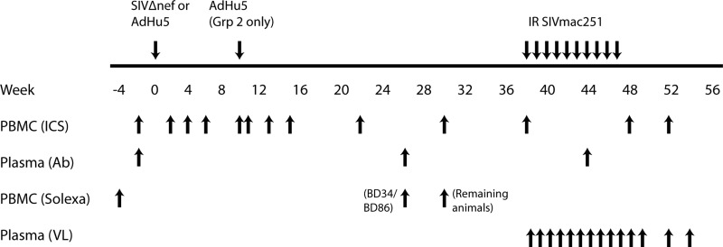
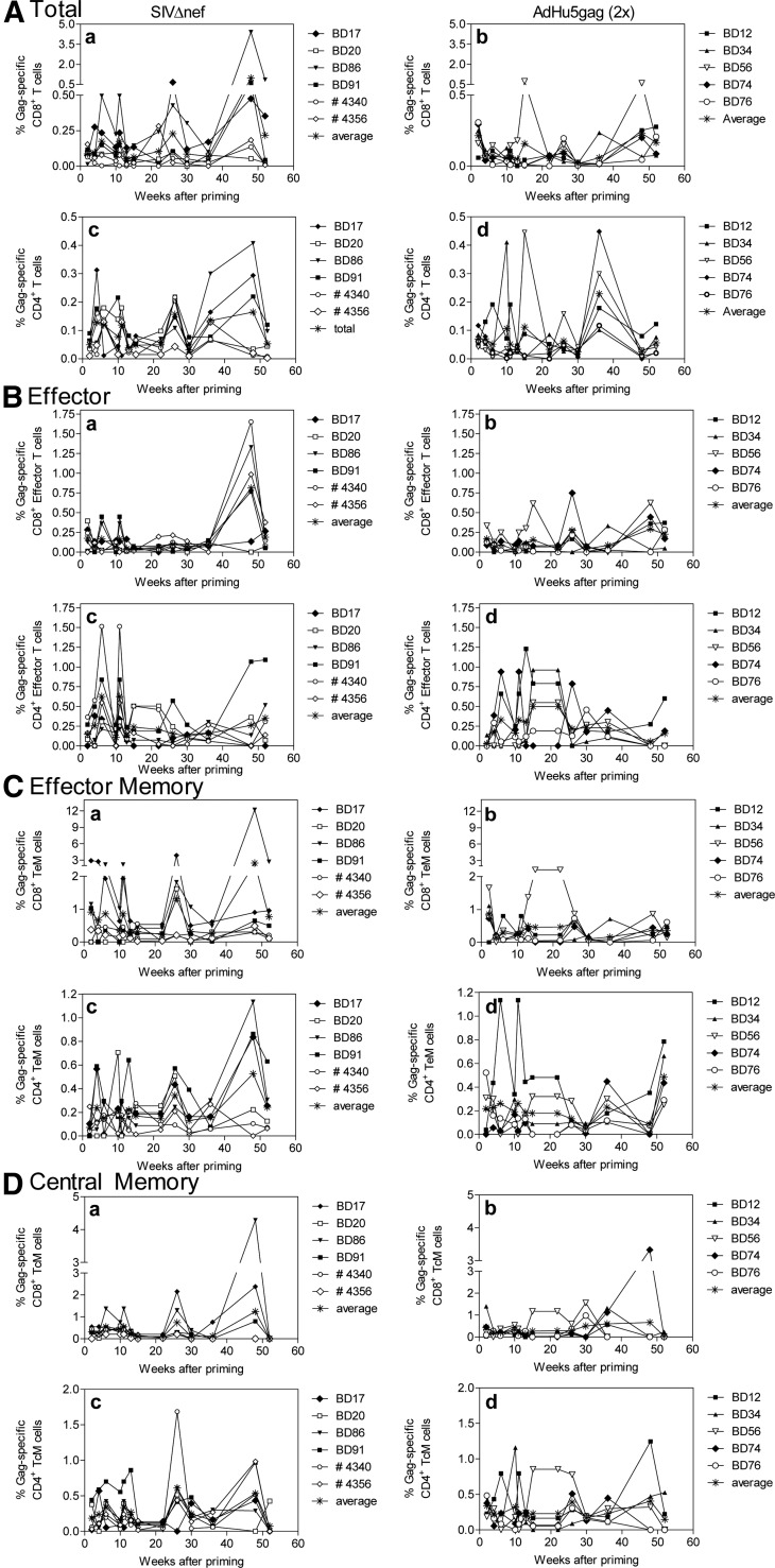
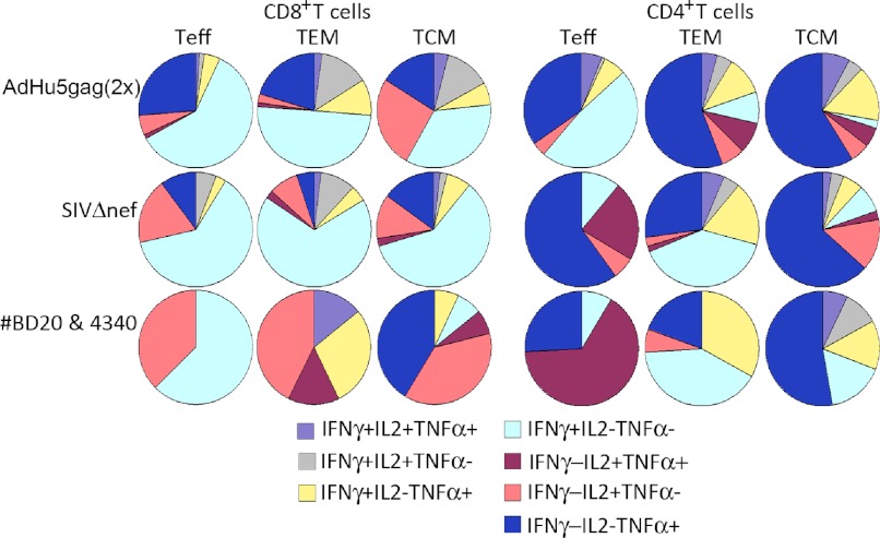
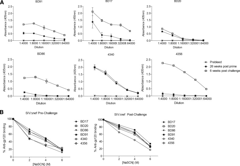
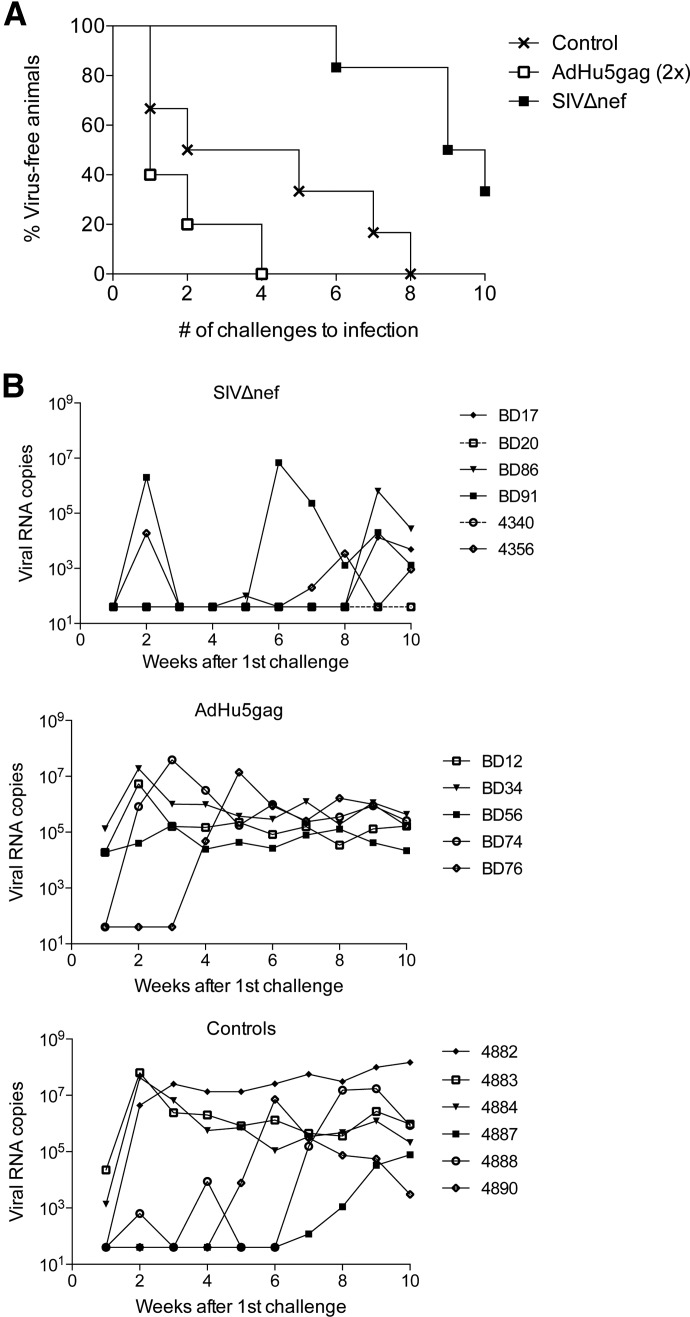
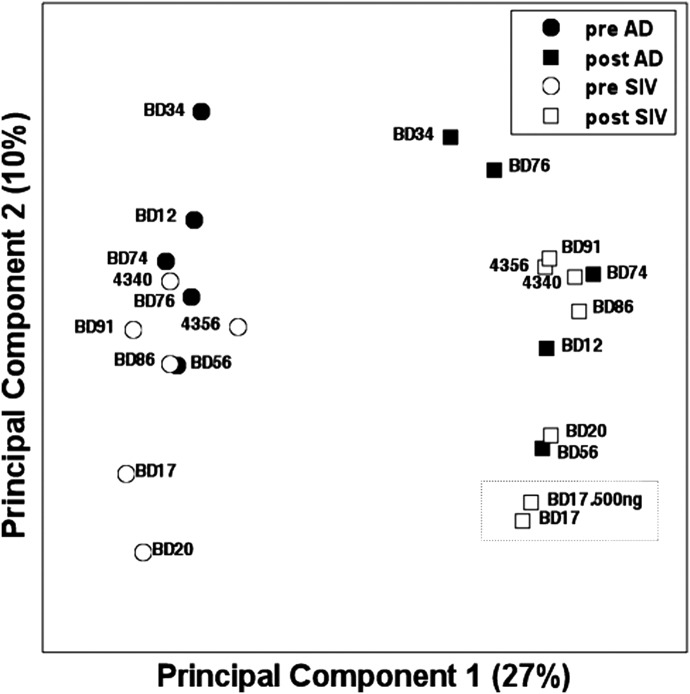
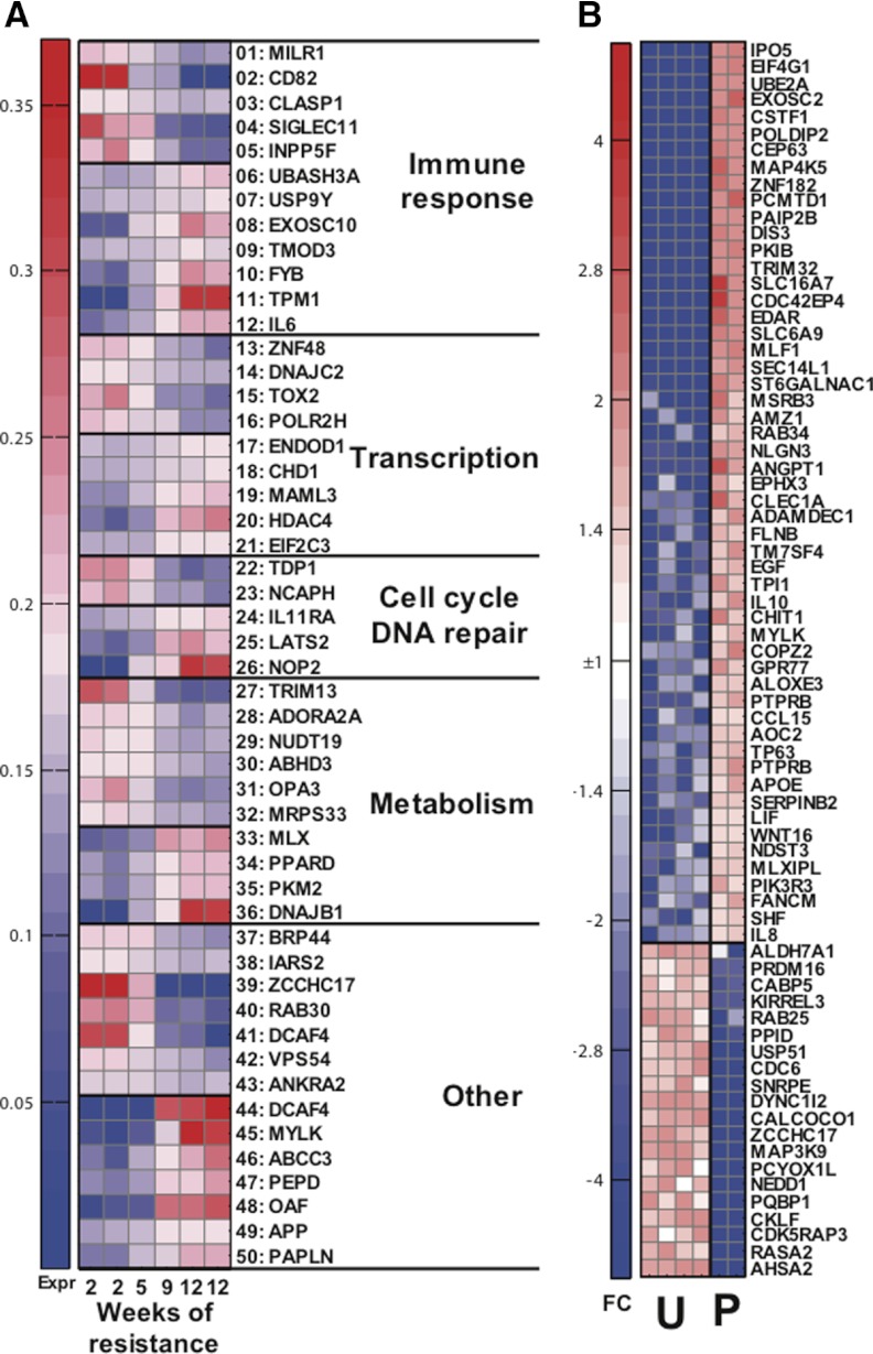
In this study, we compared the immunogenicity and protection from repeated low-dose intrarectal SIVmac251 challenge in two groups of vaccinated RMs. Animals were immunized with live SIVmac239, which had been attenuated by a deletion of the nef sequence, or they were vaccinated twice with an E1-deleted AdHu5, expressing SIVmac239gag. The vaccinated animals and a cohort of unvaccinated control animals were then challenged 10 times in weekly intervals with low doses of SIVmac251 given rectally. Our results confirm previous studies showing that whereas SIVΔnef provides some degree of protection against viral acquisition after repeated low-dose rectal SIVmac251 challenges, vaccination with an AdHu5gag vaccine designed to induce only antiviral T cell responses is ineffective. As immunological analyses of prechallenge, vaccine-induced T and B cell responses failed to reveal correlates of protection that distinguished the more susceptible from the more resistant vaccinated animals, we carried out RNA-Seq studies of paired pre- and postvaccination samples to identify transcriptional patterns that correlated with the differences in response. We show that gene expression signatures associated with the delayed SIV infection seen in some AdHu5gag recipients were largely present in prevaccination samples of those animals. In contrast, the responding SIVΔnef-immunized animals showed a predominance of vaccine-induced changes, thus enabling us to define inherited and vaccine-induced gene expression signatures and their associated pathways that may play a role in preventing SIV acquisition.
Figures
Similar articles
-
Mucosal B Cells Are Associated with Delayed SIV Acquisition in Vaccinated Female but Not Male Rhesus Macaques Following SIVmac251 Rectal Challenge.PLoS Pathog. 2015 Aug 12;11(8):e1005101. doi: 10.1371/journal.ppat.1005101. eCollection 2015 Aug. PLoS Pathog. 2015. PMID: 26267144 Free PMC article.
-
Activated CD4+CCR5+ T cells in the rectum predict increased SIV acquisition in SIVGag/Tat-vaccinated rhesus macaques.Proc Natl Acad Sci U S A. 2015 Jan 13;112(2):518-23. doi: 10.1073/pnas.1407466112. Epub 2014 Dec 30. Proc Natl Acad Sci U S A. 2015. PMID: 25550504 Free PMC article.
-
Recombinant Herpesvirus Vectors: Durable Immune Responses and Durable Protection against Simian Immunodeficiency Virus SIVmac239 Acquisition.J Virol. 2021 Jun 24;95(14):e0033021. doi: 10.1128/JVI.00330-21. Epub 2021 Jun 24. J Virol. 2021. PMID: 33910957 Free PMC article.
-
Vaccination of rhesus macaques with a vif-deleted simian immunodeficiency virus proviral DNA vaccine.Virology. 2008 May 10;374(2):261-72. doi: 10.1016/j.virol.2008.01.020. Epub 2008 Feb 7. Virology. 2008. PMID: 18261756 Free PMC article.
-
Antiviral CD8+ T cells in the genital tract control viral replication and delay progression to AIDS after vaginal SIV challenge in rhesus macaques immunized with virulence attenuated SHIV 89.6.J Intern Med. 2009 Jan;265(1):67-77. doi: 10.1111/j.1365-2796.2008.02051.x. J Intern Med. 2009. PMID: 19093961 Free PMC article. Review.
Cited by
-
Antibody responses to prime-boost vaccination with an HIV-1 gp145 envelope protein and chimpanzee adenovirus vectors expressing HIV-1 gp140.AIDS. 2016 Oct 23;30(16):2405-2414. doi: 10.1097/QAD.0000000000001224. AIDS. 2016. PMID: 27525550 Free PMC article.
-
Signatures in Simian Immunodeficiency Virus SIVsmE660 Envelope gp120 Are Associated with Mucosal Transmission but Not Vaccination Breakthrough in Rhesus Macaques.J Virol. 2015 Dec 16;90(4):1880-7. doi: 10.1128/JVI.02711-15. Print 2016 Feb 15. J Virol. 2015. PMID: 26676777 Free PMC article.
-
Using nonhuman primates to model HIV transmission.Curr Opin HIV AIDS. 2013 Jul;8(4):280-7. doi: 10.1097/COH.0b013e328361cfff. Curr Opin HIV AIDS. 2013. PMID: 23666391 Free PMC article. Review.
-
Enhancement of recombinant adenovirus vaccine-induced primary but not secondary systemic and mucosal immune responses by all-trans retinoic acid.Vaccine. 2014 Jun 5;32(27):3386-92. doi: 10.1016/j.vaccine.2014.04.028. Epub 2014 Apr 26. Vaccine. 2014. PMID: 24780251 Free PMC article.
References
-
- Hu S. L., Stamatatos L. (2007) Prospects of HIV Env modification as an approach to HIV vaccine design. Curr. HIV Res. 5, 507–513 - PubMed
-
- Roux K. H., Taylor K. A. (2007) AIDS virus envelope spike structure. Curr. Opin. Struct. Biol. 17, 244–252 - PubMed
-
- Hansen S. G., Vieville C., Whizin N., Coyne-Johnson L., Siess D. C., Drummond D. D., Legasse A. W., Axthelm M. K., Oswald K., Trubey C. M.., et al. (2009) Effector memory T cell responses are associated with protection of rhesus monkeys from mucosal simian immunodeficiency virus challenge. Nat. Med. 15, 293–299 - PMC - PubMed
Publication types
MeSH terms
Substances
Grants and funding
LinkOut - more resources
Full Text Sources
Medical
Molecular Biology Databases
Research Materials
