. 2013 Jun;9(2):259-70.
doi: 10.1007/s11302-012-9348-x. Epub 2012 Dec 28.

N-cadherin expression is regulated by UTP in schwannoma cells

Affiliations

N-cadherin expression is regulated by UTP in schwannoma cells

Tania Martiáñez et al. Purinergic Signal. 2013 Jun.

Abstract

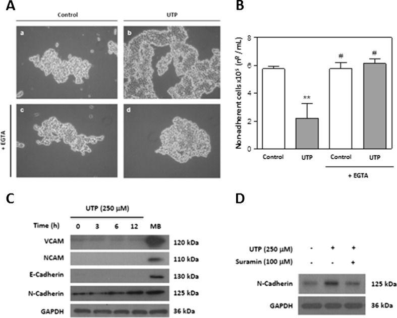
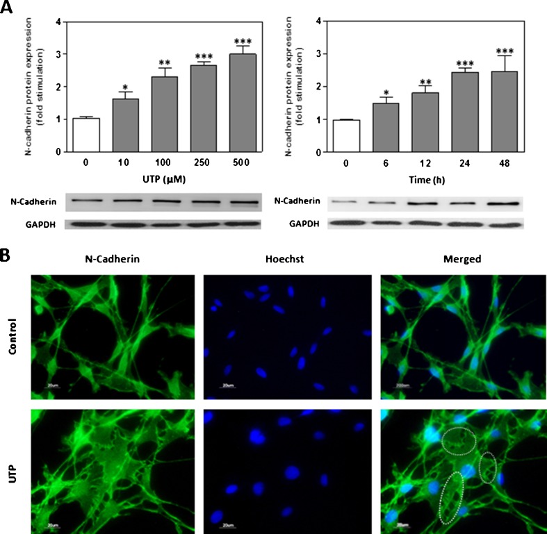
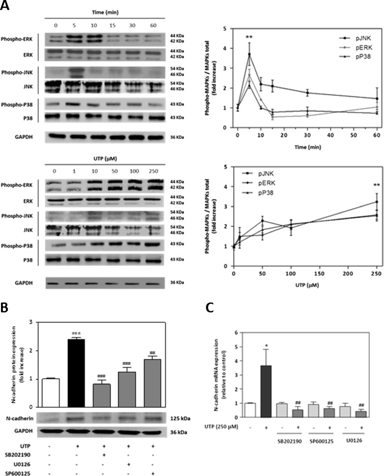
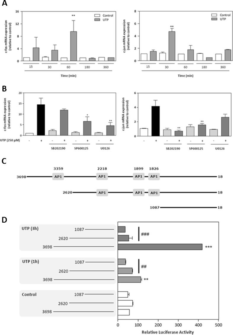
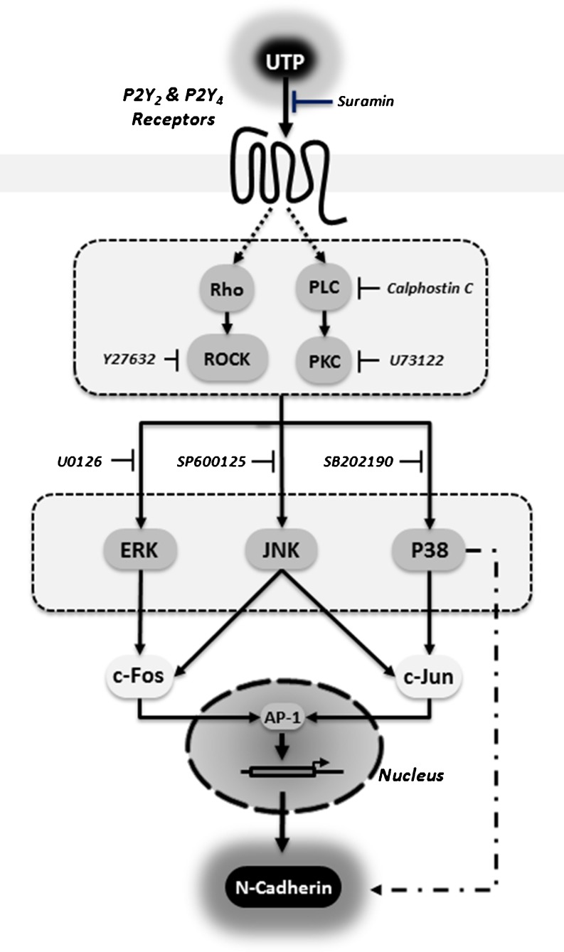
Schwann cells (SCs) are peripheral myelinating glial cells that express the neuronal Ca(2+)-dependent cell adhesion molecule, neural cadherin (N-cadherin). N-cadherin is involved in glia-glia and axon-glia interactions and participates in many key events, which range from the control of axonal growth and guidance to synapse formation and plasticity. Extracellular UTP activates P2Y purinergic receptors and exerts short- and long-term effects on several tissues to promote wound healing. Nevertheless, the contribution of P2Y receptors in peripheral nervous system functions is not completely understood. The current study demonstrated that UTP induced a dose- and time-dependent increase in N-cadherin expression in SCs. Furthermore, N-cadherin expression was blocked by the P2 purinoceptor antagonist suramin. The increased N-cadherin expression induced by UTP was mediated by phosphorylation of mitogen-activated protein kinases (MAPKs), such as Jun N-terminal kinase, extracellular-regulated kinase and p38 kinase. Moreover, the Rho kinase inhibitor Y27632, the phospholipase C inhibitor U73122 and the protein kinase C inhibitor calphostin C attenuated the UTP-induced activation of MAPKs significantly. Extracellular UTP also modulated increased in the expression of the early transcription factors c-Fos and c-Jun. We also demonstrated that the region of the N-cadherin promoter between nucleotide positions -3698 and -2620, which contained one activator protein-1-binding site, was necessary for UTP-induced gene expression. These results suggest a novel role for P2Y purinergic receptors in the regulation of N-cadherin expression in SCs.


Figures

Fig. 1
Fig. 1
Extracellular UTP increases N-cadherin expression in Schwann cells. a Photomicrographs prepared using an inverted microscope (×20), showing adhesion after the “shaking off” method involving trypsin treatment. Cells were treated with UTP (250 μM) for 5 h with (c, d) or without (b) EGTA pretreatment (1 mM, 20 min), and their appearance was compared with that of controls (a). b Non-adherent viable cells were counted after trypsin treatment using trypan blue exclusion staining. Values are expressed as the mean ± SD (n = 3). Statistical significance: **P ≤ 0.01 compared to control; #P ≤ 0.05 compared to UTP treatment. c Schwann cell protein extracts (30 μg) treated for different time periods with 250 μM UTP were resolved by SDS-PAGE and blotted with specific antibodies against VCAM and NCAM (Ca2+-independent adhesion proteins) and against N-cadherin and E-cadherin (Ca2+-independent adhesion proteins). Immunoblots are representative of three independent experiments. Mouse brain (MB) protein lysate was included as a positive control. d The P2Y-receptor antagonist suramin inhibited UTP-induced N-cadherin expression. Primary cultures of SCs were pretreated with suramin (100 μM) for 20 min before UTP (250 μM) treatment and lysates were blotted with antibodies against N-cadherin and GAPDH as a control. Immunoblots are representative of three independent experiments
Fig. 2
Fig. 2
Extracellular UTP increases N-cadherin expression in a schwannoma cell line. a Extracellular UTP increased N-cadherin expression in a dose- and time-dependent manner. Schwann cell protein extracts (30 μg) treated for different time periods manner with UTP (250 μM) or with increasing concentrations of UTP for 24 h were resolved by SDS-PAGE, blotted, and probed with an N-cadherin antibody. Fold stimulation in N-cadherin expression is expressed as the mean ± SD (six independent experiments). Statistical significance: *P ≤ 0.05, **P ≤ 0.01, ***P ≤ 0.001. b Schwann cells treated with UTP (250 μM) for 12 h were mounted on coverslips and subjected to immunocytochemistry against N-cadherin (green). Up-regulation of N-cadherin in cell–cell contacts (dotted lines) was visualised by fluorescence microscopy. Nuclei were stained with Hoechst 33342 (blue). Scale bars: 20 μm
Fig. 3
Fig. 3
N-cadherin expression depends on MAPK phosphorylation. a Western blot analysis of MAPK activation by UTP treatment. Schwannoma cells were treated with UTP (250 μM) for different times or were treated with UTP (5 min) at several concentrations. Western blots were performed using antibodies against phosphorylated and total MAPK (ERK1/2, JNK and p38). Fold increases in MAPK activities are expressed as the mean ± SD of the ratio of phosphorylated MAPK/total MAPK (n = 3, **P ≤ 0.01). Representative Western blots for each kinase are shown above the graphs. b Western blotting and c real-time PCR analysis of UTP-induced N-cadherin expression in the presence of different MAPK inhibitors. Cells were preincubated (30 min) with 10-μM selective inhibitor for each MAPK: U0126 (selective inhibitor of MEK/ERK), SP600125 (selective inhibitor of JNK); and SB202190 (selective inhibitor of p38) before UTP treatment. Values are the mean ± SD (three independent experiments). Statistical significance: *P ≤ 0.05, ***P ≤ 0.001, compared to controls; ##P ≤ 0.01, ###P ≤ 0.001 compared to UTP treatment
Fig. 4
Fig. 4
MAPK activation depends on the P2Y receptor signalling pathway. Schwannoma cells were pretreated for 30 min before UTP treatment with selective inhibitors of different proteins involved in the P2Y receptor signalling pathway: suramin (100 μM; P2Y receptor antagonist), Y27632 (10 μM; ROCK inhibitor), U73122 (10 μM; PLC inhibitor) or calphostin C (10 μM; PKC inhibitor). After extracellular UTP treatment (5 min), proteins from cell lysates were resolved by SDS-PAGE and blotted against each phosphorylated or total MAPK. Representative Western blots for each kinase are shown. Fold increases in MAPK activity are expressed as the mean ± SD of the ratio of phosphorylated MAPK/total MAPK (n = 4, *P ≤ 0.05; **P ≤ 0.01)
Fig. 5
Fig. 5
Extracellular UTP induces expression of the early genes c-Fos and c-Jun through MAPK activation. a Schwannoma cells were treated with UTP (250 μM) for five different time periods. Total RNA was subjected to real-time PCR analysis and expression of c-Fos (left) or c-Jun (right) mRNA was evaluated. Early gene expression is expressed as the fold stimulation compared to housekeeping gene GAPDH. Values are mean ± SD (n = 3, **P ≤ 0.01). b Schwannoma cells were pretreated (30 min) with 10 μM of each of the selective MAPK inhibitors SB202190, SP600125 and U0126. After UTP treatment (250 μM for 1 h), total RNA was prepared and subjected to real-time PCR analysis to determine the expression of transcripts that encoded c-Fos (left) and c-Jun (right). Early gene expression is expressed as fold stimulation compared to housekeeping gene GAPDH. Values are the mean ± SD (n = 3, *P ≤ 0.05, **P ≤ 0.01). c Schematic representation of the human N-cadherin promoter (AY512658) and two 5′-deletion mutants used, all of which were cloned in a pGL3 plasmid linked to luciferase. AP-1: AP-1 binding sites. d Schwannoma cells were either co-transfected with the complete sequence of the human N-cadherin promoter or with one of two 5′-deletion mutants that contained fewer AP-1 binding sites linked to firefly luciferase and with pGL3 that contained the coding sequence for Renilla luciferase. After 48 h, transfected RT4 cells were treated with 250 μM UTP for 1 h (grey boxes) or 3 h (black boxes), and luciferase activity was analysed. Firefly luciferase activity was normalised by comparison to levels of Renilla luciferase activity. Data represent transcriptional activities relative to the empty basic pGL3 plasmid. The results are the mean ± SD of two independent experiments performed in triplicate (n = 6, **P ≤ 0.01, ***P ≤ 0.001)
Fig. 6
Fig. 6
Schematic overview of the purinergic signalling pathway involved in P2Y receptor activation leading to N-cadherin expression in Schwannoma cell line

Similar articles

Cited by

References

    1. Fu SY, Gordon T. The cellular and molecular basis of peripheral nerve regeneration. Mol Neurobiol. 1997;14(1–2):67–116. doi: 10.1007/BF02740621. - DOI - PubMed
    1. Pereira JA, Lebrun-Julien F, Suter U. Molecular mechanisms regulating myelination in the peripheral nervous System. Trends Neurosci. 2012;35(2):123–134. doi: 10.1016/j.tins.2011.11.006. - DOI - PubMed
    1. Fannon AM, Sherman DL, Ilyina-Gragerova G, Brophy PJ, Friedrich VL, Jr, Colman DR. Novel E-cadherin-mediated adhesion in peripheral nerve: Schwann cell architecture is stabilized by autotypic adherens junctions. J Cell Biol. 1995;129(1):189–202. doi: 10.1083/jcb.129.1.189. - DOI - PMC - PubMed
    1. Wanner IB, Wood PM. N-cadherin mediates axon-aligned process growth and cell-cell interaction in rat Schwann cells. J Neurosci. 2002;22(10):4066–4079. - PMC - PubMed
    1. Wanner IB, Guerra NK, Mahoney J, Kumar A, Wood PM, Mirsky R, Jessen KR. Role of N-cadherin in Schwann cell precursors of growing nerves. Glia. 2006;54(5):439–459. doi: 10.1002/glia.20390. - DOI - PubMed

Publication types