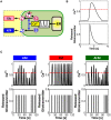
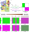
. 2012 Dec 21:6:98.
doi: 10.3389/fncom.2012.00098. eCollection 2012.

Computational quest for understanding the role of astrocyte signaling in synaptic transmission and plasticity

Affiliations

Computational quest for understanding the role of astrocyte signaling in synaptic transmission and plasticity

Maurizio De Pittà et al. Front Comput Neurosci. .

Abstract

The complexity of the signaling network that underlies astrocyte-synapse interactions may seem discouraging when tackled from a theoretical perspective. Computational modeling is challenged by the fact that many details remain hitherto unknown and conventional approaches to describe synaptic function are unsuitable to explain experimental observations when astrocytic signaling is taken into account. Supported by experimental evidence is the possibility that astrocytes perform genuine information processing by means of their calcium signaling and are players in the physiological setting of the basal tone of synaptic transmission. Here we consider the plausibility of this scenario from a theoretical perspective, focusing on the modulation of synaptic release probability by the astrocyte and its implications on synaptic plasticity. The analysis of the signaling pathways underlying such modulation refines our notion of tripartite synapse and has profound implications on our understanding of brain function.

Keywords: astrocyte modeling; astrocyte-synapse interactions; calcium encoding; calcium signaling; cortical maps; gliotransmission; metaplasticity; synaptic plasticity.


Figures

Figure 1
Figure 1
The signaling network of astrocyte-synapse interactions. (A) A simplified scheme of the different signaling pathways between synaptic terminals and astrocytes for the case of excitatory synapses in the hippocampus (see text for a detailed description). Action potentials arriving at the presynaptic terminal trigger release of glutamate, which can spill over from the synaptic cleft. Perisynaptic astrocytes take up glutamate using their plasma membrane transporters (EAATs) while glutamate, by acting on astrocytic metabotropic receptors (mGluRs), triggers Ca2+ signaling in the astrocyte. This signaling pathway includes production of IP3 and causes an increase of cytosolic Ca2+ due to efflux of this ion from the endoplasmic reticulum (ER). At some synapses, such as in the dentate gyrus, the same Ca2+ signaling pathway could also be mediated by astrocytic purinergic P2Y1 receptors, likely activated by synaptically-released ATP (see text for details). Astrocytic Ca2+ excitability can in turn lead to exocytotic release of several neuroactive substances (or “gliotransmitters”) such as glutamate (Glu), D-serine (D-ser) or ATP which can target specific receptors on pre- and post-synaptic terminals and differentially modulate synaptic transmission. Glutamate acting on presynaptic GluRs could enhance synaptic release, whereas ATP and its derivate adenosine (Adn) could depress it (red path) through presynaptic purinergic receptors (PRs). On the postsynaptic spines [depicted here by a standard RC circuit (Ermentrout and Terman, 2010)], the ensuing effect of gliotransmitters could substantially modify postsynaptic currents by enhancing activation of NMDA receptors (D-serine) or by altering expressions of AMPA receptors therein. Astrocytes could also release TNFα by Ca2+-dependent activation of the matrix metalloprotease TNFα-converting enzyme (TACE), while extracellular TNFα could in turn regulate glutamate release from the astrocyte as well as postsynaptic AMPAR expression. Moreover astrocytic Ca2+ could also propagate across different regions of the same cell or to other neighboring astrocytes by intracellular IP3 diffusion through gap junction channels (GJCs) or via extracellular ATP-dependent pathways, extending gliotransmission to some distal sites away from the considered synapse. For clarity both endocannabinoid-mediated Ca2+ signaling (Navarrete and Araque, 2008), retrograde activation of presynaptic glutamate receptors (Navarrete and Araque, 2010), regulation of postsynaptic NMDARs by presynaptic adenosine receptors (Deng et al., 2011), and the possibility for astrocyte-derived adenosine to enhance synaptic release (Panatier et al., 2011) are not included in this scheme. (B) Equivalent modeling scheme for astrocyte-synapse interactions. The astrocyte (ASTRO) constitutes a third active element of the tripartite synapse in addition to the presynaptic (PRE) and postsynaptic (POST) terminals. In its presence, the flow of input (IN) signals to the output (OUT) is no more unidirectional but presynaptically released neurotransmitter can affect astrocyte function through the interaction pathway A. In turn, the astrocyte can regulate both synaptic terminals via pathways B and C. In addition, the astrocyte could receive additional inputs from or send output to remote synapses in a heterosynaptic fashion (I/O).
Figure 2
Figure 2
Computational aspects of astrocytic Ca2+ signaling. (A) Scheme of IP3-mediated Ca2+-induced Ca2+ release in the astrocyte. Calcium and IP3 signals are controlled by synaptic glutamate through metabotropic glutamate receptor- (mGluR-) PLCβ-mediated IP3 production (see text for details). (B) IP3 and Ca2+ signals can be envisioned to encode incoming synaptic activity through frequency and amplitude of their oscillations. Astrocytes could thus encode synaptic information either by modulations of the amplitude (AM), the frequency (FM), or both (AFM) of their Ca2+ oscillations. (C) Simulated Ca2+ and IP3 patterns in response to sample synaptic glutamate release (Glu) in a model astrocyte (De Pittà et al., 2009a,b) reveals that IP3 signals could be locked in the AFM-encoding independently of the encoding mode of the associated Ca2+ signals. This feature could allow the astrocyte to optimally integrate synaptic stimuli. (D, top) Simulated Ca2+ (black trace) and IP3 (green trace) signals in the same astrocyte model as in (C), and associated rates of IP3 production (prod.) and degradation (degr.) (middle panels, dashed black lines) in response to two consecutive synaptic glutamate release events (bottom panel, Glu). The analysis of the contributions of different enzymes to IP3 signaling (solid colored traces; cyan: PLCβ ; pink: PLCδ ; orange: IP-5P; and purple: IP3-3K) reveals dynamical regulation by Ca2+ of different mechanisms of IP3 production/degradation which could ultimately underlie dynamical regulation of astrocyte processing of synaptic stimuli. (E) Simulated propagation of Ca2+ waves in a heterogeneous linear chain composed of both FM (red traces) and AFM (green traces) astrocytes reveals that encoding of synaptic activity (STIMULUS) could change according to cell location along the chain. Adapted from Goldberg et al. (2010).
Figure 3
Figure 3
Linking gliotransmitter exocytosis to various Ca2+ encoding modes. (A) Calcium-dependent glutamate and ATP exocytosis from astrocytes are both brought forth by a vesicular compartment in the astrocyte competent for regulated exocytosis. The frequency of exocytotic events is directly controlled by the shape and frequency of Ca2+ oscillations. (B) Modeling concept for an “exocytosis event” from the astrocyte. Calcium (top trace) triggers exocytosis of glutamate or ATP every time it increases beyond a certain threshold concentration value (red dashed line). The overall release can then be approximated, under proper assumptions, by an exponentially-decaying pulse of extracellular concentration of glutamate or ATP (bottom trace). (C) Distinct Ca2+ encoding patterns could translate into distinct rates of gliotransmitter exocytosis events. In this way, synaptic activity encoded by astrocytic Ca2+ signals is linked to the frequency of glutamate/ATP release from the astrocyte in a unique fashion. Adapted from De Pittà et al. (2011).
Figure 4
Figure 4
Glutamate or ATP released from astrocytes regulates transitions between facilitation and depression of synaptic transmission. (A) Conceptual framework for the regulation of synaptic release probability at basal conditions by astrocytes. Astrocyte-released glutamate increases basal synaptic release probability (U0), whereas astrocyte-released ATP/Adn generally decreases it. (B) Changes in synaptic release probability due to astrocytic gliotransmitters can be detected by variations of paired-pulse plasticity quantified by paired-pulse ratio (PPR). Paired-pulse facilitation (left, green traces) of postsynaptic currents (PSCs) corresponds to PPR values above 1 (right, green-shaded area) whereas paired-pulse depression (left, magenta traces) are associated with PPR values below 1 (right, magenta-shaded area). (C) Raster plots of simulated PSC pairs for 100 different input spike trains with same statistics colored according to the paired-pulse ratio: green for facilitation, PPR > 1; magenta for depression, PPR < 1. For increasing rates of exocytosis of gliotransmitter from the astrocyte, mimicked by increasing rates of Ca2+ crossing beyond the threshold for exocytosis (top row, red dashed line), synaptic plasticity could be progressively changed to its opposite depending on the type of gliotransmitter. Astrocytic glutamate could thus turn facilitating synapses into depressing (middle row) whereas astrocyte-derived ATP or adenosine could turn depressing synapses into facilitating (bottom row). Simulations are based on a model of astrocyte-regulation of synaptic release introduced in De Pittà et al. (2011). “Basal Synaptic Release Probability” in (B) refers to the probability of synaptic release at rest, that is when synaptic activity is assumed to be very low and the amount of neurotransmitter released upon arrival of an action potential to the presynaptic terminal is essentially independent of previous release events (Zucker and Regehr, ; De Pittà et al., 2011).
Figure 5
Figure 5
Short-term synaptic plasticity is physiologically set by the rate of gliotransmitter release from the astrocyte. (A) The rate of glutamate or ATP release from the astrocyte could differently affect basal synaptic release probability. In particular, a threshold frequency for the release of these gliotransmitters could exist (blue vertical line) beyond which a depressing synapse could turn into facilitating (right, green area) or vice versa, a facilitating synapse could become depressing (left, magenta area). Adapted from De Pittà et al. (2011). (B) In basal conditions, synaptic release is due to sporadic neuronal network firing and the possible frequency of Ca2+ fluctuations beyond the threshold for exocytosis (dashed red line) in the astrocyte (top trace). In this fashion plastic changes in paired-pulse ratio could be inherently regulated by astrocytic gliotransmitters, as shown here for the case of an originally facilitating synapse under the effect of astrocytic glutamate exocytosis (20 trials with identical input statistics). Adapted from Berry et al. (2011). The color code for the raster plot is the same as in Figure 4C.

Similar articles

Cited by

References

    1. Abbott L. F., Regehr W. G. (2004). Synaptic computation. Nature 431, 796–803 10.1038/nature03010 - DOI - PubMed
    1. Abbracchio M. P., Burnstock G., Verkhratsky A., Zimmermann H. (2009). Purinergic signalling in the nervous system: an overview. Trends Neurosci. 32, 19–29 10.1016/j.tins.2008.10.001 - DOI - PubMed
    1. Abraham W. (2008). Metaplasticity: tuning synapses and networks for plasticity. Nat. Rev. Neurosci. 9, 387–387 10.1038/nrn2356 - DOI - PubMed
    1. Abraham W., Bear M. (1996). Metaplasticity: the plasticity of synaptic plasticity. Trends Neurosci. 19, 126–130 10.1007/978-3-540-88955-7_6 - DOI - PubMed
    1. Aguado F., Espinosa-Parrilla J. F., Carmona M. A., Soriano E. (2002). Neuronal activity regulates correlated network properties of spontaneous calcium transients in astrocytes in situ. J. Neurosci. 22, 9430–9444 - PMC - PubMed

LinkOut - more resources