. 2013 Jan 8;110(2):618-23.
doi: 10.1073/pnas.1208446110. Epub 2012 Dec 24.

Deubiquitination of NF-κB by Ubiquitin-Specific Protease-7 promotes transcription

Affiliations

Deubiquitination of NF-κB by Ubiquitin-Specific Protease-7 promotes transcription

Amy Colleran et al. Proc Natl Acad Sci U S A. .

Abstract

NF-κB is the master regulator of the immune response and is responsible for the transcription of hundreds of genes controlling inflammation and immunity. Activation of NF-κB occurs in the cytoplasm through the kinase activity of the IκB kinase complex, which leads to translocation of NF-κB to the nucleus. Once in the nucleus, NF-κB transcriptional activity is regulated by DNA binding-dependent ubiquitin-mediated proteasomal degradation. We have identified the deubiquitinase Ubiquitin Specific Protease-7 (USP7) as a regulator of NF-κB transcriptional activity. USP7 deubiquitination of NF-κB leads to increased transcription. Loss of USP7 activity results in increased ubiquitination of NF-κB, leading to reduced promoter occupancy and reduced expression of target genes in response to Toll-like- and TNF-receptor activation. These findings reveal a unique mechanism controlling NF-κB activity and demonstrate that the deubiquitination of NF-κB by USP7 is critical for target gene transcription.


Conflict of interest statement

The authors declare no conflict of interest.

Figures
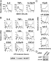
Fig. 1.
Fig. 1.
USP7 knockdown inhibits cytokine gene expression. (A) Murine bone marrow–derived macrophage was pretreated with the indicated concentration of HBX41,108 for 1 h before stimulation with LPS. mRNA levels were assessed by qPCR. Data shown are the mean ± SEM of triplicate samples. (B) RAW 264.7 macrophage was transfected with control siRNA (open circles) or siRNA directed against USP7 (filled circles). Cells were stimulated 24 h posttransfection with 100 ng/mL LPS for the indicated times before mRNA levels were assessed by qPCR. Data shown are mean ± SD of triplicate samples and representative of three independent experiments. (C) Control or USP7 knockdown cells treated with CpG, Pam-3CSK (P-CSK), poly(I:C) (pI:C), or TNFα as indicated. IL-6 mRNA levels were assessed by qPCR. (D) Immunoblot analysis of RAW 264.6 macrophage cells tansfected with USP7 or Scrambled siRNA (control) using anti-USP7 and anti-β-actin antibodies.
Fig. 2.
Fig. 2.
USP7 depletion impairs NF-κB signaling. (A) RAW 264.7 macrophage cells were transfected with USP7 siRNA or Scrambled siRNA (control) and, 24 h later, were transfected with luciferase reporter plasmids. A total of 48 h later, the cells were stimulated with LPS or left untreated. Shown is relative NF-κB luciferase activity (fold increase). (B) RAW 264.7 macrophage cells were cotransfected with USP7 dominant negative (USP7CS) and NF-κB luciferase reporter plasmids. (C) HEK293T cells were cotransfected with USP7 dominant negative (USP7CS) and NF-κB luciferase reporter plasmids and stimulated with TNFα or left untreated. (D) HEK293T cells were cotransfected with IL-23p19 promoter luciferase reporter plasmid. The cells, 24 h later, were left untreated or pretreated with the indicated concentration of HBX41,108 for 1 h before stimulation with TNFα. (E) RAW264.7 macrophage cells were cotransfected with IL-23p19 promoter luciferase reporter plasmid. The cells, 24 h later, were left untreated or pretreated with the indicated concentration of HBX41,108 for 1 h before stimulation with LPS. (F) Luciferase activity in RAW 264.7 macrophage cells cotransfected with USP7 dominant negative (USP7CS) and NF-κB or AP-1 luciferase reporter plasmids.
Fig. 3.
Fig. 3.
USP7 promotes NF-κB DNA binding. (A) RAW 264.7 macrophage cells were transfected with USP7 siRNA or scrambled siRNA (control) and 24 h later stimulated with LPS for the indicated time points before immunoblot analysis using the indicated antibodies. (B) RAW 264.7 macrophage cells were transfected with USP7 siRNA or scrambled siRNA (control) and 24 h later stimulated with LPS for the indicated time points. Nuclear and cytoplasmic fractions were analyzed by immunoblot with the indicated antibodies. (C) RAW 264.7 macrophage cells were stimulated with 100 ng/mL LPS for 6 h and followed by chromatin immunoprecipitation using anti-p65 and normal rabbit IgG (control) antibodies. Chromatin immunoprecipitations were analyzed using primers flanking the IL-6 and TNFα promoter regions.
Fig. 4.
Fig. 4.
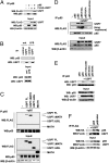
USP7 interacts with NF-κB. (A) HEK293T cells were transfected with p65 and FLAG-USP7 plasmids. Equal amounts of protein were immunoprecipitated with antibody against p65 and immunoblotted with antibodies indicated. (B) RAW 264.7 macrophage cells were stimulated with LPS and lysates immunoprecipitated with anti-USP7 antibody and immunoblotted with anti-p65 antibody. (C) HEK293T cells were transfected with p65 and full-length USP7 (USP7 FL) and USP7 deletion mutants as indicated. Equal amounts of protein were immunoprecipitated with antibody against p65 and immunoblotted for USP7. (D) HEK293T cells were transfected with WT p65, p65C197A/R198A, p65R267A, or p65R329A/R330AA, and FLAG-USP7 plasmids. Equal amounts of protein were immunoprecipitated with antibody against p65 and immunoblotted with antibodies indicated. Longer exposure of anti-USP7 immunoblot is also shown. (E) HEK293T cells were transfected with p65, p65 DNA binding mutant (p65Y36D, D39E), p65 S536A and S536E mutants, and with FLAG-USP7 plasmids. Equal amounts of protein were immunoprecipitated with p65 antibody and immunoblotted with anti-USP7 antibody. (F) HEK293T cells were transfected with p65, p65S468A, and FLAG-USP7 plasmids. Equal amounts of protein were immunoprecipitated with antibody against p65 and immunoblotted for USP7.
Fig. 5.
Fig. 5.
USP7 deubiquitinates NF-κB. (A) HEK293T cells were cotransfected with HA-ubiquitin and p65 alone or with FLAG-USP7 plasmid. Equal amounts of protein were immunoprecipitated with antibody against p65 and immunoblotted with antibody against HA. (B) HEK293T cells were transfected with HA-ubiquitin and FLAG-USP7CS and FLAG-USP7 plasmids. Cells were treated 24 h posttransfection with TNFα and whole cell lysates prepared. Equal amounts of protein were immunoprecipitated with antibody against p65 and immunoblotted with antibody against HA. (C) RAW 264.7 macrophage cells were transfected with control or USP7 or siRNA and stimulated with LPS for 60 min. MG1332 (10 μM) was added for the final 30 min. Equal amounts of protein were immunoprecipitated with anti-p65 antibody and immunoblotted with anti-ubiquitin antibody. (D) HEK293T cells were transfected with HA-ubiquitin, and 24 h posttransfection, cells were left untreated or pretreated with the indicated concentration of HBX41,108 for 60 min before treatment with TNFα for a further 60 min. MG1332 (10 μM) was added for the final 30 min. Whole cell lysates were prepared and equal amounts of protein were immunoprecipitated with antibody against p65 and immunoblotted with antibody against HA. (E) Recombinant p65 was in vitro ubiquitinated with FLAG-ubiquitin, purified, and incubated with recombinant USP7 and analyzed by immunoblotting with antibodies for FLAG, p65, and USP7. (F) HEK293T cells were transfected with empty or USP7 expression vector. After 24 h, cells were treated with cyclohexamide for the indicated times before immunoblot analysis. Relative levels of p65 protein normalized against β-actin are plotted. (G) HEK293T cells were transfected with plasmids for p65 along with FLAG-USP7 or empty vector. Equal amounts of protein were immunoprecipitated with antibody against p65 and immunoblotted with anti-K48 ubiquitin chain antibody. (H) HEK293T cells were cotransfected with HA-ubiquitin and plasmids for WT p65, p65R267A, or p65R329A/R330AA. Equal amounts of protein were immunoprecipitated with anti-p65 antibody and immunoblotted with anti-HA antibody. (I) HEK293T cells were cotransfected with HA-ubiquitin and plasmids for WT p65, p65R267A, or p65R329A/R330AA along with FLAG-USP7 or empty vector. Equal amounts of protein were immunoprecipitated with antibody against p65 and immunoblotted with antibody against HA. (J) HEK293T cells were transfected with WT p65 or p65R329A/R330AA expression plasmids as indicted along with empty or USP7 expression vector. After 24 h, cells were treated with cyclohexamide for the indicated times before immunoblot analysis. Relative levels of p65 protein normalized against β-actin are plotted.
Fig. 6.
Fig. 6.
USP7 regulates transcription through p65 ubiquitination. (A) WT or p65−/− MEFs were transfected with IL-23p19 promoter reporter constructs along with empty vector (Empty) or a USP7CS expression vector. After 24 h, cells were stimulated with TNFα (+) or left untreated (–) and luciferase activity measured. Data shown are the mean ± SEM of triplicate samples. (B) RAW 264.7 macrophage cells were cotransfected with USP7 dominant negative (USP7CS) and IL-23p19 promoter luciferase reporter plasmid (p19), and IL-23p19 promoter luciferase reporter plasmids in which the NF-κB binding sites at position –95 (p19Δκ1) or position –600 (p19Δκ2) have been mutated. After 24 h, the cells were stimulated with LPS (+) or left untreated (–) and luciferase activity measured. Data shown are the mean ± SEM of triplicate samples. (C) p65−/− MEFs stably expressing WT p65 or p65K4R were cotransfected with USP7 dominant negative (USP7CS) and NF-κB luciferase reporter plasmids. After 24 h, cells were stimulated with TNFα (+) or left untreated (–) and luciferase activity measured. Data shown are the mean ± SEM of triplicate samples. (D) HEK293T cells were transfected with NF-κB luciferase reporter plasmid and plasmids for WT p65, p65R267A, or p65R329A/R330AA as indicted along with empty vector or increasing amounts of USP7CS expression vector. Luciferase activity was measured 24 h posttransfection. Data shown are the mean ± SEM of triplicate samples.
Fig. 7.
Fig. 7.
USP7 and p65 colocalize on chromatin. RAW 264.7 macrophage cells were stimulated with LPS for the indicated time periods followed by chromatin immunoprecipitation using anti-p65 and anti-USP7 antibodies. Chromatin immunoprecipitations were analyzed using primers flanking the IL-6 and TNFα promoter regions.

Similar articles

Cited by

References

    1. Carmody RJ, Chen YH. Nuclear factor-kappaB: Activation and regulation during toll-like receptor signaling. Cell Mol Immunol. 2007;4(1):31–41. - PubMed
    1. Hayden MS, Ghosh S. Shared principles in NF-kappaB signaling. Cell. 2008;132(3):344–362. - PubMed
    1. Ghosh S, Hayden MS. New regulators of NF-kappaB in inflammation. Nat Rev Immunol. 2008;8(11):837–848. - PubMed
    1. Wan F, Lenardo MJ. The nuclear signaling of NF-kappaB: Current knowledge, new insights, and future perspectives. Cell Res. 2010;20(1):24–33. - PMC - PubMed
    1. Mao X, et al. GCN5 is a required cofactor for a ubiquitin ligase that targets NF-kappaB/RelA. Genes Dev. 2009;23(7):849–861. - PMC - PubMed

Publication types

MeSH terms

LinkOut - more resources