Review
. 2013 Jan-Feb;7(1):48-55.
doi: 10.4161/cam.22826. Epub 2012 Dec 21.

The laminin family

Affiliations
Review

The laminin family

Monique Aumailley. Cell Adh Migr. 2013 Jan-Feb.

Abstract

Laminins are large molecular weight glycoproteins constituted by the assembly of three disulfide-linked polypeptides, the α, β and γ chains. The human genome encodes 11 genetically distinct laminin chains. Structurally, laminin chains differ by the number, size and organization of a few constitutive domains, endowing the various members of the laminin family with common and unique important functions. In particular, laminins are indispensable building blocks for cellular networks physically bridging the intracellular and extracellular compartments and relaying signals critical for cellular behavior, and for extracellular polymers determining the architecture and the physiology of basement membranes.


Figures

None
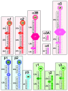
Figure 1. Known and/or predicted laminin heterotrimers. Eleven genes encode five α, three β and three γ chains in the human genome. There are two transcripts for the laminin α3 chain, one short α3A and one long α3B transcript. In theory, there are more than 50 possible heterotrimeric αβγ assemblies, but only those shown in the figure have been isolated or experimentally predicted.
None
Figure 2. Schematic representation of the highly diverging N-terminus of laminin chains. Three basic structural domains found in the N-terminus of the laminin chains are the LN (laminin N-terminal), LE (laminin-type epidermal growth (EGF) factor-like) and L4/LF (laminin IV) domains. The names (in black), location and size of the various domains are indicated for the different laminin chains. Each L4 domain corresponds to a long stretch of residues inserted between cysteine residues 3 and 4 of one LE domain. In this case the L4 and LE domains should be considered as one single domain which is emphasized by using the same color. In contrast the LF domains are located between two LE repeats as in the α3B, α5, β1 and β2 chains. When L4 or LF globular domains interrupt the arrays of repeating LE domains, they determine stretches named LEa, LEb and LEc. The numbering of the LE domains starts with 1 for each stretch (LEa1, LEa2, …; LEb1, LEb2, etc…). For example, the N-terminus of the laminin α1 chain starts with one LN domain, followed by a first array of four LE domains (LEa1 to LEa4), then one globular domain L4a (consisting of a long stretch of residue inserted within one LE domain), followed by a second array of eight LE domains (LEb1 to LEb8), another globular domain L4b (consisting of a long stretch of residue inserted within one LE domain), and finally a stretch of three LE domains (LEc1 to LEc3). In contrast the N-terminus of the laminin α3A chain consists of three LE domains only. This nomenclature adopted in 2005 is indicated to the left of each diagram. It should be noted that in the UniProt (http://uniprot.org) data bank, numbering of the LE domains is different. It is based on the total number of LE domains as indicated with the small gray lettering right to the various diagrams.
None
Figure 3. Mapping of the major functions of laminins. The laminin short arms (N-terminus) are involved in architectural function within the basement membrane, while the end of the long arm (C-terminus) is typically involved in cellular interactions.
None
Figure 4. The integrin binding site at the C-terminus of the laminin long arm. (A) The major integrin binding site on laminins is formed by the LG1 to LG3 domains and the extremity of coiled-coil fold formed by the α, β and γ chains. It is thought that the C-terminus of the γ chain is needed to stabilize a conformation of the LG1-LG3 trio compatible with integrin binding. (B) Representation of the amino acid sequences at the C-terminus of the three laminin γ chains. The γ1 and γ2 chains contains a glutamic acid residue (E, highlighted green) at the third position from the carboxyl termini. The residue is thought to be important for maintaining integrin binding activity of the structure shown in (A). The laminin γ3 chain is shorter and lacks the glutamic acid residue at the same position. Recombinant fragment engineered as shown in A and containing the γ3 chain have no integrin binding activity.

Similar articles

  • The laminins.
    Timpl R, Brown JC. Timpl R, et al. Matrix Biol. 1994 Aug;14(4):275-81. doi: 10.1016/0945-053x(94)90192-9. Matrix Biol. 1994. PMID: 7827749 Review.
  • Laminins in basement membrane assembly.
    Hohenester E, Yurchenco PD. Hohenester E, et al. Cell Adh Migr. 2013 Jan-Feb;7(1):56-63. doi: 10.4161/cam.21831. Epub 2012 Oct 17. Cell Adh Migr. 2013. PMID: 23076216 Free PMC article. Review.
  • Vascular laminins in physiology and pathology.
    Di Russo J, Hannocks MJ, Luik AL, Song J, Zhang X, Yousif L, Aspite G, Hallmann R, Sorokin L. Di Russo J, et al. Matrix Biol. 2017 Jan;57-58:140-148. doi: 10.1016/j.matbio.2016.06.008. Epub 2016 Jul 2. Matrix Biol. 2017. PMID: 27378388 Review. No abstract available.
  • Molecular analysis of laminin N-terminal domains mediating self-interactions.
    Odenthal U, Haehn S, Tunggal P, Merkl B, Schomburg D, Frie C, Paulsson M, Smyth N. Odenthal U, et al. J Biol Chem. 2004 Oct 22;279(43):44504-12. doi: 10.1074/jbc.M402455200. Epub 2004 Aug 13. J Biol Chem. 2004. PMID: 15310759
  • Laminin 332 processing impacts cellular behavior.
    Rousselle P, Beck K. Rousselle P, et al. Cell Adh Migr. 2013 Jan-Feb;7(1):122-34. doi: 10.4161/cam.23132. Epub 2012 Dec 21. Cell Adh Migr. 2013. PMID: 23263634 Free PMC article. Review.

Cited by

References

    1. Timpl R, Rohde H, Robey PG, Rennard SI, Foidart JM, Martin GR. Laminin--a glycoprotein from basement membranes. J Biol Chem. 1979;254:9933–7. - PubMed
    1. Chung AE, Jaffe R, Freeman IL, Vergnes JP, Braginski JE, Carlin B. Properties of a basement membrane-related glycoprotein synthesized in culture by a mouse embryonal carcinoma-derived cell line. Cell. 1979;16:277–87. doi: 10.1016/0092-8674(79)90005-9. - DOI - PubMed
    1. Rohde H, Wick G, Timpl R. Immunochemical characterization of the basement membrane glycoprotein laminin. Eur J Biochem. 1979;102:195–201. doi: 10.1111/j.1432-1033.1979.tb06280.x. - DOI - PubMed
    1. Engel J, Odermatt E, Engel A, Madri JA, Furthmayr H, Rohde H, et al. Shapes, domain organizations and flexibility of laminin and fibronectin, two multifunctional proteins of the extracellular matrix. J Mol Biol. 1981;150:97–120. doi: 10.1016/0022-2836(81)90326-0. - DOI - PubMed
    1. Bruch M, Landwehr R, Engel J. Dissection of laminin by cathepsin G into its long-arm and short-arm structures and localization of regions involved in calcium dependent stabilization and self-association. Eur J Biochem. 1989;185:271–9. doi: 10.1111/j.1432-1033.1989.tb15112.x. - DOI - PubMed

Publication types