

Molecular imaging of inflammation and platelet adhesion in advanced atherosclerosis effects of antioxidant therapy with NADPH oxidase inhibition
- PMID: 23239832
- PMCID: PMC3575135
- DOI: 10.1161/CIRCIMAGING.112.975193
Molecular imaging of inflammation and platelet adhesion in advanced atherosclerosis effects of antioxidant therapy with NADPH oxidase inhibition
Abstract
Background: In atherosclerosis, local generation of reactive oxygen species amplifies the inflammatory response and contributes to plaque vulnerability. We used molecular imaging to test whether inhibition of NADPH oxidase with apocynin would reduce endothelial inflammatory activation and endothelial-platelet interactions, thereby interrupting progression to high-risk plaque phenotype.
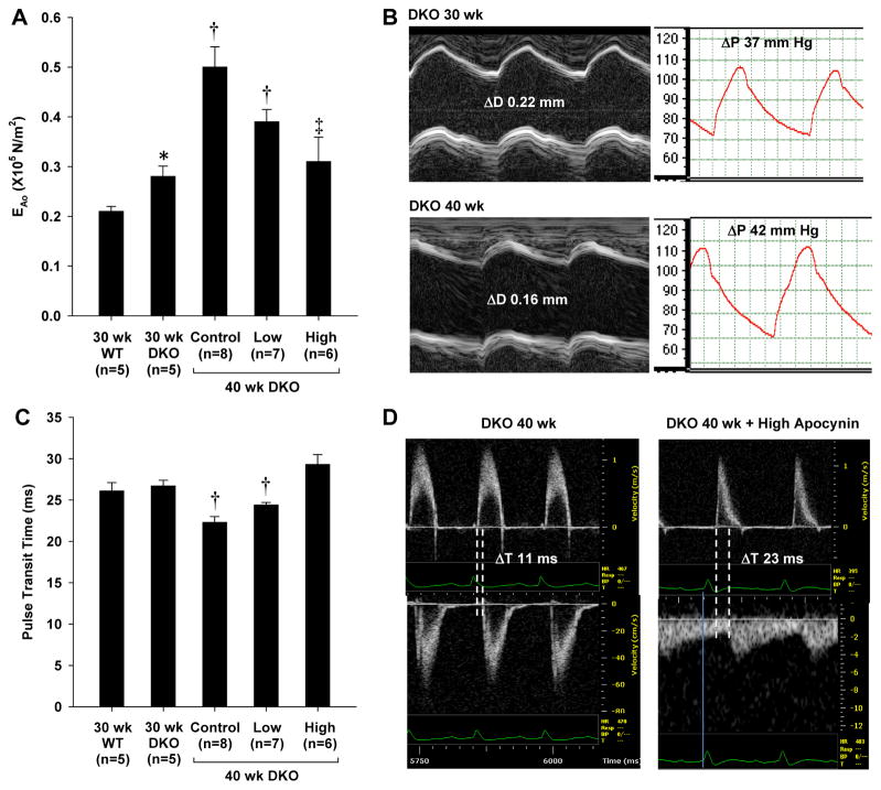
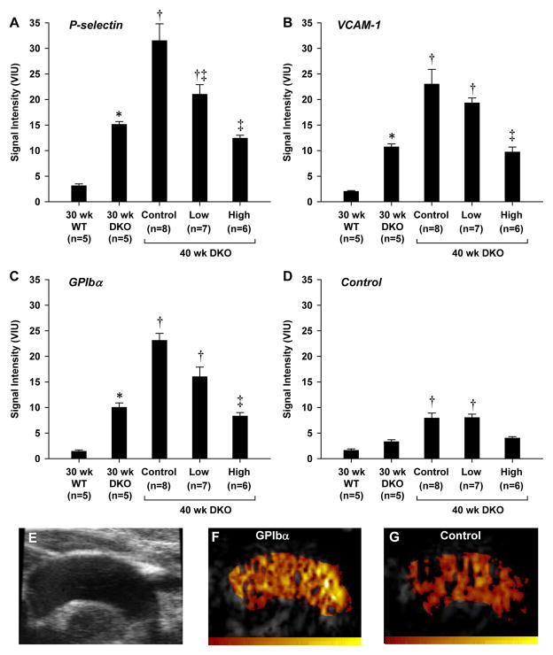
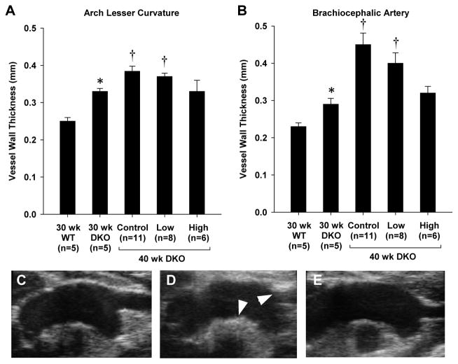
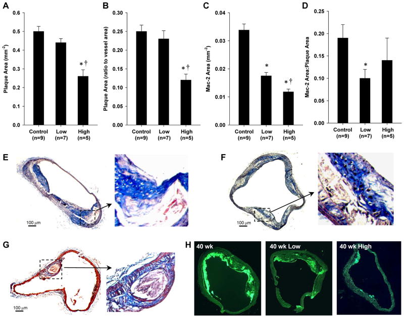
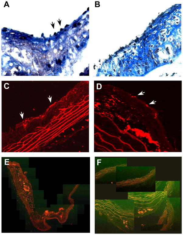
Methods and results: Mice deficient for both the low-density lipoprotein receptor and Apobec-1 were studied at 30 weeks of age and again after 10 weeks with or without apocynin treatment (10 or 50 mg/kg per day orally). In vivo molecular imaging of vascular cell adhesion molecule-1 (VCAM 1) P-selectin, and platelet glycoprotein-1bα (GPIbα) in the thoracic aorta was performed with targeted contrast-enhanced ultrasound molecular imaging. Arterial elastic modulus and pulse wave transit time were assessed using ultrahigh frequency ultrasound and invasive hemodynamic measurements. Plaque size and composition were assessed by histology. Molecular imaging in nontreated mice detected a 2-fold increase in P-selectin expression, VCAM-1 expression, and platelet adhesion between 30 and 40 weeks of age. Apocynin reduced all of these endothelial events in a dose-dependent fashion (25% and 50% reduction in signal at 40 weeks for low- and high-dose apocynin). Apocynin also decreased aortic elastic modulus and increased the pulse transit time. On histology, apocynin reduced total monocyte accumulation in a dose-dependent manner as well as platelet adhesion, although total plaque area was reduced in only the high-dose apocynin treatment group.
Conclusions: Inhibition of NADPH oxidase in advanced atherosclerosis reduces endothelial activation and platelet adhesion, which are likely responsible for the arrest of plaque growth and improvement of vascular mechanical properties.
Figures
Comment in
-
Letter by Violi et al regarding article, "molecular imaging of inflammation and platelet adhesion in advanced atherosclerosis effects of antioxidant therapy with NADPH oxidase inhibition".Circ Cardiovasc Imaging. 2013 Mar 1;6(2):e3. doi: 10.1161/CIRCIMAGING.112.000150. Circ Cardiovasc Imaging. 2013. PMID: 23512785 No abstract available.
Similar articles
-
Molecular imaging reveals rapid reduction of endothelial activation in early atherosclerosis with apocynin independent of antioxidative properties.Arterioscler Thromb Vasc Biol. 2013 Sep;33(9):2187-92. doi: 10.1161/ATVBAHA.113.301710. Epub 2013 Aug 1. Arterioscler Thromb Vasc Biol. 2013. PMID: 23908248 Free PMC article.
-
Letter by Violi et al regarding article, "molecular imaging of inflammation and platelet adhesion in advanced atherosclerosis effects of antioxidant therapy with NADPH oxidase inhibition".Circ Cardiovasc Imaging. 2013 Mar 1;6(2):e3. doi: 10.1161/CIRCIMAGING.112.000150. Circ Cardiovasc Imaging. 2013. PMID: 23512785 No abstract available.
-
Assessment of Novel Antioxidant Therapy in Atherosclerosis by Contrast Ultrasound Molecular Imaging.J Am Soc Echocardiogr. 2018 Nov;31(11):1252-1259.e1. doi: 10.1016/j.echo.2018.07.017. Epub 2018 Sep 11. J Am Soc Echocardiogr. 2018. PMID: 30213420 Free PMC article.
-
The role of the methoxyphenol apocynin, a vascular NADPH oxidase inhibitor, as a chemopreventative agent in the potential treatment of cardiovascular diseases.Curr Vasc Pharmacol. 2008 Jul;6(3):204-17. doi: 10.2174/157016108784911984. Curr Vasc Pharmacol. 2008. PMID: 18673160 Review.
-
Vascular cell adhesion molecule-1 expression and signaling during disease: regulation by reactive oxygen species and antioxidants.Antioxid Redox Signal. 2011 Sep 15;15(6):1607-38. doi: 10.1089/ars.2010.3522. Epub 2011 May 11. Antioxid Redox Signal. 2011. PMID: 21050132 Free PMC article. Review.
Cited by
-
Platelets and von Willebrand factor in atherogenesis.Blood. 2017 Mar 16;129(11):1415-1419. doi: 10.1182/blood-2016-07-692673. Epub 2017 Feb 7. Blood. 2017. PMID: 28174163 Free PMC article. Review.
-
Ultrasound imaging for risk assessment in atherosclerosis.Int J Mol Sci. 2015 Apr 29;16(5):9749-69. doi: 10.3390/ijms16059749. Int J Mol Sci. 2015. PMID: 25938969 Free PMC article. Review.
-
Molecular Ultrasound Imaging.Nanomaterials (Basel). 2020 Sep 28;10(10):1935. doi: 10.3390/nano10101935. Nanomaterials (Basel). 2020. PMID: 32998422 Free PMC article. Review.
-
Arterial Platelet Adhesion in Atherosclerosis-Prone Arteries of Obese, Insulin-Resistant Nonhuman Primates.J Am Heart Assoc. 2021 May 4;10(9):e019413. doi: 10.1161/JAHA.120.019413. Epub 2021 Apr 21. J Am Heart Assoc. 2021. PMID: 33880941 Free PMC article.
-
Molecular Imaging of VWF (von Willebrand Factor) and Platelet Adhesion in Postischemic Impaired Microvascular Reflow.Circ Cardiovasc Imaging. 2018 Nov;11(11):e007913. doi: 10.1161/CIRCIMAGING.118.007913. Circ Cardiovasc Imaging. 2018. PMID: 30571316 Free PMC article.
References
-
- Madamanchi NR, Vendrov A, Runge MS. Oxidative stress and vascular disease. Arterioscler Thromb Vasc Biol. 2005;25:29–38. - PubMed
-
- Wolin MS. Interactions of oxidants with vascular signaling systems. Arterioscler Thromb Vasc Biol. 2000;20:1430–1442. - PubMed
-
- Kunsch C, Medford RM. Oxidative stress as a regulator of gene expression in the vasculature. Circ Res. 1999;85:753–766. - PubMed
-
- Freedman JE. Oxidative stress and platelets. Arterioscler Thromb Vasc Biol. 2008;28:s11–16. - PubMed
Publication types
MeSH terms
Substances
Grants and funding
- R01 DK063508/DK/NIDDK NIH HHS/United States
- RC1 HL100659/HL/NHLBI NIH HHS/United States
- RC1-HL100659/HL/NHLBI NIH HHS/United States
- P01 HL031950/HL/NHLBI NIH HHS/United States
- P01 HL078784/HL/NHLBI NIH HHS/United States
- R01-DK063508/DK/NIDDK NIH HHS/United States
- P01-HL78784/HL/NHLBI NIH HHS/United States
- R01 HL111969/HL/NHLBI NIH HHS/United States
- R01-HL42846/HL/NHLBI NIH HHS/United States
- T32 HL094294/HL/NHLBI NIH HHS/United States
- T32-HL094294/HL/NHLBI NIH HHS/United States
- R01 HL078610/HL/NHLBI NIH HHS/United States
- R01-HL111969/HL/NHLBI NIH HHS/United States
- P01-HL31950/HL/NHLBI NIH HHS/United States
- R01-HL078610/HL/NHLBI NIH HHS/United States
- R01HL101972/HL/NHLBI NIH HHS/United States
- R01 HL042846/HL/NHLBI NIH HHS/United States
- R01 HL101972/HL/NHLBI NIH HHS/United States
LinkOut - more resources
Full Text Sources
Other Literature Sources
Medical
Miscellaneous
