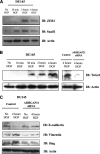
. 2013 Jan 25;288(4):2179-89.
doi: 10.1074/jbc.M112.432716. Epub 2012 Dec 12.

ARHGAP21 protein, a new partner of α-tubulin involved in cell-cell adhesion formation and essential for epithelial-mesenchymal transition

Affiliations

ARHGAP21 protein, a new partner of α-tubulin involved in cell-cell adhesion formation and essential for epithelial-mesenchymal transition

Karin S A Barcellos et al. J Biol Chem. .

Abstract

Cell-cell adhesions and the cytoskeletons play important and coordinated roles in cell biology, including cell differentiation, development, and migration. Adhesion and cytoskeletal dynamics are regulated by Rho-GTPases. ARHGAP21 is a negative regulator of Rho-GTPases, particularly Cdc42. Here we assess the function of ARHGAP21 in cell-cell adhesion, cell migration, and scattering. We find that ARHGAP21 is localized in the nucleus, cytoplasm, or perinuclear region but is transiently redistributed to cell-cell junctions 4 h after initiation of cell-cell adhesion. ARHGAP21 interacts with Cdc42, and decreased Cdc42 activity coincides with the appearance of ARHGAP21 at the cell-cell junctions. Cells lacking ARHGAP21 expression show weaker cell-cell adhesions, increased cell migration, and a diminished ability to undergo hepatocyte growth factor-induced epithelial-mesenchymal transition (EMT). In addition, ARHGAP21 interacts with α-tubulin, and it is essential for α-tubulin acetylation in EMT. Our findings indicate that ARHGAP21 is a Rho-GAP involved in cell-cell junction remodeling and that ARHGAP21 affects migration and EMT through α-tubulin interaction and acetylation.


Figures
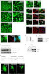
FIGURE 1.
FIGURE 1.
Localization of ARHGAP21 at cell-cell junctions during adhesion formation. A, shown is immunofluorescence analysis of MDCK cells stained with an antibody against ARHGAP21 (green) after calcium restoration at 0, 1, 4, 7 and 24 h; images were taken using a 60× oil immersion objective. The arrow indicates ARHGAP21 localization at cell-cell adhesion sites. Note that ARHGAP21 was detected at cell-cell junctions 4 h after calcium restoration in MDCK cells. B, shown is an enlarged image of MDCK cells 4 h after calcium restoration stained with an antibody against ARHGAP21 (green) and F-actin (phalloidin-TRITC); co-localization of ARHGAP21 and actin is observed in the merged image. C, shown is Cdc42 Rho-GTPase activity during cell-cell adhesion formation at 0, 1, 2, 3, 4, 5, and 7 h after calcium restoration in MDCK cells. Cdc42 Rho-GTPase activity drastically reduced after ARHGAP21 localization at the cell-cell junctions. IB, immunoblot. D, shown is a schematic representation of ARHGAP21 and its domains and of the ARHGAP21 regions cloned in fusion to GFP. E, F, and G, MDCK cells were transfected with plasmids encoding ARHGAP21 constructs fused to GFP (PDZ-ARHGAP21, PH+GAP-ARHGAP21, Cter-ARHGAP21); the PDZ-ARHGAP21 domain was at the cell-cell contacts 4 h after calcium addition. H, shown is immunofluorescence analysis of DU145 cells stained with antibody against ARHGAP21 (green) 4 and 24 h after calcium restoration; images were acquired with 100× oil immersion objective. Note that ARHGAP21 was detected at cell-cell junctions 4 h after calcium restoration in DU145 cells. I, shown is an enlarged image of DU145 cells 4 h after calcium restoration stained with antibody against ARHGAP21 (green) and E-cadherin (red) which shows that both proteins are not co-localized at cell-cell contacts. J, shown is an enlarged image of DU145 cells 4 h after calcium restoration stained with antibody against ARHGAP21 (green) and JAM-A (red). K, shown is an enlarged image of DU145 cells 4 h after calcium restoration stained with antibody against ARHGAP21 (green) and α-catenin (red); the arrows indicate that both proteins were not co-localized at cell-cell contacts. L, shown is immunofluorescence analysis of DU145 ARHGAP21 siRNA cells stained with antibodies against ARHGAP21 (green) and α-catenin (red), indicating that α-catenin is still located at the cell membrane in ARHGAP21-depleted cells. M and N, quantitative RT-PCR and Western blotting analyses of ARHGAP21 expression in DU145 cells submitted to control siRNA or to ARHGAP21 siRNA transfection show efficiency of ARHGAP21 inhibition. O, shown is endogenous ARHGAP21 co-immunoprecipitated with Cdc42. Total extracts from 4 and 24 h after calcium restoration in DU145 cells were submitted to immunoprecipitation with an anti-ARHGAP21 antibody followed by Western blot analysis using anti-Cdc42 and anti-ARHGAP21 antibody. Bar, 10 μm.
FIGURE 2.
FIGURE 2.
ARHGAP21 role in cell aggregation. Shown is analysis of ARHGAP21 knockdown SW480 cells and control-transfected SW480 cells in a quantitative and functional adhesion assay, the hanging drop aggregation assay, at time points of 0, 1, and 3 h with triturated and non-triturated cells. A, shown is Western blot analysis of SW480 cells submitted to control shRNA or to ARHGAP21 shRNA transfection immunoblotted (IB) with anti-ARHGAP21 and anti-actin antibodies (as a control for equal sample loading). B and C, graphs show the percentage of cells in clusters of 0–10 cells (gray), 11–50 cells (dark gray), and >50 cells (white) at the time points indicated with and without trituration, respectively. D—K, photographs are representative fields at 1 and 3 h with and without trituration. D, control cells at 1 h not triturated. E, shRNA cells at 1 h not triturated. F, control cells at 1 h triturated. G, shRNA cells at 1 h triturated. H, control cells at 3 h not triturated. I, shRNA cells at 3 h not triturated. J, control cells at 3 h triturated. K, shRNA cells at 3 h triturated. Note that ARHGAP21 shRNA cells do not present clusters after trituration. Objective 20×.
FIGURE 3.
FIGURE 3.
ARHGAP21 knockdown increases cell migration and ARHGAP21 interacts with α-tubulin. A, SW480 ARHGAP21 shRNA and control shRNA cell migration were detected using Oris Cell Migration 96-well plate. Images from the central well region were captured; cells are visualized in black with the empty area in white. B, the graph shows the migration rates of control and ARHGAP21 shRNA cells, with an increased migration in the shRNA cells (p = 0.0035). C, shown is an immunofluorescence analysis of DU145 cells in a wound-healing assay stained with antibody against ARHGAP21 (green) at 0 and 6 h of migration. DAPI staining indicates nuclear area. D, shown is endogenous ARHGAP21 co-immunoprecipitated (IP) with α-tubulin. Total extracts from independent samples of DU145 cells were submitted to immunoprecipitation with an anti-ARHGAP21 antibody (lanes IP1 and IP2) followed by Western blot (IB) analysis using anti-α-tubulin and anti-ARHGAP21 antibody. Lanes TE1 and TE2 correspond to a Western blot performed on total cell extracts. E, DU145 cell extracts were submitted to immunoprecipitation with an anti-α-tubulin antibody followed by Western blot analysis using anti-ARHGAP21 and anti-α-tubulin antibody. F, DU145 cells were transfected with the ARHGAP21 siRNA or control siRNA. After 24 h cells were seeded in sextuplicate onto a 96-well filter plate. HGF was added below the filter as a chemo-attractant 24 h later. After 20 h the migration was stopped, and the cells were fixed and analyzed. The percentage of migration was calculated relative to control. An increased migration of DU145 ARHGAP21 siRNA compared with the control cells was observed. The migration of control siRNA cells was increased proportionally to HGF amount. However, the migration of ARHGAP21 siRNA cells was not modulated by HGF treatment. *, p < 0.05. Bar, 10 μm.
FIGURE 4.
FIGURE 4.
ARHGAP21 knockdown inhibits EMT induced by HGF. A, live cell imaging shows colonies of DU145 cells on collagen responding to HGF stimulation. ARHGAP21 siRNA and control siRNA cells were starved in medium with 0.5% of FBS for 24 h before the HGF stimulation. Colonies were filmed for 10 h. Images from DU145 cells not stimulated with HGF and ARHGAP21/control siRNA-transfected cells stimulated with HGF are exemplified here. Those images were obtained from two independent experiments for each condition (ARHGAP21 siRNA and control siRNA) and used as representative data. B, the colonies were graduated according to the intensity of scattering response. No scattering, hardly any ICS and RF; Light scattering, showing ICS/RF but no cells break away; Semi-scattering, ICS/RF with at least one cell that completely breaks away; Full scattering, ICS/RF with two or more cells breaking from colony. Data were collected from 24 colonies for ARHGAP21 siRNA and 28 colonies for control-siRNA over 3 independent experiments. ARHGAP21 inhibition results in a diminishing of cell ability to undergo EMT in response to HGF signaling. Bar, 50 μm.
FIGURE 5.
FIGURE 5.
ARHGAP21 depletion affects the expression of proteins EMT markers during EMT induced by HGF. A, Western blotting (IB) shows ZEB1 and Snail1 expression in DU145 cells non-treated or induced with HGF for 30 min, 2 h, and 4 h. B, shown is expression of Twist1 in DU145 cells non-treated or induced with HGF for 10 min, 30 min, 2 h, and 4 h and control and ARHGAP21 siRNA stimulated with 4 h HGF. C, Western blotting shows E-cadherin, vimentin, and slug expression in control and ARHGAP21 DU145 cells non-treated or induced with HGF for 4 h. The Western blotting confirm that ARHGAP21 inhibition results in a diminishing of cells ability to undergo EMT in response to HGF signaling. Anti-β-actin blotting was used as loading control in A, B, and C.
FIGURE 6.
FIGURE 6.
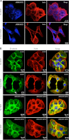
ARHGAP21 translocates to the nucleus after HGF stimulus. A, shown is immunofluorescence analysis of DU145 cells stained with antibody against ARHGAP21 (blue) and F-actin (phalloidin-TRITC) with or without HGF 2 h stimulation. Note that ARHGAP21 localizes at perinuclear areas in the absence of HGF and in the nucleus after HGF stimulation. B, shown is an immunofluorescence image of DU145 cells with no HGF and 2 h after HGF stimulus stained with antibody against β-catenin (green) and F-actin (phalloidin-TRITC). Images were acquired with a 60× oil immersion objective lens.
FIGURE 7.
FIGURE 7.
ARHGAP21 knockdown inhibits α-tubulin acetylation. A, Western blotting (IB) shows acetylated-α-tubulin expression in DU145 cells, no HGF, induced with HGF for 10 min and 3 h; total α-tubulin was used as a control of the reaction. B, shown is expression of acetylated and total α-tubulin proteins in DU145 siRNA control and siRNA ARHGAP21 cells with no HGF and induced with HGF for 3 h. C, the graph shows HDAC activity in cytoplasmic extracts of control and ARHGAP21 siRNA DU145 cells, with no difference in HDAC activity between the samples (p > 0.05); bars represent analysis of colorimetric assay as mean ± S.D. of three independent experiments versus control, set as 100%.

Similar articles

Cited by

References

    1. Niessen C. M., Gottardi C. J. (2008) Molecular components of the adherens junction. Biochim. Biophys. Acta 1778, 562–571 - PMC - PubMed
    1. Arthur W. T., Noren N. K., Burridge K. (2002) Regulation of Rho family GTPases by cell-cell and cell-matrix adhesion. Biol. Res. 35, 239–246 - PubMed
    1. Fukata M., Kaibuchi K. (2001) Rho-family GTPases in cadherin-mediated cell-cell adhesion. Nat. Rev. Mol. Cell Biol. 2, 887–897 - PubMed
    1. Kim S. H., Li Z., Sacks D. B. (2000) E-cadherin-mediated cell-cell attachment activates Cdc42. J. Biol. Chem. 275, 36999–37005 - PubMed
    1. Van Aelst L., Symons M. (2002) Role of Rho family GTPases in epithelial morphogenesis. Genes Dev. 16, 1032–1054 - PubMed

Publication types

LinkOut - more resources