Review
. 2012;88(10):554-82.
doi: 10.2183/pjab.88.554.

My journey into the world of sphingolipids and sphingolipidoses

Affiliations
Review

My journey into the world of sphingolipids and sphingolipidoses

Konrad Sandhoff. Proc Jpn Acad Ser B Phys Biol Sci. 2012.

Abstract

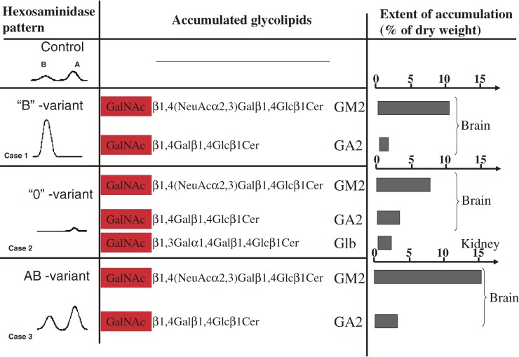
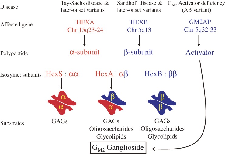
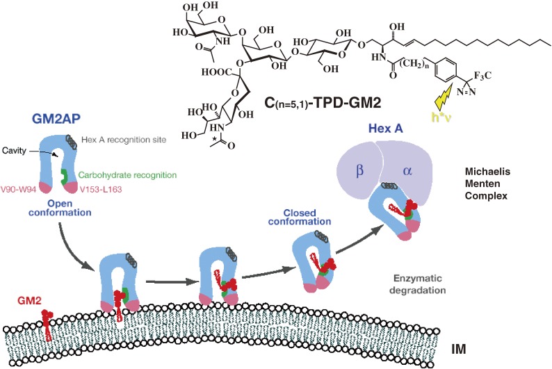
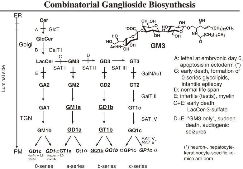
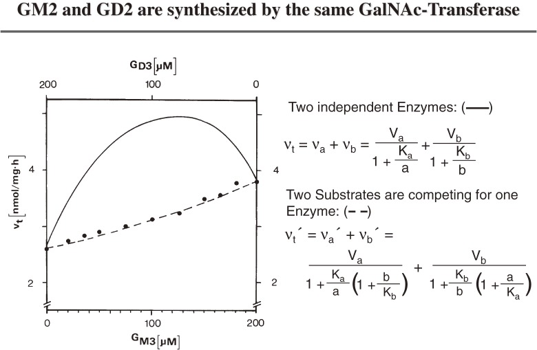
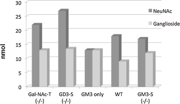
Analysis of lipid storage in postmortem brains of patients with amaurotic idiocy led to the recognition of five lysosomal ganglioside storage diseases and identification of their inherited metabolic blocks. Purification of lysosomal acid sphingomyelinase and ceramidase and analysis of their gene structures were the prerequisites for the clarification of Niemann-Pick and Farber disease. For lipid catabolism, intraendosomal vesicles are formed during the endocytotic pathway. They are subjected to lipid sorting processes and were identified as luminal platforms for cellular lipid and membrane degradation. Lipid binding glycoproteins solubilize lipids from these cholesterol poor membranes and present them to water-soluble hydrolases for digestion. Biosynthesis and intracellular trafficking of lysosomal hydrolases (hexosaminidases, acid sphingomyelinase and ceramidase) and lipid binding and transfer proteins (GM2 activator, saposins) were analyzed to identify the molecular and metabolic basis of several sphingolipidoses. Studies on the biosynthesis of glycosphingolipids yielded the scheme of Combinatorial Ganglioside Biosynthesis involving promiscuous glycosyltransferases. Their defects in mutagenized mice impair brain development and function.


Figures

Figure 1.
Figure 1.
Biochemical analysis of brains with infantile amaurotic idiocy. Lipid and hexosaminidase analysis in the brain tissue of the three first cases revealed puzzling results in 1967 and spring of 1968 (summarized in: ref. 249). - The tissue of case 1 with TSD (called “B”-variant) was deficient in HexA activity and had increased HexB activity, a finding which was confirmed in 1968 by an additional case250) and the observation by Okada and O’Brian.251) - Brain and visceral organs of case 2 –characterized by the additional storage of globoside (Glb) in visceral tissues– was completely deficient in both major hexosaminidases, A and B, and was left with a minor activity of HexS.26,250,252,253) This one we named “0”-variant of GM2-gangliosidosis and it was later called Sandhoff disease. - Apparently contradicting the above findings, the brain tissue of case 3 contained both, HexA and HexB activity, even at elevated levels despite a huge accumulation of GM2 and GA2 (later called AB-variant after the defect of a cofactor –GM2AP– was discovered, see below).
Figure 2.
Figure 2.
The GM2 Gangliosidoses and the β-hexosaminidase system (modified after ref. 25). Three polypeptides, each encoded in a different gene, are needed for the degradation of ganglioside GM2: the α- and β-subunit of the A isoenzyme and the activator protein which binds the ganglioside and presents it to the enzyme. GAGs, glycosaminoglycans.
Figure 3.
Figure 3.
Mechanism of GM2AP-Liftase. Model for the interaction of GM2-activator protein with luminal lysosomal membranes in the degradation of ganglioside GM2 (modified after ref. 36). GM2AP interacts with the membrane by dipping the two exposed hydrophobic loops, V90-W94 and V153-L163, into the apolar part of the membrane. Ganglioside GM2 is recognized by specific sites at the rim of the cavity. In the open protein conformation, the large hydrophobic area reaching from the apolar phase of the membrane to the activator’s cavity lowers the energy barrier for lipids leaving the membrane in an upward direction. After the ceramide tail has moved inside the activator’s cavity, the inward pointing orientation for the hydrophobic loop V153-L163 is favoured and the conformation changes to the closed form.254) This folding in of the hydrophobic loop leaves a more polar patch close to the membrane. The activator is then anchored only by the loop V90-W94. It may now rotate slightly upwards to expose all polar patches more fully to the solvent, and it may also leave the membrane and interact with the degrading enzyme. The photoaffinity label analogues C(n=5,1)-TPD-GM2 were photoincorporated specifically into the region V153-L163.
Figure 4.
Figure 4.
Residual catabolic activity correlates with clinical forms of GM2 gangliosidoses (modified after ref. 53). A) Steady-state substrate concentration as a function of enzyme concentration and activity.51) The model underlying this theoretical calculation assumes influx of the substrate into the lysosomal compartment at a constant rate (vi) and its subsequent utilization by the catabolic enzyme (for details see text). Blue line = [S]eq steady-state substrate concentration; ……… = theoretical threshold of enzyme activity; - - - - - - - = critical threshold value, taking limited solubility of substrate into account; red line = turnover rate of substrate (flux rate). B) In order to experimentally verify the basic assumptions on which this model rests, studies were performed in cell culture.53) The radiolabeled substrate ganglioside GM2 was added to cultures of skin fibroblasts with different activities of beta-hexosaminidase A and its uptake and turnover measured. The correlation between residual enzyme activity and the turnover rate of the substrate was essentially as predicted: degradation rate of ganglioside GM2 increased steeply with residual activity, to reach the control level at a residual activity of approximately 10–15% of normal. All cells with an activity above this critical threshold had a normal turnover. Comparison of the results of these feeding studies with the clinical status of the donor of each cell line basically confirmed our notions but also revealed the limitations of the cell culture approach.
Figure 5.
Figure 5.
Sap-B stimulates the enzymatic hydrolysis of micellar sulfatides with long acyl chains. Derivates of sulfatide were hydrolyzed by human arylsulfatase A in the absence or presence of Sap-B (0.1 nmol) in a total incubation volume of 50 µl. The degradation rates given are means from six experiments. Experimental values were all within a range of 20 per cent of the mean. Sulfatides contain different acyl residues in their ceramide moieties: C18-stearoyl, C8-octanoyl, C6-hexanoyl and C2-acetyl-residues.68)
Figure 6.
Figure 6.
Human Prosaposin. The cDNA of human prosaposin codes for a sequence of 524 (or 527) amino acids including a signal peptide of 16 amino acids.92,94) The precursor is proteolytically processed in the endolysosomal compartment to 4 domains, saposin A, B, C and D.102) Cystein residues are marked by vertical bars, N-glycosylation sites by arrow heads. Disease causing mutations affecting human Sap-A,255) Sap-B,64,65) Sap-C256) and prosaposin67,96,257) have been described. Murine models of prosaposin deficiency77) and deficiencies of individual saposins have been investigated: Sap-A,78,80,258) Sap-B,76) Sap-C79) and Sap-D.80,105,259)
Figure 7.
Figure 7.
Extracellular metabolism of glucosylceramide (GlcCer) and formation of the lipid-bound envelope (LBE) (modified after ref. 260). PM: plasma membrane, Cer: ceramide, consisting of sphingosine (black) and long-chain ω-hydroxyl fatty acid (blue). Linoleic acid (green) is found esterified to the ω-hydroxyl moiety. Catabolic blocks in β-glucocerebrosidase- and SAP-C deficiency are indicated by the arrows with slashes through them.
Figure 8.
Figure 8.
Principles of lysosomal sphingolipid catabolism and membrane digestion. Proposed topology of endocytosis and lysosomal degradation.130) A section of the plasma membrane is internalized by way of coated pits or caveolae. These membrane patches include glycosphingolipids (GSL, red) and receptors such as EGFR (epidermal growth factor receptor, blue). These vesicles fuse with the early endosomes which mature to late endosomes. Endosomal perimeter membranes form invaginations, controlled by ESCRT proteins,129) which bud off, forming intra-endosomal vesicles. Lipid sorting occurs at this stage. The pH of the lumen is at about 5. At this pH, acid sphingomyelinase is active and degrades sphingomyelin of the intra-endosomal vesicles to ceramide, whereas the perimeter membrane is protected against the action of ASM by the glycocalix facing the lumen. This reduction of the sphingomyelin level, coupled with the increase in ceramide, facilitates the binding of cholesterol to NPC-2 and its transport to the perimeter membrane of the late endosome where it is transferred to NPC-1.261) This protein enables the export of cholesterol through the glycocalix, eventually reaching cholesterol binding proteins in the cytosol. Ultimately, late endosomes fuse with lysosomes. The GSLs are in intra-lysosomal vesicles facing the lumen of the lysosome and are degraded by hydrolases with the assistance of LLBPs. The products of this degradation are exported to the cytosol or loaded on CD1b immunoreceptors and exported to the plasma membrane for antigen presentation. Gradients of pH in the lysosol, and intra-endo-lysosomal vesicle content of cholesterol (Chol), BMP, sphingomyelin (SM, hypothetical) and ceramide (Cer; hypothetical) are shown (modified after ref. 83).
Figure 9.
Figure 9.
Scheme of ganglioside biosynthesis. Major gangliosides occurring on neurons in adult mammalian brain are underlined. Enzyme defects in knockout mice are indicated. A, ceramide glucosyltransferase −/−; B, galactosyltransferase I −/−; C, sialyltransferase I −/−; D, sialyltransferase II −/−; E, GalNAc-transferase −/−; “GM3 only” mouse = D + E; “LacCer only” mouse = C + E. GD1α, GT1α, GQ1bα and GP1cα with a sialic acid residue α2,6-glycosidically linked to N-acetylgalactosamine are formed by a yet uncharacterized sialyltransferase, whereas GD1c, GT1a, GQ1b and GP1c are formed by SAT V T (modified after ref. 211).
Figure 10.
Figure 10.
Competition between ganglioside GM3 and GD3 in the GalNAc-transferase assay.210) GM3 and GD3 were used as acceptors for GalNAc-transferase in various partial concentrations, keeping total substrate at 200 micro M. Total reaction velocities as experimentally determined (●) or as calculated for the different models are plotted versus the partial substrate concentrations. ——, vt calculated from upper Eq. (two different enzymes); - - - - -, vt calculated from lower Eq. (one enzyme).
Figure 11.
Figure 11.
Total lipid-bound sialic acid and ganglioside levels in brain tissue of mice with engineered defects in ganglioside biosynthesis.219) Data refer to 10 mg of wet weight brain tissue; abbreviations are given in Fig. 9.

Similar articles

Cited by

References

    1. Jatzkewitz H. (1958) Zwei Typen von Cerebrosid-Schwefelsäureestern als sog. Prälipoide und Speichersubstanzen bei der Leukodystrophy, Typ Scholz (Metachromatische Form der diffusen Sklerose). Hoppe-Seyler’s Z. Physiol. Chem. 311, 279–282 - PubMed
    1. Jatzkewitz H. (1964) Eine neue Methode zur quantitativen Ultramikrobestimmung der Sphingolipoide aus Gehirn. Hoppe-Seyler’s Z. Physiol. Chem. 336, 25–39 - PubMed
    1. Jatzkewitz H., Mehl E. (1969) Cerebroside-sulphatase and arylsulphatase A deficiency in metachromatic leukodystrophy (ML). J. Neurochem. 16, 19–28 - PubMed
    1. Sandhoff, K. (1965) Die Amaurotische Idiotie des Menschen als Störung im Glykosphingolipidstoffwechsel. Thesis, University of Munich.
    1. Jatzkewitz H., Sandhoff K. (1963) On a biochemically special form of infantile amaurotic idiocy. Biochim. Biophys. Acta 70, 354–356 - PubMed

Publication types

Substances