. 2012 Dec 26;109(52):21354-9.
doi: 10.1073/pnas.1218272110. Epub 2012 Dec 7.

Arl13b regulates endocytic recycling traffic

Affiliations

Arl13b regulates endocytic recycling traffic

Duarte C Barral et al. Proc Natl Acad Sci U S A. .

Abstract

Intracellular recycling pathways play critical roles in internalizing membrane and fluid phase cargo and in balancing the inflow and outflow of membrane and cell surface molecules. To identify proteins involved in the regulation of endocytic recycling, we used an shRNA trafficking library and screened for changes in the surface expression of CD1a antigen-presenting molecules that follow an endocytic recycling route. We found that silencing of the ADP-ribosylation factor (Arf)-like small GTPase Arl13b led to a decrease in CD1a surface expression, diminished CD1a function, and delayed CD1a recycling, suggesting that Arl13b is involved in the regulation of endocytic recycling traffic. Arl13b appears to be required for the major route of endocytic trafficking, causing clustering of early endosomes and leading to the accumulation of endocytic cargo. Moreover, Arl13b colocalized with markers of the endocytic recycling pathway followed by CD1a, namely Arf6 and Rab22a. We also detected an interaction between Arl13b and the actin cytoskeleton. Arl13b was previously implicated in cilia formation and function. Our present results indicate a previously unidentified role for Arl13b in endocytic recycling traffic and suggest a link between Arl13b function and the actin cytoskeleton.


Conflict of interest statement

The authors declare no conflict of interest.

Figures
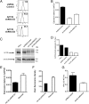
Fig. 1.
Fig. 1.
Arl13b silencing leads to a decrease in CD1a surface expression and function. (A) HeLa:CD1a stable transfectant cells were transduced with lentiviruses encoding Arl13b-targeting shRNA (E3 and E4) or control (Mission) and selected in puromycin for 1 wk. CD1a surface levels were analyzed by flow cytometry (black lines). Gray lines correspond to isotype controls. The number in each panel represents the geometric mean fluorescence intensity. Results are representative of at least two independent experiments. (B) Arl13b mRNA levels were analyzed by quantitative RT-PCR. Error bars represent SD of triplicate measurements, and the results are representative of at least three independent experiments. (C) Arl13b protein expression levels from total cell lysates were analyzed by Western blot using anti-Arl13b affinity-purified Ab. α-tubulin was used as an internal loading control. (D) Quantification of band intensities from C using ImageJ software. (E and F) HeLa:CD1a:shRNA cells were transfected with Arl13b (“rescued”) or an empty vector (“Arl13b shRNA E4”) and selected in blasticydin. Arl13b mRNA levels were analyzed by quantitative RT-PCR (E), and CD1a surface levels were analyzed by flow cytometry (F). Error bars represent the SE of four cell lines for rescued and two lines for Arl13b shRNA E4. Analysis of Arl13b mRNA levels (E) was done in triplicate. (G) Cells transduced with Arl13b-targeting shRNA or control (Mission) were incubated with 0.11 μg/mL sonicated antigen and CD8-2 T cells, which recognize DDM presented by CD1a. After 22 h, the release of IFN-γ was measured by ELISA. Error bars represent SE of two Arl13b-targeting shRNAs and two control samples, each analyzed in triplicate. The results are representative of three independent experiments.
Fig. 2.
Fig. 2.
Arl13b silencing impairs CD1a recycling and results in early endosome clustering. (A) HeLa cells were transiently cotransfected with CD1a and Arl13b-targeting shRNA (E3 and E4) or control (Mission), selected in puromycin, and incubated with anti-CD1a Ab on ice and then at 37 °C to allow for internalization. After the surface-bound Ab was stripped in a brief acidic wash, cells were incubated at 37 °C for different periods to allow for recycling. Cells were then stained with anti-mouse secondary Ab and analyzed by flow cytometry. The results (in arbitrary units) are normalized for recycling at time 0. The results are representative of three independent experiments. (B–E) HeLa:CD1a (B and C) or HeLa:CD1b (E) stable transfectant cells or HeLa untransfected cells (D) were transfected with Arl13b-targeting shRNA (Lower) or control (Upper) and selected in puromycin. (B) Cells were fixed, permeabilized, and stained with anti-EEA1 primary Ab, followed by Alexa Fluor 546-conjugated anti-mouse secondary Ab. (C) Cells were serum-starved for 30 min and then incubated with anti-CD1a mAb for 30 min on ice. Cells were then washed and incubated for 15 min at 37 °C with Alexa Fluor 488-conjugated transferrin (green). After the surface-bound Ab was stripped in a brief acidic wash, cells were fixed, permeabilized, and labeled with Alexa Fluor 546-conjugated anti-mouse to detect anti-CD1a (red). (D) Cells were serum-starved for 30 min and incubated with Alexa Fluor 488-conjugated transferrin (green) and Alexa Fluor 546-conjugated dextran (red) for 15 min. After washing, cells were chased for 20 min and then fixed. (E) Cells were incubated with anti-CD1b mAb for 30 min on ice, and then washed and incubated for 30 min at 37 °C. After the surface-bound Ab was stripped as in B, cells were fixed, permeabilized, stained for EEA1 with a rabbit polyclonal Ab (green), and labeled with Alexa Fluor 546-conjugated anti-mouse to detect anti-CD1b (red). Arrows indicate cargo clustering. (Scale bars: 10 μm.)
Fig. 3.
Fig. 3.
Arl13b-EGFP colocalizes with CD1a, Rab22a, and Arf6 in HeLa cells. (A) HeLa cells were transiently transfected with Arl13b-EGFP and analyzed by confocal microscopy. Arrows indicate Arl13b-decorated tubular structures. (B) HeLa cells were cotransfected with CD1a and Arl13b-EGFP and incubated with anti-CD1a mAb for 30 min on ice. Cells were then washed and incubated for 30 min at 37 °C. After the surface-bound Ab was stripped in a brief acidic wash, cells were fixed, permeabilized, and labeled with Alexa Fluor 546-conjugated anti-mouse secondary Ab to detect anti-CD1a (red). (C) HeLa cells were transiently cotransfected with EGFP-Rab22a Q64L and Arl13b-FLAG, fixed, permeabilized, and stained with Cy3-conjugated mouse monoclonal anti-FLAG Ab (red). (D) HeLa cells were transiently cotransfected with Arl13b-EGFP and HA-Arf6 T27N, fixed, permeabilized, and stained with anti-HA tag polyclonal Ab followed by Alexa Fluor 546-conjugated anti-rabbit secondary Ab (red). (Scale bars: 10 μm.)
Fig. 4.
Fig. 4.
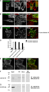
Arl13b interacts with actin. (A–C) HeLa cells were transfected with Arl13b-EGFP for 24 h, treated with 5 µM of cytochalasin D for 45 min (C) or untreated, fixed, permeabilized, and stained with anti–α-tubulin Ab (A) or Alexa Fluor 568-conjugated phalloidin, which labels filamentous actin (B and C) (red). Arrowheads indicate actin filaments that colocalize with Arl13b tubules. Arrows indicate plasma membrane accumulations of actin and Arl13b. (D and E) HeLa:CD1a stable transfectant cells were transiently transfected with Arl13b-EGFP for 24 h, treated with cytochalasin D for 45 min at the indicated concentrations (0.2 µM in E), fixed, permeabilized, and stained with anti-CD1a mAb (red). The number of Arl13b tubules per cell was counted (n = 95) and grouped into cells with >10 and <10 tubules per cell (D). Arrows indicate Arl13b and CD1a colocalization in tubules (E). Results are representative of two independent experiments. (F and G) HeLa and NIH 3T3 cell lysates were immunoprecipitated with mouse monoclonal anti–β-actin (F) or rabbit polyclonal anti-Arl13b (G) Ab, separated by SDS/PAGE, and immunoblotted with anti-Arl13b (F) or anti–β-actin (G) Ab, followed by HRP-conjugated anti-rabbit or anti-mouse secondary Ab, respectively. Rabbit and mouse IgG were used as negative controls. (Scale bars: 10 μm.)
Fig. 5.
Fig. 5.
Arl13b 1–193 truncation mutant displays a punctate distribution and colocalizes with transferrin. HeLa cells were transfected with Arl13b 1–193-EGFP, starved, pulsed with Alexa Fluor 546-conjugated transferrin (red) for 30 min and fixed. (Scale bar: 10 μm.)

Similar articles

Cited by

References

    1. Grant BD, Donaldson JG. Pathways and mechanisms of endocytic recycling. Nat Rev Mol Cell Biol. 2009;10(9):597–608. - PMC - PubMed
    1. Barral DC, et al. CD1a and MHC class I follow a similar endocytic recycling pathway. Traffic. 2008;9(9):1446–1457. - PMC - PubMed
    1. Weigert R, Yeung AC, Li J, Donaldson JG. Rab22a regulates the recycling of membrane proteins internalized independently of clathrin. Mol Biol Cell. 2004;15(8):3758–3770. - PMC - PubMed
    1. Brown FD, Rozelle AL, Yin HL, Balla T, Donaldson JG. Phosphatidylinositol 4,5-bisphosphate and Arf6-regulated membrane traffic. J Cell Biol. 2001;154(5):1007–1017. - PMC - PubMed
    1. Garg S, et al. Lysosomal trafficking, antigen presentation, and microbial killing are controlled by the Arf-like GTPase Arl8b. Immunity. 2011;35(2):182–193. - PMC - PubMed

Publication types

MeSH terms