. 2013 Jan;41(2):817-26.
doi: 10.1093/nar/gks1182. Epub 2012 Dec 4.

Promoter cross-talk via a shared enhancer explains paternally biased expression of Nctc1 at the Igf2/H19/Nctc1 imprinted locus

Affiliations

Promoter cross-talk via a shared enhancer explains paternally biased expression of Nctc1 at the Igf2/H19/Nctc1 imprinted locus

Bokkee Eun et al. Nucleic Acids Res. 2013 Jan.

Abstract

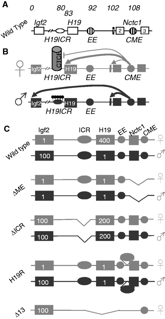
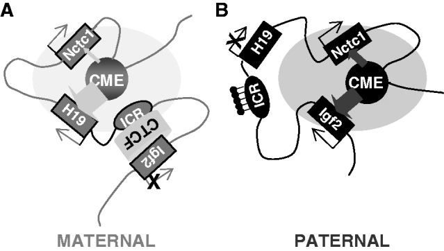
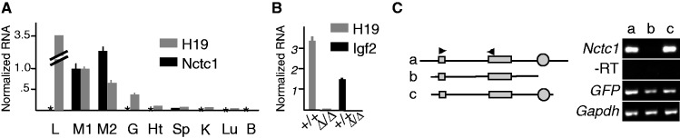
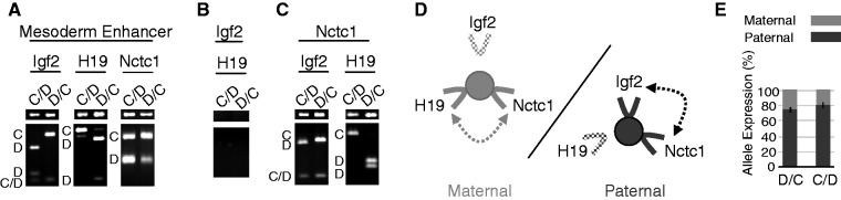
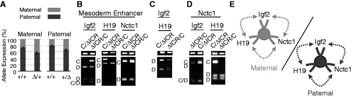
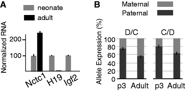
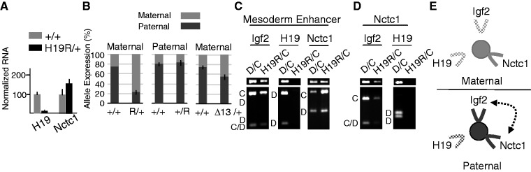
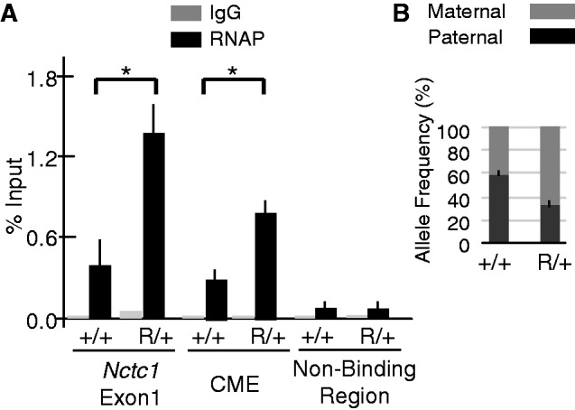
Developmentally regulated transcription often depends on physical interactions between distal enhancers and their cognate promoters. Recent genomic analyses suggest that promoter-promoter interactions might play a similarly critical role in organizing the genome and establishing cell-type-specific gene expression. The Igf2/H19 locus has been a valuable model for clarifying the role of long-range interactions between cis-regulatory elements. Imprinted expression of the linked, reciprocally imprinted genes is explained by parent-of-origin-specific chromosomal loop structures between the paternal Igf2 or maternal H19 promoters and their shared tissue-specific enhancer elements. Here, we further analyze these loop structures for their composition and their impact on expression of the linked long non-coding RNA, Nctc1. We show that Nctc1 is co-regulated with Igf2 and H19 and physically interacts with the shared muscle enhancer. In fact, all three co-regulated genes have the potential to interact not only with the shared enhancer but also with each other via their enhancer interactions. Furthermore, developmental and genetic analyses indicate functional significance for these promoter-promoter interactions. Altogether, we present a novel mechanism to explain developmental specific imprinting of Nctc1 and provide new information about enhancer mechanisms and about the role of chromatin domains in establishing gene expression patterns.


Figures

Figure 1.
Figure 1.
The Igf2/H19/Nctc1 locus. (A) Organization of the wild-type locus. Open rectangles denote the Igf2, H19 and Nctc1 genes. Open oval denotes the H19ICR. Striped circles denote the endoderm (EE) and CME. Numbers above the line indicate kilobase relative to the Igf2 isoform 1 transcriptional start site. (B) The insulator model for imprinted expression at the Igf2/H19 locus. On the maternal (gray) chromosome, the H19ICR is not methylated and binds the transcriptional insulator, CTCF, preventing activation of the distal Igf2 promoters. On the paternal (black) chromosome, methylation of the CpGs (black lollipops) within the H19ICR prevents CTCF binding and thus allows enhancer activation of Igf2. In addition, H19ICR epigenetic changes at the adjacent H19 promoter prevent its expression (32). (C) Structures of the ΔME, ΔICR, H19R and Δ13 mutant alleles. Numbers inside the gene boxes indicate the approximate relative expression levels in muscle cells for H19 and Igf2 on these maternal (gray) and paternal (black) chromosomes as determined in the references cited above. ΔME (8) carries a deletion that removes the shared CME (41) and exons 1 and 2 of the Nctc1 gene. ΔICR (32) carries a deletion that removes the 2.4-kb H19ICR but leaves the adjacent H19 promoter intact. H19R (29) carries an insertion of the 2.4-kb H19ICR at +10 kb, between the endodermal (EE) and mesodermal enhancers (CME). Δ13 (33) carries a deletion that removes the entire H19 RNA-coding region plus 10 kb of upstream sequences including the H19 promoter and the H19ICR.
Figure 2.
Figure 2.
Parent-of-origin specific structures mediate gene expression at the Igf2/H19/Nctc1 imprinted locus in muscle cells. (A) On the maternal chromosome, a CTCF-dependent insulator organizes the DNA loops between distal cis-regulatory elements to favor H19 expression and to prevent interactions between the Igf2 promoters and the shared downstream CME. Recent work from Nativio et al. (17) demonstrates a critical importance for cohesin in establishing these maternal-specific chromosomal structures. Also, see Zhang et al. (25) for detailed mechanisms describing maternal ICR–CTCF–Igf2 interactions. (B) Paternal-specific methylation of CpGs within the ICR prevents CTCF binding, resulting in DNA loop structures that favor Igf2 promoter interactions with the shared enhancer. The loss of CTCF binding also results in a spread of DNA methylation and heterochromatin from the ICR into the adjacent H19 promoter region to H19 transcription. Here, we propose that Nctc1 levels are regulated by competition with H19 and Igf2 promoters for activation by the transcriptional complexes assembling at the shared enhancer. For simplicity, this model does not describe Igf2 differentially methylated regions (DMRs) located near the Igf2 promoters. DMR1 is an muscle-specific repressor of Igf2 expression that is required for complete full postnatal repression of both maternal and paternal chromosomes (62). DMR2 is a tissue non-specific positive regulatory element (63). In muscle, deletion of DMR2 results in modest decreases in Igf2. The participation of these two elements in DNA looping structures has been extensively analyzed (12,13,64) but to date, only in endodermal cells.
Figure 3.
Figure 3.
Co-regulation of Igf2, H19 and Nctc1 in muscle cells. (A) Tissue-specific expression of the Nctc1 gene. RNAs isolated from wild-type neonates were analyzed by qRT-PCR for Nctc1 and H19. Expression in hind limb muscle (M1) was set to 1. Other tissues analyzed were liver (L), tongue (M2), gut (G), heart (Ht), spleen (Sp), kidney (K), lung (Lu) and Brain (B). Asterisk denotes no detectable Nctc1. (B) The ΔME deletion affects Igf2 and H19 expression only in tissues where Nctc1 is expressed. RNAs from +/+ and from ΔME/ΔME primary myocytes were analyzed by qRT-PCR for expression of H19 and Igf2 normalized to glyceraldehyde 3-phosphate dehydrogenase (GAPDH). Mutant cells display >1000-fold decreases. No effect of the deletion was seen in liver, gut, heart, kidney, lung and brain. (C) Expression of Nctc1 depends on the CME. ΔME/ΔME primary myoblasts were transfected with DNA constructs a, b, or c and a plasmid carrying GFP. After 24-h growth in differentiation media, RNAs were prepared and analyzed by qRT-PCR for Nctc1, GAPDH and GFP. On construct maps, Nctc1 exons 1 and 2 are depicted as filled rectangles and the CME is shown as a filled circle.
Figure 4.
Figure 4.
Long-range interactions between the Igf2, H19 and Nctc1 promoters and the shared muscle enhancer and Nctc1 imprinting on wild-type maternal and paternal chromosomes. (A–C) Chromatin was prepared from wild-type primary myocyte cultures and analyzed for long-range DNA interactions by 3C. (A) Interactions between the mesoderm enhancer and the Igf2, the H19 or the Nctc1 promoter regions. (B) Interactions between the Igf2 and H19 promoters. (C) Interactions between the Nctc1 promoter and the Igf2 or H19 promoter regions. In each case, the narrow top gel displays the PCR amplicon indicative of the DNA loop formation. The bottom gel displays these amplicons after restriction enzyme digestion to reveal the allelic origin(s) of the products as marked along the left margin: C, castaneus; D, domesticus and C/D, castaneus and domesticus. In describing genotypes, we use the convention: maternal allele/paternal allele. Primers and the amplicon sizes before and after digestion are described in Supplementary Table S1 and Supplementary Figure S2. For each analysis at least three independent chromatin preparations were assayed. (D) Summary of long-range interactions on maternal (left, gray) or paternal (right, black) chromosomes. On both chromosomes, transcripitionally active promoters (solid lines) interact with the CME (filled circle) and with each other while silenced promoters (stippled lines) do not interact. (E) Parent-of-origin specific expression of Nctc1. To quantitate the effects of parental origin, allele-specific expression was quantitated for RNAs isolated from D/C and from C/D neonates. Error bars show standard deviations.
Figure 5.
Figure 5.
Nctc1 imprinting and chromatin loop structures are altered on ΔICR chromosomes. (A) Parent-of-origin specific expression of Nctc1. To quantitate the effects of maternal inheritance, allele-specific expression was measured for RNAs isolated from D/C and of ΔICR/C neonates. To quantitate the effects of paternal inheritance, C/D and C/ΔICR pups were compared. (B–D) Chromatin was prepared from primary myocytes isolated from mice carrying a paternal (C/ΔICR) or a maternal deletion (ΔICR/C) of the H19ICR and analyzed for long-range DNA interactions by 3C. (B) Interactions between the mesoderm enhancer and the Igf2, the H19 or the Nctc1 promoter regions. (C) Interactions between the Igf2 and H19 promoters. (D) Interactions between the Nctc1 promoter and the Igf2 or H19 promoter regions. (E) Summary of long-range interactions on maternal (left, gray) or paternal (right, black) ΔICR chromosomes. On both chromosomes, Igf2, H19 and Nctc1 promoters are all active and interact with the CME (filled circle) and with each other. See Figure 4 for additional details.
Figure 6.
Figure 6.
Developmental regulation of Nctc1, H19 and Igf2 gene expression and of Nctc1 imprinting. (A) RNAs were prepared from hind limbs of neonatal (p3) and of adult (12 weeks) animals and analyzed by qRT-PCR for Nctc1, H19 and Igf2. The relative expression for each gene in neonates was set to 100. (B) To ascertain the effect of development on Nctc1 imprinting, allelic frequencies were measured in RNAs isolated from D/C and C/D wild-type animals as described in Figure 4. Error bars show standard deviations.
Figure 7.
Figure 7.
Nctc1 expression, imprinting and chromatin loop structures are altered on maternal H19R chromosomes. (A) Transcription of H19 and Nctc1 in H19R/+ muscle cells was quantitated by qRT-PCR and normalized to GAPDH RNA levels. Expression of each gene in wild-type (+/+) cells is set at 100. (B) Parent-of-origin specific expression of Nctc1 on H19R and on Δ13 chromosomes. To quantitate the effects of maternal inheritance of H19R, allele-specific expression was measured for RNAs isolated from D/C and of H19R/C neonates. To quantitate the effects of paternal inheritance of H19R, C/D and C/ΔICR pups were compared. To quantitate the effects of maternal inheritance of Δ13, allele-specific expression was measured for RNAs isolated from D/C and of Δ13/C neonates. (C–D) Chromatin was prepared from primary myocytes isolated from wild-type mice (D/C) and from mice carrying a maternally inherited copy of the H19R insertion mutation (H19R/C) and analyzed by 3C. (C) Interactions between the mesoderm enhancer and the Igf2, the H19 or the Nctc1 promoter regions. (D) Interactions between the Nctc1 promoter and the Igf2 or H19 promoter regions. (E) Summary of long-range interactions on maternal (top, gray) or paternal (bottom, black) H19R chromosomes. Maternal inheritance of the ICR insertion prevents H19 promoter interactions with the CME (filled circle) and with the Nctc1 promoter. See Figure 4 for additional details.
Figure 8.
Figure 8.
Allele-specific Ser-5(P)-RNAP II binding at the Nctc1 gene in wild-type versus H19R/+ mice. Primary myoblasts were isolated from wild-type and H19R/+ mice and differentiated in culture for 24 h. (A) Total Ser-5(P)-RNAP association at the Nctc1 gene. ChIP analysis was conducted to measure binding at both Nctc1 exon1 and the CME in +/+ and in H19R/+ mutant cells. An intergenic region between H19 and Igf2 was assayed as a control for non-specific binding. (B) Allele-specific frequency of Ser-5(P)-RNAP binding at the Nctc1 gene. ChIP samples for Nctc1 exon 1 from panel A were further analyzed for allele frequency content using DNA melting analysis. Error bars represent standard errors. P < 0.05, n = 3.

Similar articles

Cited by

References

    1. Bulger M, Groudine M. Functional and mechanistic diversity of distal transcriptional enhancers. Cell. 2011;144:327–339. - PMC - PubMed
    1. Shen Y, Yue F, McCleary D, Ye Z, Edsall L, Kuan S, Wagner U, Dixon J, Lee L, Lobanenkov V, et al. A map of the cis-regulatory sequences in the mouse genome. Nature. 2012;488:116–120. - PMC - PubMed
    1. Li G, Ruan X, Auerbach R, Sandhu K, Zheng M, Wang P, Poh H, Goh Y, Lim J, Zhang J, et al. Extensive promoter-centered chromatin interactions provide a topological basis for transcription regulation. Cell. 2012;148:84–98. - PMC - PubMed
    1. Wallace J, Felsenfeld G. We gather together: insulators and genome organization. Curr. Opin. Genet. Dev. 2007;17:400–417. - PMC - PubMed
    1. Ainscough JF-X, Dandola L, Surani MA. Appropriate expression of the mouse H19 gene utilises three or more distinct enhancer regions spread over more than 130 kb. Mech. Dev. 2000;91:365–368. - PubMed

Publication types