. 2012 Dec 26;109(52):21426-31.
doi: 10.1073/pnas.1209673109. Epub 2012 Dec 3.

RNAi screening identifies mediators of NOD2 signaling: implications for spatial specificity of MDP recognition

Affiliations

RNAi screening identifies mediators of NOD2 signaling: implications for spatial specificity of MDP recognition

Simone Lipinski et al. Proc Natl Acad Sci U S A. .

Abstract

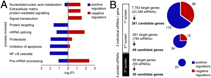
The intracellular nucleotide-binding oligomerization domain-2 (NOD2) receptor detects bacteria-derived muramyl dipeptide (MDP) and activates the transcription factor NF-κB. Here we describe the regulatome of NOD2 signaling using a systematic RNAi screen. Using three consecutive screens, we identified a set of 20 positive NF-κB regulators including the known pathway members RIPK2, RELA, and BIRC4 (XIAP) as well as FRMPD2 (FERM and PDZ domain-containing 2). FRMPD2 interacts with NOD2 via leucine-rich repeats and forms a complex with the membrane-associated protein ERBB2IP. We demonstrate that FRMPD2 spatially assembles the NOD2-signaling complex, hereby restricting NOD2-mediated immune responses to the basolateral compartment of polarized intestinal epithelial cells. We show that genetic truncation of the NOD2 leucine-rich repeat domain, which is associated with Crohn disease, impairs the interaction with FRMPD2, and that intestinal inflammation leads to down-regulation of FRMPD2. These results suggest a structural mechanism for how polarity of epithelial cells acts on intestinal NOD-like receptor signaling to mediate spatial specificity of bacterial recognition and control of immune responses.


Conflict of interest statement

The authors declare no conflict of interest.

Figures

Fig. 1.
Fig. 1.
Systematic siRNA screening identifies modulators of NOD2 signaling. (A) Enrichment of candidate genes in different functional groups (GO terms) after the primary screen. (B) Screening procedure and number of candidate genes at different screening stages. Primary and secondary screens were performed with three individual siRNAs per gene. Pools of four siRNAs were used for the third screen. Candidate genes with an enhancing or inhibitory effect on NF-κB activation are indicated in blue or red, respectively.
Fig. 2.
Fig. 2.
Confirming FRMPD2 as a positive regulator of NOD2-mediated NF-κB activation. (A) Validation of 20 candidate genes in Caco-2 cells using a luciferase assay. Cells were transfected and stimulated with MDP according to previous assay procedures. (B) Functional interaction network of candidate proteins including members of the NOD2-signaling pathway (gray boxes) based on data from STRING 8.3 (highest confidence; P > 0.9) and BioGRID. Dashed lines represent manually added interactions based on publications not yet integrated into databases. Nodes represent candidate genes. Colored in blue are candidate genes from the TNF-α counterscreen that did not share canonical NF-κB activation. Out of these candidates, those whose fold inductions fell below the cutoff level in the Caco-2 screen are in bold. (C) IL-8 release of MDP-stimulated HEK293 cells transfected with siRNA of the indicated genes and NOD2 plasmids. IL-8 concentration was normalized against Renilla luciferase. (D) KC release of Frmpd2 and control. siRNA-transfected and MDP-stimulated IECs from WT and Nod2 KO mice (n = 3 each group) cultured in a 3D collagen matrix. (E) Western blot (WB) analysis of MDP-mediated IκB-α degradation in HEK293 cells transfected with control and FRMPD2 siRNAs together with plasmids encoding NOD2. (F) MDP-induced luciferase activity in HEK293 cells at 48 h after transfection with negative control or RIPK2 siRNAs, plasmids encoding NOD2, FRMPD2, or mock control, and luciferase reporter constructs. (G) Fluorescence microscopy of polarized Caco-2 cells transfected with plasmids encoding FRMPD2-DsRed and ERBB2IP-myc [stained with antibody to myc (green); Upper] or with NOD2-GFP and FRMPD2-DsRed constructs (Lower). (Scale bars, 10 µm.) Arrowheads indicate colocalization. (H) Coimmunoprecipitation (Co-IP) of FLAG- and GFP-tagged proteins from whole-cell lysates of HEK293 cells with or without MDP stimulation (10 µg/mL for 90 min), followed by WB analysis of NOD2 (anti-FLAG), FRMPD2 (anti-GFP), and ERBB2IP (anti-ERBB2IP). (I) Co-IP of endogenous FRMPD2 with overexpressed NOD2-FLAG from Caco-2 cells followed by WB analysis with antibody to FRMPD2 (anti-FRMPD2) or NOD2 (anti-FLAG). Data in A, C, D, and F show mean and SD of at least three in-plate replicates; *P < 0.05, **P < 0.01, and ***P < 0.001.
Fig. 3.
Fig. 3.
FRMPD2 controls NOD2 signaling from the basolateral membrane in intestinal epithelial cells. (A) Fluorescence microscopy of paraffin sections from colonic epithelial biopsy samples stained using anti-NOD2 (green) and anti-FRMPD2 (red) antibodies (Upper). Secondary Abs only were used as control (Lower). Images are representative of at least three independent experiments or n = 5 individuals. (Scale bar, 10 µm.) (B) Cross-sectional xy, xz, and yz images acquired by confocal fluorescence microscopy of FRMPD2-dsRED– and NOD2-GFP–transfected Caco-2 cells grown on transwell inserts for induction of polarized monolayers. Cells were fixed and stained with antibody to the basolateral marker Na+/K+-ATPase (purple). Arrowheads indicate sites of lateral colocalization. (Scale bar, 10 µm.) (C) KC ELISA of small intestinal mucosal explants of culture supernatants. Intestinal segments from WT and Nod2 KO mice were incubated in either calcium-free and EDTA-containing or normal RPMI 1640 media and stimulated with MDP (10 µg/mL, 24 h). (D) Real-time SYBR Green PCR on BD-1, BD-2, lysozyme, and IL-8 transcripts normalized to GAPDH from polarized Caco-2 cells treated with control or FRMPD2 siRNA in response to either apical or basal MDP stimulation (10 µg/mL, 12 h). (E) WB analysis of cellular distribution of NOD2 and RIPK2 protein in response to MDP stimulation in polarized Caco-2 monolayers at 72 h after transfection with FRMPD2 siRNA or control. (F) Fluorescence microscopy of RIPK2-, FRMPD2-dsRED–, and NOD2-GFP–transfected Caco-2 cells grown on transwell inserts for induction of polarized monolayers. Cells were fixed and stained with antibody to RIPK2 (purple). (Scale bar, 10 µm.) Data in C and D show mean and SD of at least three in-plate replicates; *P < 0.05.
Fig. 4.
Fig. 4.
The FERM-PDZ2 domain of FRMPD2 serves as a membrane scaffold for the LRR of NOD2 and recruits RIPK2 to the membrane. (A) Luciferase assay of MDP-induced NF-κB activation in HEK293 cells after transfection with FRMPD2 siRNAs or negative control, plasmids encoding NOD2, GFP-FERM-PDZ domains, GFP-FRMPD2-full length, or GFP-control, and luciferase reporter constructs. (B) WB analysis of cellular distribution of NOD2 CARD, NBD, or LRR domains dependent on GFP-control or GFP-FERM-PDZ2 cotransfection in HEK293 cells. The black arrowhead indicates the shift of the LRR domain from the cytosolic to the membrane fraction when coexpressed with FERM-PDZ2. (C) WB analysis of cellular distribution of RIPK2 in HEK293 cells cotransfected with NOD2 or empty vector, GFP-control, or GFP-FERM-PDZ2. (D) Co-IP of WT and mutant (L1007fsinsC) NOD2 with endogenous RIPK2 in HEK293 cells transfected with FLAG-NOD2 or empty vectors. (E) Real-time TaqMan PCR on FRMPD2 transcripts normalized to ACTB from Caco-2 cells treated with 25 ng/mL TNF-α or IFN-γ for 2 or 8 h. (F) Real-time TaqMan PCR on FRMPD2 transcripts normalized to ACTB from sigmoid colon biopsies from healthy individuals (n = 21), normal and inflamed intestinal mucosa of patients with CD (n = 22 and n = 21), and diseased controls (n = 7 and n = 11). (G) Real-time SYBR Green PCR on Frmpd2 transcripts normalized to Actb from colon biopsies of untreated C57BL/6J WT mice (n = 6) and DSS-treated mice (n = 6, 4% DSS for 6 d). Data in A and EG show mean and SD of at least three in-plate replicates; *P < 0.05 and **P < 0.01.
Fig. 5.
Fig. 5.
Spatial specificity of MDP-induced NOD2 signaling in polarized IECs. Upon basolateral MDP stimulation, NOD2 is recruited to the plasma membrane by FRMPD2, which is facilitated by ERBB2IP. Subsequently, RIPK2 joins the complex and enables NF-κB activation and induction of the proinflammatory chemokine IL-8 and antimicrobial peptide BD-2 (Left). The CD-associated genetic variant of NOD2 (L1007fsinsC) leads to a truncated LRR of NOD2. In response to MDP stimulation, the interaction of FRMPD2 with NOD2 L1007fsins variant C is impaired. This potentially leads to a disturbed compartmentalization of NOD2 and failure of NF-κB activation and target gene induction (Right).

Comment in

  • FRMBP2 directs NOD2 to the membrane.
    Kabi A, McDonald C. Kabi A, et al. Proc Natl Acad Sci U S A. 2012 Dec 26;109(52):21188-9. doi: 10.1073/pnas.1219395110. Epub 2012 Dec 17. Proc Natl Acad Sci U S A. 2012. PMID: 23248319 Free PMC article. No abstract available.

Similar articles

Cited by

References

    1. Maekawa T, Kufer TA, Schulze-Lefert P. NLR functions in plant and animal immune systems: So far and yet so close. Nat Immunol. 2011;12(9):817–826. - PubMed
    1. Lange C, et al. Defining the origins of the NOD-like receptor system at the base of animal evolution. Mol Biol Evol. 2011;28(5):1687–1702. - PubMed
    1. Ogura Y, et al. Nod2, a Nod1/Apaf-1 family member that is restricted to monocytes and activates NF-kappaB. J Biol Chem. 2001;276(7):4812–4818. - PubMed
    1. Girardin SE, et al. Nod2 is a general sensor of peptidoglycan through muramyl dipeptide (MDP) detection. J Biol Chem. 2003;278(11):8869–8872. - PubMed
    1. Mo J, et al. Pathogen sensing by nucleotide-binding oligomerization domain-containing protein 2 (NOD2) is mediated by direct binding to muramyl dipeptide and ATP. J Biol Chem. 2012;287(27):23057–23067. - PMC - PubMed

Publication types

MeSH terms

Substances

LinkOut - more resources