. 2013 Mar;43(3):734-46.
doi: 10.1002/eji.201242914. Epub 2013 Jan 18.

Dendritic cell modification as a route to inhibiting corneal graft rejection by the indirect pathway of allorecognition

Affiliations
Free PMC article

Dendritic cell modification as a route to inhibiting corneal graft rejection by the indirect pathway of allorecognition

Adnan Khan et al. Eur J Immunol. 2013 Mar.
Free PMC article

Abstract

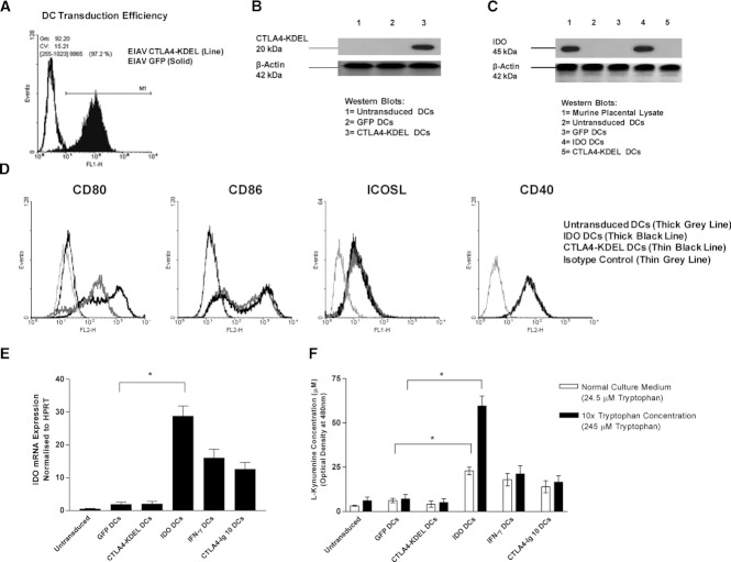
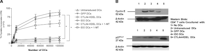
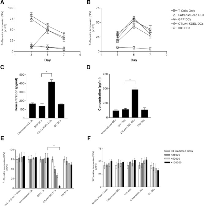
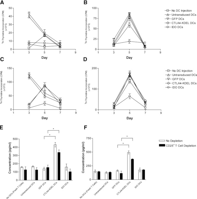
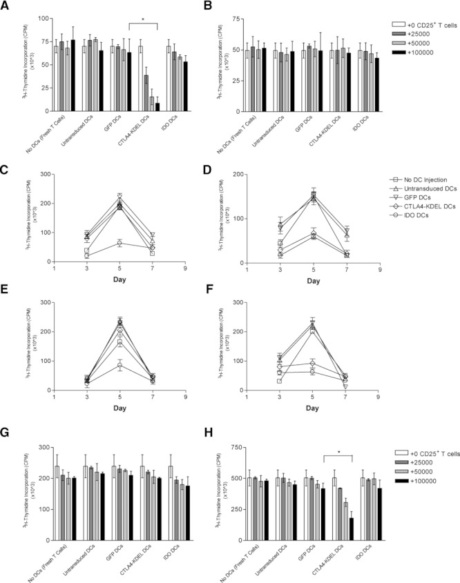
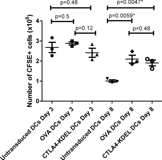
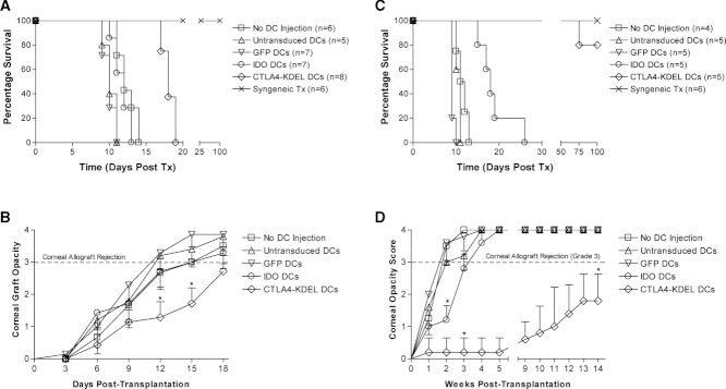
Dendritic cell (DC) modification is a potential strategy to induce clinical transplantation tolerance. We compared two DC modification strategies to inhibit allogeneic T-cell proliferation. In the first strategy, murine DCs were transduced with a lentiviral vector expressing CTLA4-KDEL, a fusion protein that prevents surface CD80/86 expression by retaining the co-stimulatory molecules within the ER. In the second approach, DCs were transduced to express the tryptophan-catabolising enzyme IDO. CTLA4-KDEL-expressing DCs induced anergy in alloreactive T cells and generated both CD4(+) CD25(+) and CD4(+) CD25(-) Treg cells (with direct and indirect donor allospecificity and capacity for linked suppression) both in vitro and in vivo. In contrast, T-cell unresponsiveness induced by IDO(+) DCs lacked donor specificity. In the absence of any immunosuppressive treatment, i.v. administration of CTLA4-KDEL-expressing DCs resulted in long-term survival of corneal allografts only when the DCs were capable of indirect presentation of alloantigen. This study demonstrates the therapeutic potential of CTLA4-KDEL-expressing DCs in tolerance induction.


Figures

Figure 1
Figure 1
DC transduction with EIAV-CTLA4-KDEL or EIAV-IDO. BM-derived BALB/c DCs were transduced with EIAV on day 6 of culture prior to LPS stimulation on day 8. (A) DC transduction efficiency was assessed by GFP expression using flow cytometry 72 h after transduction with EIAV-GFP or EIAV-CTLA4-KDEL (control). Expression of the (B) 20 kDa CTLA4-KDEL protein and (C) 45 kDa IDO protein in DC lysates was determined by western blotting 72 h after transduction with EIAV-CTLA-KDEL or EIAV-IDO, respectively, and compared with expression in EIAV-GFP-transduced or untransduced DCs and, in the case of IDO, murine placenta (positive control). Expression of the 42 kDa β-actin housekeeping protein was measured as a loading control. (D) Flow cytometry histograms show the surface expression of CD80, CD86, ICOSL, and CD40 on untransduced DCs, and DCs 72 h after transduction with EIAV-CTLA4-KDEL or EIAV-IDO (control). The results shown in (A–D) are representative of three independent experiments. (E–F) Untransduced, immature DCs were treated on day 7 with either IFN-γ or CTLA4-Ig. The DC culture media was supplemented with L-tryptophan on day 6 (final concentration, 245 μM), followed by LPS stimulation on day 8. (E) DCs were harvested on day 9 for quantitative PCR analysis to assess IDO mRNA expression. (F) IDO activity was assessed by a kynurenine assay using DC culture supernatants that were either supplemented with tryptophan or unsupplemented. Results are shown as the mean ± SD of triplicate wells and are representative of three independent experiments performed. * p < 0.05, two-tailed t-test.
Figure 2
Figure 2
Inhibition of allogeneic T-cell proliferation after DC expression of CTLA4-KDEL or IDO. BALB/c (H-2d) DCs were transduced on day 6 of culture with EIAV-GFP (control), EIAV-CTLA4-KDEL or EIAV-IDO and stimulated with LPS on day 8 of culture. All DC populations were cultured on day 9 with fully MHC-disparate, spleen-derived C3H/He CD4+ T cells. Where indicated, 250 μM 1-methyl-D,L-tryptophan (1-MT), in combination with excess tryptophan, was added to the medium at the start of coculture. (A) Increasing numbers of both EIAV transduced and untransduced DC populations (0–105) were co-cultured with C3H/He-derived CD4+ T cells. Proliferation of CD4+ T cells was detected by thymidine incorporation on day 5 of the MLR, and the results are shown as the mean ± SD of triplicate wells and are representative of three independent experiments. *p < 0.05, two-tailed t-test. (B) Expression of the 52 kDa cyclin E protein and p27Kip1 protein (and the 42 kDa β-actin protein), in the lysates of CD4+ T cells incubated with either the transduced or untransduced DCs described was determined by western blotting on day 4 of co-culture. Data shown are representative of three independent experiments performed.
Figure 3
Figure 3
Induction of T-cell anergy and regulation in vitro with indirect donor allospecificity. CBK (H-2k + Kb) DCs were transduced with either EIAV-GFP (control), EIAV-CTLA4-KDEL or EIAV-IDO, followed by stimulation with LPS. The transduced (and untransduced) DCs were subsequently incubated in vitro with CBA/Ca-derived CD4+ T cells. After 10 days, T cells were harvested and rechallenged in vitro with (A) donor CBK DCs or (B) third-party B10.A (H-2k + Dd) DCs, and CD4+ T-cell proliferation was assessed by thymidine incorporation on days 3, 5 and 7. (C–D) Culture supernatant from the donor DC rechallenge assay was harvested for detection of the immunoregulatory cytokines (C) TGF-β and (D) IL-10 by ELISA. (E–F) In addition, after 10 days of the primary DC-CD4+ T-cell coculture, T cells were also harvested, irradiated and added (0–105 CD4+ T cells) to a primary MLR between freshly isolated CBA CD4+ T cells and (E) donor CBK DCs or (F) third-party B10.A DCs. T-cell proliferation was assessed by thymidine incorporation after 5 days. All results are shown as the mean ± SD of triplicate wells and are representative of three independent experiments performed. *p < 0.05, two-tailed t-test.
Figure 4
Figure 4
Induction of T-cell anergy in vivo with indirect donor allospecificity and production of immunoregulatory cytokines. 2.5 × 10⁶ CBK DCs (either untransduced, or transduced with EIAV-GFP, EIAV-CTLA4-KDEL or EIAV-IDO) were injected i.v. into CBA/Ca mice. (A–D) After 10 days, CD4+ T cells purified from the spleens of injected mice were rechallenged in vitro with (A) donor CBK DCs or (B) third-party B10.A DCs, and CD4+ T-cell proliferation was assessed by thymidine incorporation on days 3, 5 and 7. The purified CD4+ T cells were also rechallenged with (C) donor CBK DCs or (D) third-party B10.A DCs in the presence of 100 U/mL exogenous IL-2. (E–F) Culture supernatant from donor DC rechallenge assays (including those from CD4+CD25+ T-cell-depleted rechallenge assays) was harvested for detection of the immunoregulatory cytokines (E) TGF-β and (F) IL-10 by ELISA. All results are shown as the mean ± SD of triplicate wells and are representative of three independent experiments performed. *p < 0.05, two-tailed t-test.
Figure 5
Figure 5
Generation of Treg cells in vivo with indirect donor allospecificity and capacity for linked suppression. 2.5 × 10⁶ CBK DCs (either untransduced, or transduced with EIAV-GFP (control), EIAV-CTLA4-KDEL or EIAV-IDO) were injected i.v. into C3H/He mice. (A–B) After 10 days, CD4+CD25+ T cells purified from the spleens were irradiated and added (0–105 CD4+CD25+ T cells) to a primary MLR between freshly isolated CBA/Ca-derived CD4+ T cells and (A) donor CBK DCs or (B) third-party B10.A DCs. T-cell proliferation was assessed by thymidine incorporation after 5 days. (C–D) CD4+ T cells purified from the spleens of injected mice were also rechallenged in vitro with (C) (BALB/c × C57BL/6)F1 DCs expressing only third-party MHC or (D) (C57BL/6 × CBA/Ca)F1 DCs expressing both donor (Kb presented in the context of I-Ak/I-Ek) and third-party MHC, and CD4+ T-cell proliferation was assessed by thymidine incorporation on days 3, 5 and 7. (E–F) Rechallenge assays were repeated using CD4+ T cells that were depleted of CD4+CD25+ T cells prior to rechallenge with (E) (BALB/c × C57BL/6)F1 DCs or (F) (C57BL/6 × CBA/Ca)F1 DCs. (G–H) In addition, CD4+CD25+ T cells purified from the spleens of injected mice were irradiated and added (0–105 CD4+CD25+ T cells) to new primary MLRs between freshly isolated CBA/Ca-derived CD4+ T cells and (G) (BALB/c × C57BL/6)F1 DCs or (H) (C57BL/6 × CBA/Ca)F1 DCs and T-cell proliferation assessed by thymidine incorporation after 5 days. All results are shown as the mean ± SD of triplicate wells and are representative of three independent experiments performed. *p < 0.05, two-tailed t-test.
Figure 6
Figure 6
Enhanced survival of equine infectious anaemia virus (EIAV)-transduced DCs in vivo. 1 × 10⁷ CFSE-labelled C3H DCs (either untransduced, or transduced with EIAV CTLA4-KDEL or EIAV OVA) were injected intravenously into female syngeneic mice. The spleens from these mice were harvested on days 3 and 8 post-injection and the total number of CFSE-labelled DCs present assessed by flow cytometric analysis. Each symbol represents an individual animal and bars represent the mean ± SD; data shown are representative of three independent experiments performed. *p < 0.05, two-tailed t-test.
Figure 7
Figure 7
Prolongation of corneal graft survival following administration of modified DCs. BALB/c mice were either untreated (n = 6) or given 2.5 × 10⁶ C3H/He DCs intravenously. The DCs were either untransduced (n = 5), or transduced 72 h earlier ex vivo with equine infectious anaemia virus (EIAV)-GFP (control, n = 7), EIAV-IDO (n = 7) or EIAV-CTLA4-KDEL (n = 8). Ten days later, the BALB/c mice received a complete MHC-disparate C3H/He corneal graft or a syngeneic BALB/c graft (n = 6). (A) Data were plotted using the Kaplan–Meyer method and differences in graft survival were analysed using a log-rank test. (B) Corneal graft opacity scores were plotted and statistical differences between CTLA4-KDEL- and GFP (control)-expressing DCs calculated using the Mann–Whitney U test. * p < 0.008. (C) Using the same protocol, BALB/c mice were either untreated (n = 4) or given 2.5 × 10⁶ (CBA × BALB/c)F1 DCs intravenously. The DCs were either untransduced (n = 5), or transduced 72 h earlier ex vivo with EIAV-GFP (n = 5), EIAV-IDO (n = 5) or EIAV-CTLA4-KDEL (n = 5). 10 days later, BALB/c mice received a complete MHC-disparate CBA/Ca corneal graft or a syngeneic BALB/c graft (n = 6). Data were analysed as above. (D) Corneal graft opacity scores were plotted and statistical differences between CTLA4-KDEL- or IDO-expressing DCs and GFP-expressing DCs calculated using the Mann–Whitney U Test. *p < 0.008. Data shown are representative of three independent experiments performed.

Comment in

Similar articles

Cited by

References

    1. Morelli AE, Thomson AW. Tolerogenic dendritic cells and the quest for transplant tolerance. Nat. Rev. Immunol. 2007;7:610–621. - PubMed
    1. Mirenda V, Berton I, Read J, Cook T, Smith J, Dorling A, Lechler RI. Modified dendritic cells coexpressing self and allogeneic major histocompatability complex molecules: an efficient way to induce indirect pathway regulation. J. Am. Soc. Nephrol. 2004;15:987–997. - PubMed
    1. Andreakos E, Smith C, Monaco C, Brennan FM, Foxwell BM, Feldmann M. Ikappa B kinase 2 but not NF-kappa B-inducing kinase is essential for effective DC antigen presentation in the allogeneic mixed lymphocyte reaction. Blood. 2003;101:983–991. - PubMed
    1. Yoshimura S, Bondeson J, Brennan FM, Foxwell BM, Feldmann M. Antigen presentation by murine dendritic cells is nuclear factor-kappa B dependent both in vitro and in vivo. Scand. J. Immunol. 2003;58:165–172. - PubMed
    1. Munro S, Pelham HR. A C-terminal signal prevents secretion of luminal ER proteins. Cell. 1987;48:899–907. - PubMed

Publication types

MeSH terms

LinkOut - more resources