. 2013 Jan 18;288(3):1739-49.
doi: 10.1074/jbc.M112.404657. Epub 2012 Dec 3.

Rapid nucleotide exchange renders Asp-11 mutant actins resistant to depolymerizing activity of cofilin, leading to dominant toxicity in vivo

Affiliations

Rapid nucleotide exchange renders Asp-11 mutant actins resistant to depolymerizing activity of cofilin, leading to dominant toxicity in vivo

Nobuhisa Umeki et al. J Biol Chem. .

Abstract

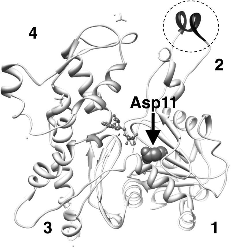
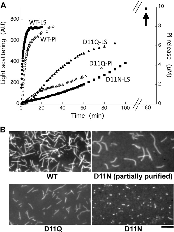
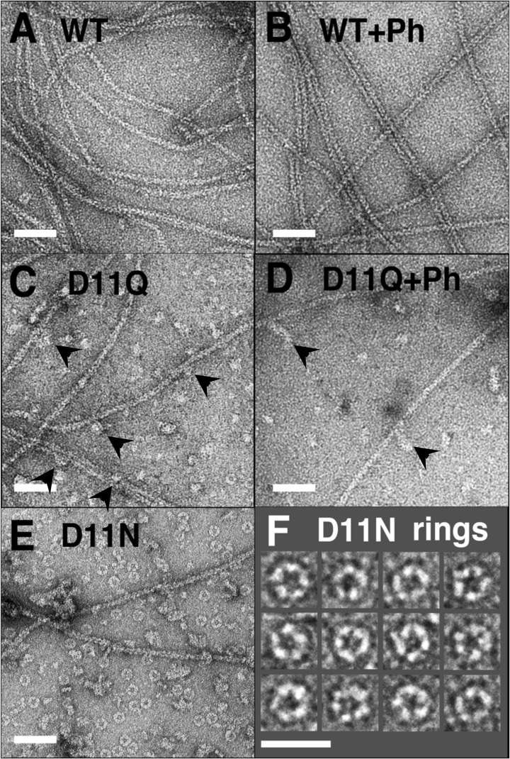
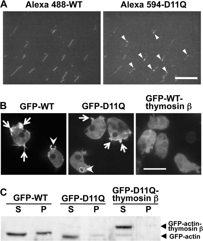
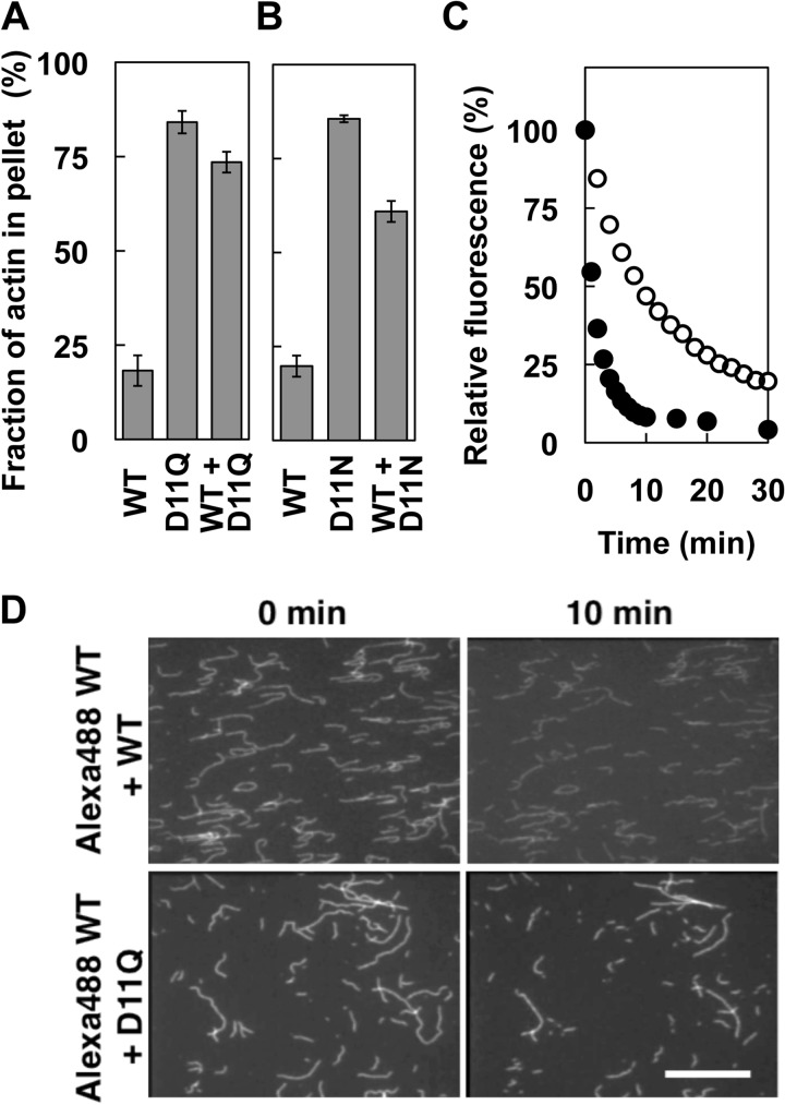
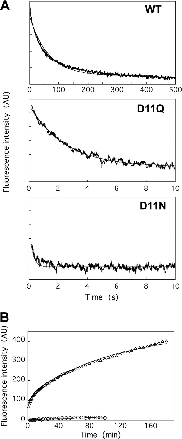
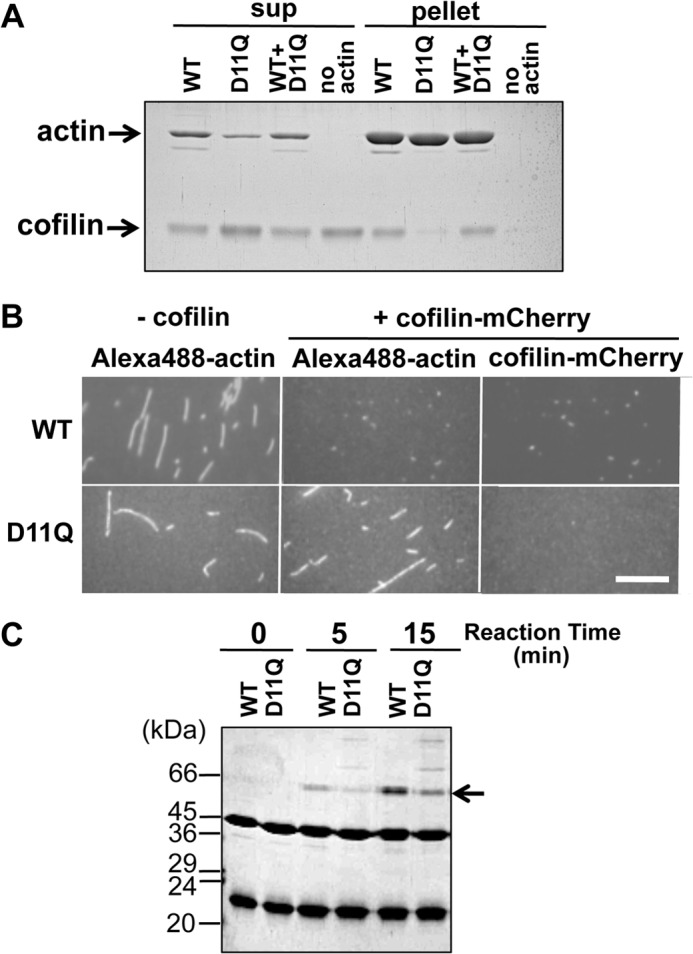
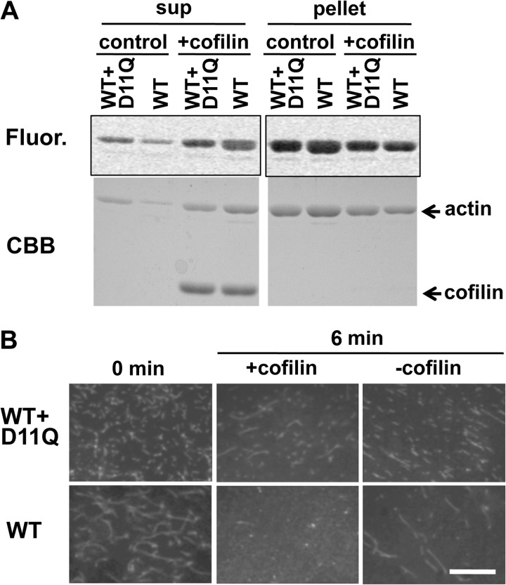
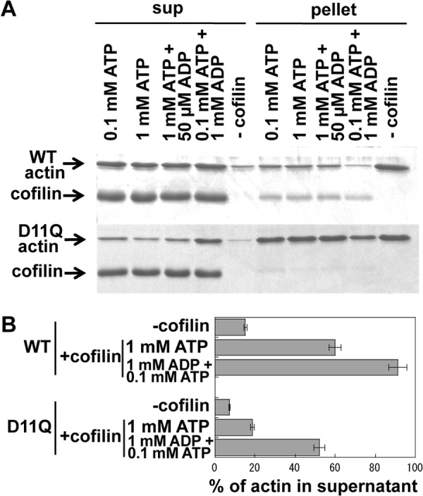
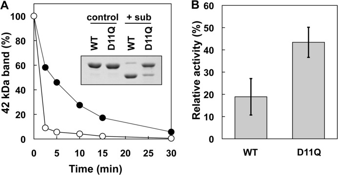
Conserved Asp-11 of actin is a part of the nucleotide binding pocket, and its mutation to Gln is dominant lethal in yeast, whereas the mutation to Asn in human α-actin dominantly causes congenital myopathy. To elucidate the molecular mechanism of those dominant negative effects, we prepared Dictyostelium versions of D11N and D11Q mutant actins and characterized them in vitro. D11N and D11Q actins underwent salt-dependent reversible polymerization, although the resultant polymerization products contained small anomalous structures in addition to filaments of normal appearance. Both monomeric and polymeric D11Q actin released bound nucleotides more rapidly than the wild type, and intriguingly, both monomeric and polymeric D11Q actins hardly bound cofilin. The deficiency in cofilin binding can be explained by rapid exchange of bound nucleotide with ATP in solution, because cofilin does not bind ATP-bound actin. Copolymers of D11Q and wild type actins bound cofilin, but cofilin-induced depolymerization of the copolymers was slower than that of wild type filaments, which may presumably be the primary reason why this mutant actin is dominantly toxic in vivo. Purified D11N actin was unstable, which made its quantitative biochemical characterization difficult. However, monomeric D11N actin released nucleotides even faster than D11Q, and we speculate that D11N actin also exerts its toxic effects in vivo through a defective interaction with cofilin. We have recently found that two other dominant negative actin mutants are also defective in cofilin binding, and we propose that the defective cofilin binder is a major class of dominant negative actin mutants.


Figures

FIGURE 1.
FIGURE 1.
Conserved Asp-11 indicated by an arrow and shown in space filling representation in the atomic structure of actin in filaments (Protein Data Bank code 3G37 (56)). In this structure, DNase loop is modeled as a helix and is darkly colored in the dotted circle. Numbers show the subdomains.
FIGURE 2.
FIGURE 2.
Polymerization of WT and Asp-11-mutant actins. A, polymerization of WT (filled circles), D11Q (filled triangles), and D11N (filled squares) actin solutions. Final concentration of actin was 10 μm, and polymerization was monitored by the increase of light scattering at 360 nm (left abscissa). In parallel, release of phosphate from polymerizing WT (open circles) and D11Q (open triangles) actin was monitored using the EnzCheck phosphate assay kit (right abscissa). Arrow indicates light scattering of D11N actin polymer at 160 min. B, fluorescence micrograph of WT, D11Q, and D11N actin filaments stained by rhodamine-phalloidin overnight at 5 °C. For D11N actin, the partially purified fraction from Q-Sepharose column chromatography and the purified fraction by a depolymerization/polymerization cycle are shown. Bar, 10 μm. AU, arbitrary units.
FIGURE 3.
FIGURE 3.
Electron micrographs of negatively stained actin polymers. WT (A and B), D11Q (C and D), and D11N (E and F) actins were polymerized in F-buffer in the absence (A, C, E, and F) or presence (B and D) of 20 μm phalloidin (Ph) for 2 h, diluted, and stained with uranyl acetate. Arrowheads indicate oligomeric structures in D11Q polymers that appear associated along the length of filaments. F is a gallery of D11N rings. Bars, 50 nm, except for F (25 nm).
FIGURE 4.
FIGURE 4.
Copolymerization of WT and D11Q actin. A, filaments obtained by copolymerization of WT actin labeled with Alexa-Fluor 488 and D11Q actin labeled with Alexa-Fluor 594. The two fluorophores were observed in the green (left) and red fluorescence (right) channels, respectively. Arrowheads indicate puncta of Alexa-Fluor 594-D11Q actin within or along copolymers. Bar, 20 μm. B, fluorescence micrograph of Dictyostelium cells expressing GFP-WT actin, GFP-D11Q actin, and GFP-WT actin fused with thymosin β. Arrows indicate the accumulation of GFP-actin along cell peripheries and thin projections, and arrowheads indicate enrichment around macropinocytic cups. Bar, 20 μm. C, Western blotting analysis of cells expressing GFP-WT actin, GFP-D11Q actin, or GFP-D11Q actin fused with thymosin β. Triton-soluble (S) and -insoluble (P) fractions were separated by SDS-PAGE and probed with anti-GFP antibodies.
FIGURE 5.
FIGURE 5.
Depolymerization of Asp-11-mutant actins. A and B, latrunculin-induced depolymerization of D11Q (A) and D11N (B) actins. Solutions of polymers of WT, mutant, and a 1:1 mixture of both (concentration of total actin was 5 μm in all samples) were ultracentrifuged with or without preincubation with 60 μm latrunculin A for 10 min. The supernatant and pellet fractions were analyzed by SDS-PAGE, and the factions of actin in pellets were calculated by densitometry. Error bars indicate standard deviation of three independent measurements, and Student's t test demonstrated that the difference between WT and D11Q, WT and WT + D11Q, WT and D11N, and WT and WT + D11N are all significant, with p values < 0.00006. C, latrunculin-induced depolymerization of pyrene-labeled WT actin copolymerized with the same concentration of unlabeled WT (filled circles) or D11Q (open circles) actin, respectively, and assayed as in A. D, depolymerization of individual actin filaments. Alexa-Fluor 488-labeled WT actin copolymerized with the same concentration of unlabeled WT or D11Q actin was immobilized on a heavy meromyosin-coated surface and imaged immediately and 10 min after flushing with F-buffer. Bar, 20 μm.
FIGURE 6.
FIGURE 6.
Nucleotide release from WT and Asp-11-mutant actins. A, release of ϵ-ATP from monomeric actin was assayed using a stopped flow apparatus. An actin solution dialyzed against G-buffer containing 0.2 mm ϵ-ATP was rapidly mixed with an equal volume of G-buffer containing 1 mm Ca-ATP. The averages of 3, 7, and 7 traces of WT, D11Q, and D11N actins, respectively are shown, and the fine solid line shows fitting with single exponentials. B, exchange of filament-bound ATP with exogenous ϵ-ATP, as assayed by an increase in fluorescence following the addition of 0.1 mm ϵ-ATP to solutions of WT (circles) or D11Q (triangles) actin filaments dialyzed against F-buffer containing 0.1 mm ATP and then treated with Dowex resin to remove free ATP. Solid lines show fitting with single (WT) and double (D11Q) exponentials. AU, arbitrary units.
FIGURE 7.
FIGURE 7.
Cofilin binding. A, cosedimentation of 5 μm WT, D11Q, and 1:1 mixture polymers with 2.5 μm cofilin at pH 6.5. Supernatant (sup) and pellet fractions after ultracentrifugation were analyzed by SDS-PAGE. Densitometric analyses of three sets of data showed that 49.5 ± 4.7, 0.57 ± 0.09, and 42.8 ± 2.1% of cofilin cosedimented with WT, D11Q, and WT+D11Q filaments, respectively. B, fluorescence microscopic observation of binding of cofilin-mCherry to WT or D11Q actin filaments labeled with Alexa-Fluor 488. Bar, 15 μm. C, cofilin binding to monomeric actin. Binding of 14 μm cofilin to 7 μm monomeric WT or D11Q actin in G-buffer, detected by cross-linking with 40 mm 1-ethyl-3-(3-dimethylaminopropyl) carbodiimide for 5 min, followed by SDS-PAGE. Arrow shows the position of the cross-linked actin-cofilin. Average of three independent measurements indicated that the cross-linking of D11Q actin was 47 ± 15% slower than WT actin, and this difference is statistically significant with p < 0.05 by Student's t test. Higher molecular weight ladders formed in D11Q-cofilin cross-link reactions were formed even when D11N or D11Q actin, but not WT actin, was treated with 1-ethyl-3-(3-dimethylaminopropyl) carbodiimide in G-buffer in the absence of cofilin.
FIGURE 8.
FIGURE 8.
Cofilin-induced depolymerization. A, 5 μm WT actin filaments and a 1:1 mixture of WT and D11Q actin polymers were treated with 10 μm cofilin at pH 8.3, and after incubation for 15 min, the mixtures were subjected to ultracentrifugation followed by SDS-PAGE of the supernatant (sup) and pellet fractions. 2.5 μm WT actin was labeled with Alexa-Fluor 488 (Fluor). Fluorogram visualized WT subunits only and Coomassie Brilliant Blue (CBB) stained both WT and mutant actins. B, fluorescence microscopic observation of cofilin-induced depolymerization of Alexa-Fluor 488-labeled WT filaments and 1:1 copolymer of labeled WT and unlabeled D11Q actin. Bar, 20 μm.
FIGURE 9.
FIGURE 9.
Effects of ADP on cofilin-mediated depolymerization of actin filaments. WT or D11Q actin filaments in F-buffer containing 0.1 mm ATP were diluted to 5 μm in F-buffer that contained 2 mm Hepes, pH 7.4, and various concentrations of nucleotides. After 30 min of incubation, concentrated Hepes buffer, pH 8.35, and cofilin were added to a final concentration of 10 mm and 10 μm, respectively. After incubation for 15 min, the mixtures were subjected to ultracentrifugation, and supernatant (sup) and pellet fractions were analyzed by SDS-PAGE. A is representative of three independent sets experiments. B shows the average and standard deviation of the three sets of data. The difference between cofilin-induced depolymerization of WT actin and D11Q actin in the presence of 1 mm ATP, as well as that of D11Q actin between 1 mm ATP and 1 mm ADP, were statistically significant by Student's t test (p < 0.001).
FIGURE 10.
FIGURE 10.
Effects of D11Q mutation on the conformation of the DNase loop in monomeric actin. A, time course of the subtilisin (sub) cleavage of monomeric WT and D11Q actins in G-buffer, as assayed by SDS-PAGE and densitometry of the stained gel. Inset, SDS-PAGE of WT and D11Q actins at 0 (control) and 2.5 min (+sub) of incubation with 1 μg/ml subtilisin. B, inhibitory effect of WT and D11Q actins on the activity of DNase I. Student's t test on three independent sets of data indicated that the difference is significant with p = 0.016.

Similar articles

Cited by

References

    1. Wertman K. F., Drubin D. G., Botstein D. (1992) Systematic mutational analysis of the yeast ACT1 gene. Genetics 132, 337–350 - PMC - PubMed
    1. An H. S., Mogami K. (1996) Isolation of 88F actin mutants of Drosophila melanogaster and possible alterations in the mutant actin structures. J. Mol. Biol. 260, 492–505 - PubMed
    1. Noguchi T. Q., Toya R., Ueno H., Tokuraku K., Uyeda T. Q. (2010) Screening of novel dominant negative mutant actins using glycine targeted scanning identifies G146V actin that cooperatively inhibits cofilin binding. Biochem. Biophys. Res. Commun. 396, 1006–1011 - PubMed
    1. Drummond D. R., Hennessey E. S., Sparrow J. C. (1991) Characterisation of missense mutations in the Act88F gene of. Drosophila melanogaster. Mol. Gen. Genet. 226, 70–80 - PubMed
    1. van Wijk E., Krieger E., Kemperman M. H., De Leenheer E. M., Huygen P. L., Cremers C. W., Cremers F. P., Kremer H. (2003) A mutation in the γ-actin 1 (ACTG1) gene causes autosomal dominant hearing loss (DFNA20/26). J. Med. Genet. 40, 879–884 - PMC - PubMed

Publication types

MeSH terms

LinkOut - more resources