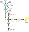
Involvement of autophagy in coronavirus replication
- PMID: 23202545
- PMCID: PMC3528273
- DOI: 10.3390/v4123440
Involvement of autophagy in coronavirus replication
Abstract
Coronaviruses are single stranded, positive sense RNA viruses, which induce the rearrangement of cellular membranes upon infection of a host cell. This provides the virus with a platform for the assembly of viral replication complexes, improving efficiency of RNA synthesis. The membranes observed in coronavirus infected cells include double membrane vesicles. By nature of their double membrane, these vesicles resemble cellular autophagosomes, generated during the cellular autophagy pathway. In addition, coronavirus infection has been demonstrated to induce autophagy. Here we review current knowledge of coronavirus induced membrane rearrangements and the involvement of autophagy or autophagy protein microtubule associated protein 1B light chain 3 (LC3) in coronavirus replication.
Figures


Similar articles
-
Membrane heist: Coronavirus host membrane remodeling during replication.Biochimie. 2020 Dec;179:229-236. doi: 10.1016/j.biochi.2020.10.010. Epub 2020 Oct 25. Biochimie. 2020. PMID: 33115667 Free PMC article. Review.
-
Biogenesis and dynamics of the coronavirus replicative structures.Viruses. 2012 Nov 21;4(11):3245-69. doi: 10.3390/v4113245. Viruses. 2012. PMID: 23202524 Free PMC article. Review.
-
An autophagy-independent role for LC3 in equine arteritis virus replication.Autophagy. 2013 Feb 1;9(2):164-74. doi: 10.4161/auto.22743. Epub 2012 Nov 26. Autophagy. 2013. PMID: 23182945 Free PMC article.
-
Characteristics of the Life Cycle of Porcine Deltacoronavirus (PDCoV) In Vitro: Replication Kinetics, Cellular Ultrastructure and Virion Morphology, and Evidence of Inducing Autophagy.Viruses. 2019 May 18;11(5):455. doi: 10.3390/v11050455. Viruses. 2019. PMID: 31109068 Free PMC article.
-
Properties of Coronavirus and SARS-CoV-2.Malays J Pathol. 2020 Apr;42(1):3-11. Malays J Pathol. 2020. PMID: 32342926 Review.
Cited by
-
Potential Fast COVID-19 Containment With Trehalose.Front Immunol. 2020 Jul 7;11:1623. doi: 10.3389/fimmu.2020.01623. eCollection 2020. Front Immunol. 2020. PMID: 32733488 Free PMC article. Review.
-
SARS-CoV-2 Exploits Non-Canonical Autophagic Processes to Replicate, Mature, and Egress the Infected Vero E6 Cells.Pathogens. 2022 Dec 14;11(12):1535. doi: 10.3390/pathogens11121535. Pathogens. 2022. PMID: 36558869 Free PMC article.
-
Autophagy and Viral Infection.Adv Exp Med Biol. 2019;1209:55-78. doi: 10.1007/978-981-15-0606-2_5. Adv Exp Med Biol. 2019. PMID: 31728865 Free PMC article. Review.
-
Host cell autophagy promotes BK virus infection.Virology. 2014 May;456-457:87-95. doi: 10.1016/j.virol.2014.03.009. Epub 2014 Apr 2. Virology. 2014. PMID: 24889228 Free PMC article.
-
Host Factors in Coronavirus Replication.Curr Top Microbiol Immunol. 2018;419:1-42. doi: 10.1007/82_2017_25. Curr Top Microbiol Immunol. 2018. PMID: 28643204 Free PMC article. Review.
References
-
- Lai M.M.C., Perlman S., Anderson L.J. Coronaviridae. In: Knipe D.M., Howley P.M., editors. Fields Virology. Lippincott Williams and Wilkins; Philidelphia, PA, USA: 2007. pp. 1305–1327.
-
- Snijder E.J., van Tol H., Roos N., Pedersen K.W. Non-structural proteins 2 and 3 interact to modify host cell membranes during the formation of the arterivirus replication complex. J. Gen. Virol. 2001;82:985–994. - PubMed
Publication types
MeSH terms
Grants and funding
LinkOut - more resources
Full Text Sources

