. 2012 Aug;1(8):615-26.
doi: 10.5966/sctm.2012-0032. Epub 2012 Jul 27.

1α,25-dihydroxyvitamin D3 modulates the hair-inductive capacity of dermal papilla cells: therapeutic potential for hair regeneration

Affiliations

1α,25-dihydroxyvitamin D3 modulates the hair-inductive capacity of dermal papilla cells: therapeutic potential for hair regeneration

Noriyuki Aoi et al. Stem Cells Transl Med. 2012 Aug.

Abstract

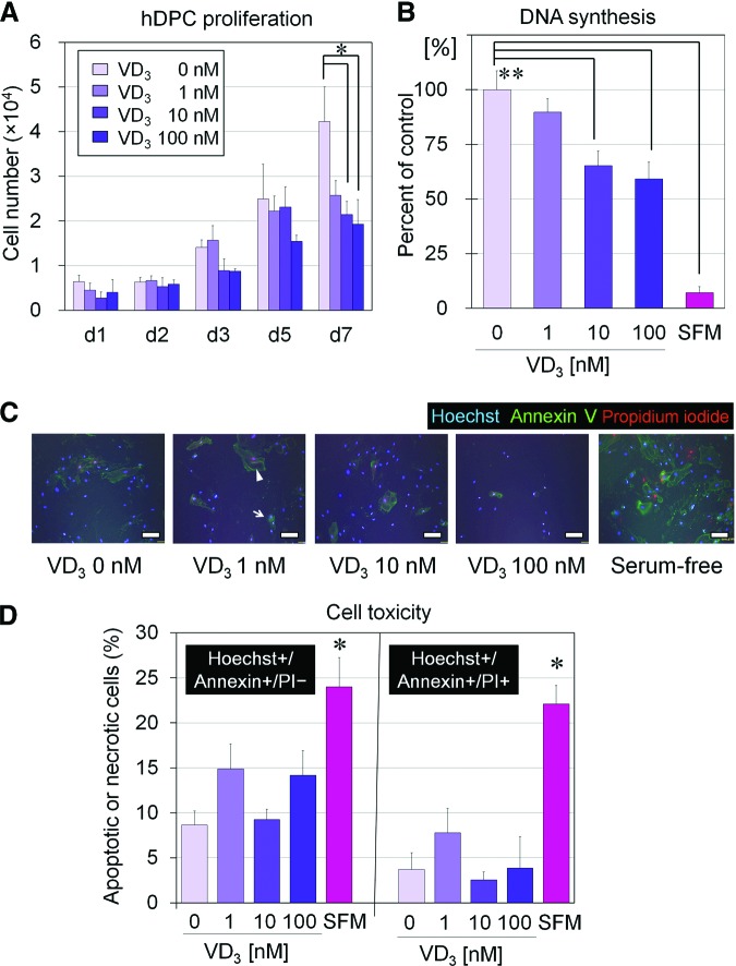
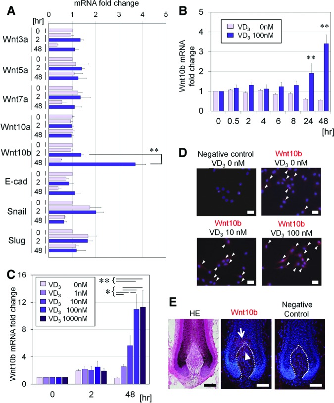
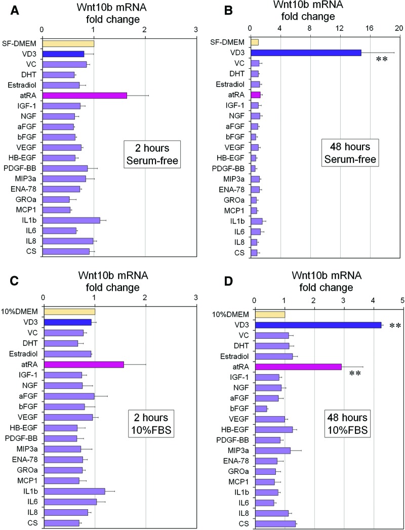
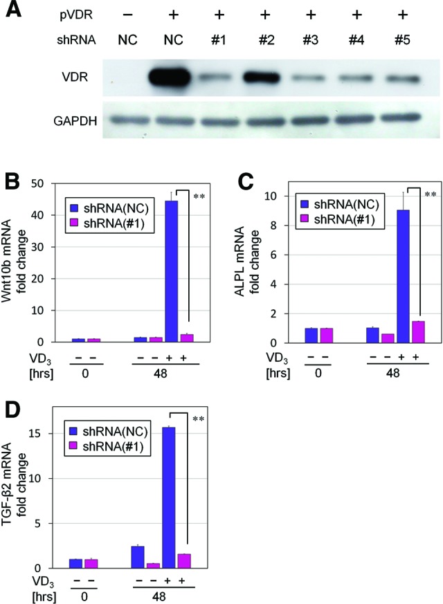
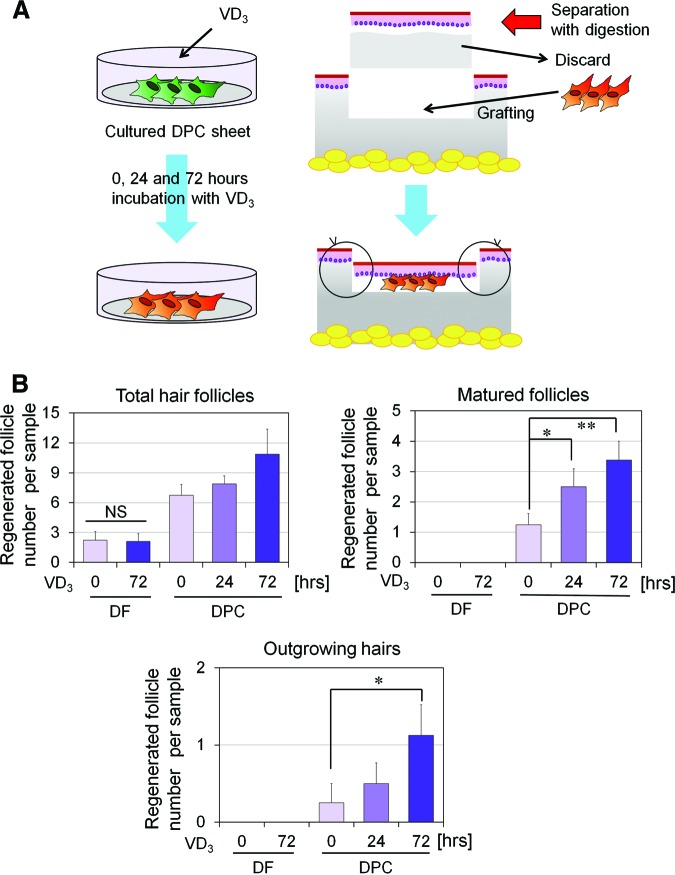
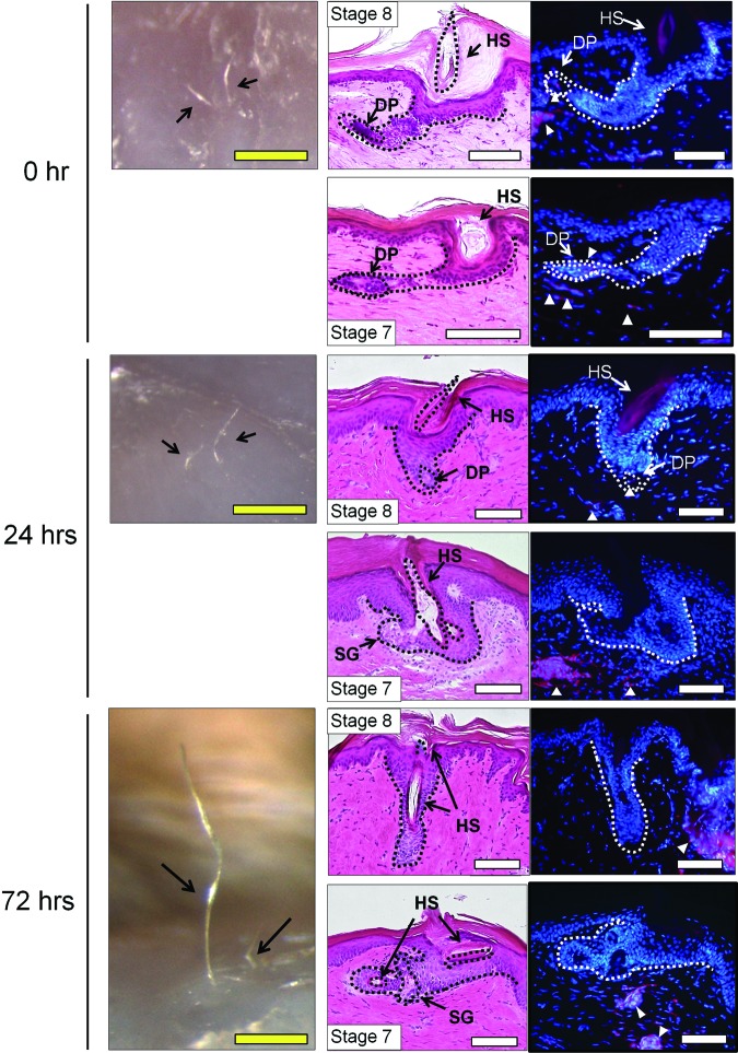
Dermal papilla cells (DPCs) have the potential to induce differentiation of epithelial stem cells into hair, and Wnt signaling is deeply involved in the initiation process. The functional limitation of expanded adult DPCs has been a difficult challenge for cell-based hair regrowth therapy. We previously reported that 1α,25-dihydroxyvitamin D(3) (VD(3)) upregulates expression of transforming growth factor (TGF)-β2 and alkaline phosphatase (ALP) activity, both features of hair-inducing human DPCs (hDPCs). In this study, we further examined the effects and signaling pathways associated with VD(3) actions on DPCs. VD(3) suppressed hDPC proliferation in a dose-dependent, noncytotoxic manner. Among the Wnt-related genes investigated, Wnt10b expression was significantly upregulated by VD(3) in hDPCs. Wnt10b upregulation, as well as upregulation of ALPL (ALP, liver/bone/kidney) and TGF-β2, by VD(3) was specific in hDPCs and not detected in human dermal fibroblasts. Screening of paracrine or endocrine factors in the skin indicated that all-trans retinoic acid (atRA) upregulated Wnt10b gene expression, although synergistic upregulation (combined atRA and VD(3)) was not seen. RNA interference with vitamin D receptor (VDR) revealed that VD(3) upregulation of Wnt10b, ALPL, and TGF-β2 was mediated through the genomic VDR pathway. In a rat model of de novo hair regeneration by murine DPC transplantation, pretreatment with VD(3) significantly enhanced hair folliculogenesis. Specifically, a greater number of outgrowing hair shafts and higher maturation of regenerated follicles were observed. Together, these data suggest that VD(3) may promote functional differentiation of DPCs and be useful in preserving the hair follicle-inductive capacity of cultured DPCs for hair regeneration therapies.


Figures

Figure 1.
Figure 1.
VD3 effects on proliferation of cultured hDPCs. (A): Proliferation of hDPCs cultured for 1–7 days (d1, d2, d3, d5, and d7) in Dulbecco's modified Eagle's medium (DMEM)/10% fetal bovine serum (FBS) supplemented with various concentrations of VD3 (0, 1, 10, or 100 nM) (n = 3). *, Significant differences from VD3-free (0 nM) medium (p < .05). (B): DNA synthesis (5-bromo-2′-deoxyuridine incorporation) compared from hDPCs cultured in DMEM/10% FBS in the presence of various concentration of VD3 (0, 1, 10, or 100 nM) or in DMEM/SFM (n = 4). **, Significant differences from the control (VD3-free [0 nM] serum-containing medium) (p < .01). (C): Representative images of immunocytochemical staining of cultured hDPCs with Hoechst 33342, Annexin V, and PI. Arrow and arrowhead show representative images of an apoptotic cell and a necrotic cell, respectively. Scale bars = 50 μm. (D): Percentage of Hoechst+/Annexin+/PI− (apoptotic) and Hoechst+/Annexin+/PI+ (necrotic) cells cultured in media with each VD3 concentration. SFM showed a significantly higher apoptotic or necrotic cell number compared with VD3-free medium. However, there were no significant differences among hDPCs cultured with different VD3 doses (n = 4). *, Significant difference from VD3-free medium (p < .05). Abbreviations: d, day; hDPC, human dermal papilla cell; PI, propidium iodide; SFM, serum-free medium; VD3, 1α,25-dihydroxyvitamin D3.
Figure 2.
Figure 2.
Effect of VD3 on expression of Wnt-related genes in cultured human dermal papilla cells (hDPCs). (A): Gene expression of Wnt3a, Wnt5a, Wnt7a, Wnt10a, Wnt10b, E-cad, snail, and slug was examined in hDPCs cultured in Dulbecco's modified Eagle's medium (DMEM)/10% fetal bovine serum (FBS) for 0, 2, or 48 hours, with or without VD3 (100 nM). Data are shown as fold change compared with baseline (n = 4). **, Significant differences between pairs (p < .01). (B): Kinetic analysis (0, 2, 4, 6, 8, 24, or 48 hours) of Wnt10b mRNA relative expression in hDPCs cultured in DMEM/10% FBS, supplemented with or without VD3 (100 nM). Data are shown as fold changes compared with the baseline expression (n = 4). **, Significant differences between conditions (with and without VD3), at each incubation time point (p < .01). (C): Dose-dependent effects (0, 1, 10, 100, or 1,000 nM) of VD3 on Wnt10b mRNA expression, in hDPCs cultured for 0, 2, or 48 hours (n = 4). Significant differences between groups, at each incubation period, are shown as * (p < .05) or ** (p < .01). (D): Representative images of immunocytochemical staining for Wnt10b in hDPCs cultured with various concentrations of VD3 (0, 10, or 100 nM) for 3 days. Goat IgG was used as a negative control. Arrowheads indicate Wnt10b protein expression. Scale bars = 50 μm. (E): Representative serial sections of the intact human scalp hair follicles. Shown are HE staining (left), immunohistochemical staining for Wnt10b (middle), and negative control with goat IgG (right). Wnt10b was expressed in the top portion of human dermal papilla (arrowhead) and the adjacent hair matrix (arrow). Scale bars = 100 μm. Abbreviations: E-cad, E-cadherin; HE, hematoxylin/eosin; VD3, 1α,25-dihydroxyvitamin D3.
Figure 3.
Figure 3.
VD3 effects on gene expression of Wnt10b, ALPL, and TGF-β2 in hDPCs and hDFs. Gene expression of Wnt10b (A), ALPL (B), and TGF-β2 (C) was examined in both hDPCs and hDFs cultured in Dulbecco's modified Eagle's medium/serum-free medium, supplemented with or without VD3 (100 nM) for 0, 2, or 48 hours. Data are shown as the ratio of the respective gene expression to GAPDH mRNA expression (n = 4). *, Significant difference between pairs (p < .05). DPCs showed much higher expression of the three genes and upregulated expression of Wnt10b and TGF-β2 after a 48-hour incubation with VD3. Abbreviations: ALPL, ALP, liver/bone/kidney; GAPDH, glyceraldehyde-3-phosphate dehydrogenase; hDF, human dermal fibroblast; hDPC, human dermal papilla cell; TGF, transforming growth factor; VD3, 1α,25-dihydroxyvitamin D3.
Figure 4.
Figure 4.
Influence of keratinocyte-conditioned medium components and other factors on Wnt10b mRNA expression in human dermal papilla cells (hDPCs). (A, B): Wnt10b mRNA expression of hDPCs cultured in SF-DMEM with supplementation of each factor for 2 (A) or 48 (B) hours. Data are shown as fold change compared with control medium (SF-DMEM) (n = 3). **, Significant differences from control medium (p < .01). (C, D): Also shown is Wnt10b mRNA expression of hDPCs cultured in DMEM/10% FBS with supplementation of each factor for 2 (C) or 48 (D) hours. Data are shown as fold change compared with control medium (10% DMEM) (n = 3). **, Significant differences from control media (p < .01). Supplements and concentrations used were as follows: aFGF (100 ng/ml), bFGF (100 ng/ml), IL-1β (100 ng/ml), IL-6 (100 ng/ml), IL-8 (100 ng/ml), VEGF (100 ng/ml), PDGF-BB (100 ng/ml), NGF (100 ng/ml), HB-EGF (100 ng/ml), MIP (100 ng/ml), MCP (100 ng/ml), IGF (100 ng/ml), ENA (100 ng/ml), GROa (100 ng/ml), VD3 (100 nM), VC (100 μM), CS (100 μM), atRA (10 nM), 17β-estradiol (10 nM), and DHT (10 nM). Abbreviations: aFGF, acidic fibroblast growth factor; atRA, all-trans retinoic acid; bFGF, basic fibroblast growth factor; CS, cholesterol sulfate; DHT, dihydrotestosterone; ENA-78, epithelial cell-derived neutrophil-activating peptide-78; FBS, fetal bovine serum; GROa, growth-related oncogene-α; HB-EGF, heparin-binding epidermal growth factor-like growth factor; IGF-1, insulin-like growth factor 1; IL, interleukin; MCP1, monocyte chemotactic protein 1; MIP3a, macrophage inflammatory protein-3α; NGF, nerve growth factor; PDGF-BB, platelet-derived growth factor-BB; SF-DMEM, serum-free Dulbecco's modified Eagle's medium; VC, ascorbic acid; VD3, 1α,25-dihydroxyvitamin D3; VEGF, vascular endothelial growth factor.
Figure 5.
Figure 5.
Knockdown effects of prepared shRNAs on VDR expression. (A): Each designed shRNA (negative control and shRNAs 1–5) was inserted into the cloning site of pSIREN-retroQ ZsGreen vectors. Both pVDR and an individual knockdown vector were cotransfected into HEK293 cells according to the combinations shown in the list. Western blotting revealed the knockdown efficacy of each shRNA on VDR protein expression. VDR expression was well knocked down by shRNAs 1, 3, 4, and 5. (B–D): Quantitative real-time polymerase chain reaction for gene expression of Wnt10b (B), ALPL (C), and TGF-β2 (D). Human dermal papilla cells cultured in serum-free Dulbecco's modified Eagle's medium, with or without the supplementation of VD3 (100 nM), were treated with NC or VDR knocking down shRNA 1 for 48 hours (n = 4). Data are shown as fold changes compared with the baseline expression at 0 hours. **, Significant differences between pairs (p < .01). Abbreviations: GAPDH, glyceraldehyde-3-phosphate dehydrogenase; NC, negative control short hairpin RNA; pVDR, flag-tagged human vitamin D receptor-expressing vector; shRNA, short hairpin RNA; TGF, transforming growth factor; VD3, 1α,25-dihydroxyvitamin D3; VDR, vitamin D receptor.
Figure 6.
Figure 6.
Hair folliculogenesis induced by transplantation of murine DPCs (mDPCs) or murine DFs (mDFs) pretreated with VD3 for 0, 24, or 72 hours. (A): Hair regeneration experiment using transplantation of a cultured mDPC construct in F344 rats. Cultured cell sheets of mDPCs (or mDFs as a control) were prepared and treated with VD3 for 0, 24, or 72 hours. After VD3 pretreatment, the mDPC (or mDF) sheet was fragmented, and the small fragment was transplanted into the nonhairy sole skin of F344 rats, using the hemivascularized sandwich method. Split-thickness skin (150–300 μm in thickness) was sliced off and digested with dispase, to separate the epidermis from the dermal compartment. The dermal compartment was discarded, and the epidermis was replaced on the remaining deep dermis after a cultured mDPC or mDF sheet fragment was grafted. Eight weeks after the transplantation, hair folliculogenesis was evaluated. (B): Hair regeneration induced by mDPC or mDF transplantation. Hair folliculogenesis of each sample was histologically evaluated (detailed data are summarized in supplemental online Table 3). Regenerated hair follicles were classified by maturity into stages 1–8. The numbers of total regenerated hair follicles (top left), matured (stage 6–8) follicles (top right), and outgrowing hairs (bottom) are shown. Significant differences among groups are shown as * (p < .05) or ** (p < .01). Abbreviations: DF, dermal fibroblast; DPC, dermal papilla cell; NS, not significant; VD3, 1α,25-dihydroxyvitamin D3.
Figure 7.
Figure 7.
Representative macroscopic and histological images of regenerated hair follicles via transplantation of murine dermal papilla cells (mDPCs) pretreated with 1α,25-dihydroxyvitamin D3 (VD3) for 0, 24, or 72 hours. Left column shows representative macroscopic views 8 weeks after transplantation of an mDPC sheet fragment pretreated with VD3 for 0 (top), 24 (center), and 72 (bottom) hours. Arrows indicate hair regrowth on the skin. Middle and right columns show histological serial sections stained with hematoxylin/eosin (middle) or Hoechst 33342 (right). Two representative samples for each group are shown. Arrowheads indicate DiI-labeled grafted mDPCs. Scale bars = 500 μm (yellow), 100 μm (white). Abbreviations: DP, dermal papilla; HS, hair shaft; SG, sebaceous gland.

Similar articles

Cited by

References

    1. Jahoda CA. Induction of follicle formation and hair growth by vibrissa dermal papillae implanted into rat ear wounds: Vibrissa-type fibres are specified. Development. 1992;115:1103–1109. - PubMed
    1. Jahoda CA, Reynolds AJ, Oliver RF. Induction of hair growth in ear wounds by cultured dermal papilla cells. J Invest Dermatol. 1993;101:584–590. - PubMed
    1. Reynolds AJ, Jahoda CA. Cultured dermal papilla cells induce follicle formation and hair growth by transdifferentiation of an adult epidermis. Development. 1992;115:587–593. - PubMed
    1. Lichti U, Weinberg WC, Goodman L, et al. In vivo regulation of murine hair growth: Insights from grafting defined cell populations onto nude mice. J Invest Dermatol. 1993;101:124S–129S. - PubMed
    1. Weinberg WC, Goodman LV, George C, et al. Reconstitution of hair follicle development in vivo: Determination of follicle formation, hair growth, and hair quality by dermal cells. J Invest Dermatol. 1993;100:229–236. - PubMed

Publication types

MeSH terms