. 2012 Dec;122(12):4555-68.
doi: 10.1172/JCI64896. Epub 2012 Nov 26.

IL-13-induced airway mucus production is attenuated by MAPK13 inhibition

Affiliations

IL-13-induced airway mucus production is attenuated by MAPK13 inhibition

Yael G Alevy et al. J Clin Invest. 2012 Dec.

Abstract

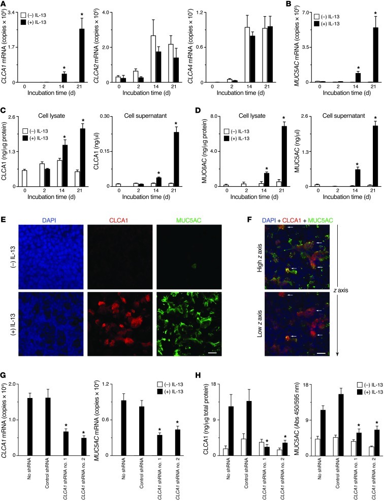
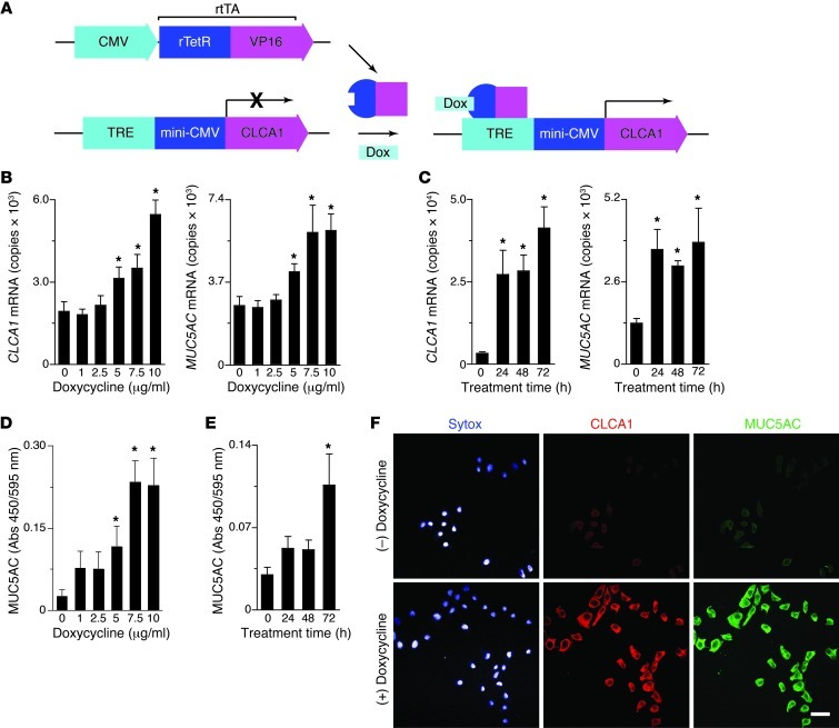
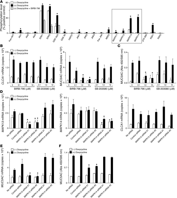
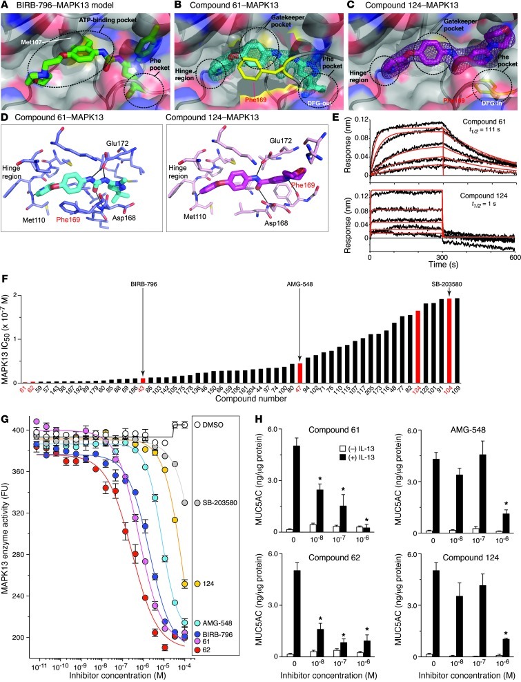
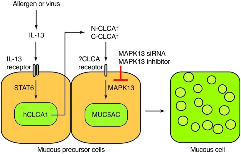
Increased mucus production is a common cause of morbidity and mortality in inflammatory airway diseases, including asthma, chronic obstructive pulmonary disease (COPD), and cystic fibrosis. However, the precise molecular mechanisms for pathogenic mucus production are largely undetermined. Accordingly, there are no specific and effective anti-mucus therapeutics. Here, we define a signaling pathway from chloride channel calcium-activated 1 (CLCA1) to MAPK13 that is responsible for IL-13-driven mucus production in human airway epithelial cells. The same pathway was also highly activated in the lungs of humans with excess mucus production due to COPD. We further validated the pathway by using structure-based drug design to develop a series of novel MAPK13 inhibitors with nanomolar potency that effectively reduced mucus production in human airway epithelial cells. These results uncover and validate a new pathway for regulating mucus production as well as a corresponding therapeutic approach to mucus overproduction in inflammatory airway diseases.


Figures

Figure 1
Figure 1. CLCA1 is necessary for IL-13–stimulated mucus production in human airway epithelial cells.
(A) hTECs were incubated with IL-13 (50 ng/ml) under submerged conditions for 2 days and then air-liquid interface conditions for up to 21 days, and cell lysates were analyzed for CLCA1, CLCA2, and CLCA4 mRNA levels by real-time qPCR assay. CLCA3 mRNA was undetectable under these conditions (data not shown). (B) Corresponding levels of MUC5AC mRNA. (C) Corresponding levels of CLCA1 in cell lysate and apical cell supernatant determined by ELISA. (D) Corresponding levels of MUC5AC determined by ELISA. (E) Corresponding immunocytochemistry for DAPI, CLCA1, and MUC5AC using confocal microscopy. Scale bar: 50 μm. (F) Corresponding immunocytochemistry at a more apical (high z axis) and subjacent (low z axis) cellular location. Arrows indicate the same reference cells for high and low z axis. Scale bar: 50 μm. (G) Levels of CLCA1 and MUC5AC mRNA in hTECs that were transduced with lentivirus encoding CLCA1 or control shRNA. (H) For conditions in E, corresponding cellular levels of CLCA1 and MUC5AC by ELISA. All values represent mean ± SEM. *P < 0.05, increase from corresponding no IL-13 treatment control or decrease from no shRNA treatment.
Figure 2
Figure 2. CLCA1 expression is sufficient for mucus production in lung epithelial cells.
(A) Diagram for constructs used to establish an NCI-H292 cell line carrying rtTA- and CLCA1-expression cassettes. (B) Doxycycline dose response for mucin gene expression in the NCI-H292-rtTA-CLCA1 cell line. Cells were treated with doxycycline (0–10 μg/ml) for 48 hours, and CLCA1 and MUC5AC mRNA levels were determined by real-time qPCR assay. (C) Time course for mucin gene expression in NCI-H92-rrTA-CLCA1 cells. Cells were treated with doxycycline (7.5 μg/ml) for 0 to 72 hours. (D) Dose response for mucus protein levels in NCI-H292-rtTA-CLCA1 cells. Cells were treated with doxycycline for 48 hours, and MUC5AC levels were determined by cell-based ELISA. (E) Time course for mucus protein levels in NCI-H92-rrTA-CLCA1 cells. Cells were treated with doxycycline (7.5 μg/ml for 0 to 72 hours), and MUC5AC levels were determined by ELISA. (F) Representative immunocytochemistry for NCI-H92-rrTA-CLCA1 cells treated with doxycycline (7.5 μg/ml for 48 hours) and then incubated with Sytox nuclear stain and immunostained for CLCA1 and MUC5AC. Scale bar: 100 μm. (BE) Values represent mean ± SEM. *P < 0.05, increase from corresponding no doxycycline treatment control.
Figure 3
Figure 3. MAPK13 is necessary for CLCA1-driven mucus production in lung epithelial cells.
(A) NCI-H292-rtTA-CLCA1 cells were treated with doxycycline (7.5 μg/ml for 18 hours) with or without BIRB-796 (10 μM), and cell lysates were analyzed by phospho-MAPK antibody array. The box indicates results for MAPK values. Values represent the percentage of positive control (mean ± SEM). *P < 0.05, increase from corresponding no doxycycline control. (B) Cells were treated as in A at indicated concentrations of BIRB-796, SB-203580, and vehicle control, and levels of CLCA1 and MUC5AC mRNA were determined by real-time qPCR assay. (C) Corresponding MUC5AC levels determined by cell-based ELISA. (D) Cells were transfected with or without control or MAPK13 or MAPK14 siRNA, (25 nM) and treated with or without doxycycline (7.5 μg/ml for 48 hours), and levels of MAPK13, MAPK14, and CLCA1 mRNA were determined by real-time qPCR. (E) Corresponding levels of MUC5AC mRNA for conditions in D. (F) Corresponding MUC5AC levels determined by cell-based ELISA for conditions in D. (BF) Values represent mean ± SEM. *P < 0.05, decrease from corresponding no drug treatment or no siRNA control.
Figure 4
Figure 4. MAPK13 is necessary for IL-13–driven mucus production in human airway epithelial cells.
(A) hTECs were incubated with IL-13 for 21 days, as described in Figure 1, with or without BIRB-796 or SB-203580, and levels of CLCA1 and MUC5AC mRNA were determined by real-time qPCR assay. (B) Corresponding MUC5AC levels determined by cell-based ELISA for conditions in A. (C) hTECs were transduced with lentivirus encoding MAPK13, MAPK14, or control shRNA and then were treated with IL-13 for 21 days, as described in Figure 1. Cell lysates were analyzed for CLCA1, MAPK13, and MAPK14 mRNA levels using real-time qPCR assay. (D) Corresponding levels of MUC5AC mRNA. (E) Corresponding MUC5AC levels determined by cell-based ELISA. (AE) Values represent mean ± SEM. *P < 0.05, decrease from corresponding no drug treatment or shRNA control condition.
Figure 5
Figure 5. Evidence of a CLCA1 to MAPK13 signaling pathway for IL-13–stimulated mucus production in COPD.
(A) Levels of IL13, CLCA1, MAPK13, and MUC5AC mRNA in lung tissues from lung transplant donors without COPD (n = 4) and recipients with very severe COPD (n = 9). (B) Levels of CLCA1 and MUC5AC determined by ELISA from lung samples obtained as in A. Values represent mean of triplicate values for each subject. (A and B) For box-and-whisker plots, whiskers represent range, the plus sign indicates mean values, and boxes indicate 25th to 75th percentiles. *P < 0.05, difference from non-COPD control. (C) Representative photomicrographs of corresponding lung sections subjected to immunostaining for CLCA1 and MUC5AC. Scale bar: 100 μm. (D) Levels of MAPK activation based on analysis of phospho-MAPK antibody array for lung samples obtained as in A. Values represent percentage of positive control (mean ± SEM). *P < 0.05.
Figure 6
Figure 6. Discovery and validation of potent MAPK13 inhibitors.
(A) Superposition of BIRB-796 into the MAPK13 binding pocket. The position of the gatekeeper Met107 is shown in a space-filling model. Circles indicate ATP-binding and Phe pockets. (B) Crystal structure of compound 61 (cyan) bound to MAPK13 in DFG-out mode. Difference electron density for the compound following rigid-body refinement is shown in mesh. Circles indicate binding pocket features. Phe169 (of the DFG sequence) is highlighted in yellow. (C) Crystal structure of compound 124 (magenta) bound to MAPK13 in DFG-in mode. (D) MAPK13 binding pockets for compound 61 (cyan) and compound 124 (magenta). All residues that make van der Waals contacts or polar contacts are shown. Residues that make direct hydrogen bonds (black lines) with inhibitors are labeled in black. (E) BioLayer interferometry sensorgrams for compound 61 and 124 binding to MAPK13. Processed data are shown as black lines with global kinetic fits overlaid as red lines. (F) MAPK13 inhibitory activity in a screen of test compounds (numbered from 43–205 and arranged from most to least potent) using IMAP assay. Values represent IC50 for compounds with values <20 εM. Red-colored bars indicate compounds selected for detailed analysis. (G) Concentration response for MAPK13 inhibition for indicated test compounds. Values represent fluorescence polarization (mP) from IMAP assay. (H) hTECs were cultured with or without IL-13 for 21 days with the indicated compounds, and MUC5AC levels were determined by ELISA. (G and H) Values represent mean ± SEM. *P < 0.05, decrease from no drug treatment condition.
Figure 7
Figure 7. Scheme for CLCA1/MAPK13 signaling to mucus production.
The CLCA1/MAPK13 signaling pathway is initiated when environmental stimuli (e.g., allergen or virus) stimulate IL-13 production and subsequent activation of IL-13 receptor on the surface of mucous precursor cells. IL-13 receptor activation leads to STAT6 activation and consequent induction of CLCA1 gene expression. The CLCA1 protein is secreted as N-terminal and C-terminal CLCA1 (N-CLCA1 and C-CLCA1) peptide fragments that might activate a putative CLCA1 receptor and in turn MAPK13 to drive a consequent increase in mucin (especially MUC5AC) gene expression and mucus production in airway mucous cells.

Similar articles

Cited by

References

    1. Kuyper LM, et al. Characterization of airway plugging in fatal asthma. Am J Med. 2003;115(1):6–11. - PubMed
    1. Hogg JC, et al. The nature of small-airway obstruction in chronic obstructive pulmonary disease. N Engl J Med. 2004;350(26):2645–2653. doi: 10.1056/NEJMoa032158. - DOI - PubMed
    1. Brito-Mutunayagam R, Appleton SL, Wilson DH, Ruffin RE, Adams RJ. Global initiative for chronic obstructive lung disease stage 0 is associated with excess FEV1 decline in a representative population sample. Chest. 2010;138(3):605–613. doi: 10.1378/chest.09-2607. - DOI - PubMed
    1. Rose MC, Voynow JA. Respiratory tract mucin genes and mucin glycoproteins in health and disease. Physiol Rev. 2006;86(1):245–278. doi: 10.1152/physrev.00010.2005. - DOI - PubMed
    1. Wills-Karp M, et al. Interleukin-13: central mediator of allergic asthma. Science. 1998;282(5397):2258–2261. doi: 10.1126/science.282.5397.2258. - DOI - PubMed

Publication types

MeSH terms