. 2012;7(11):e49886.
doi: 10.1371/journal.pone.0049886. Epub 2012 Nov 21.

A novel function for SNAP29 (synaptosomal-associated protein of 29 kDa) in mast cell phagocytosis

Affiliations

A novel function for SNAP29 (synaptosomal-associated protein of 29 kDa) in mast cell phagocytosis

Jordan Wesolowski et al. PLoS One. 2012.

Abstract

Mast cells play a critical role in the innate immune response to bacterial infection. They internalize and kill a variety of bacteria and process antigen for presentation to T cells via MHC molecules. Although mast cell phagocytosis appears to play a significant role during bacterial infection, little is known about the proteins involved in its regulation. In this study, we demonstrate that the SNARE protein SNAP29 is involved in mast cell phagocytosis. SNAP29 is localized in the endocytic pathway and is transiently recruited to Escherichia coli (E. coli)-containing phagosomes. Interestingly, overexpression of SNAP29 significantly increases the internalization and killing of E. coli, while it does not affect mast cell exocytosis of inflammatory mediators. To our knowledge, these data are the first to demonstrate a novel function of SNAP29 in mast cell phagocytosis and have implications in protection against bacterial infection.


Conflict of interest statement

Competing Interests: The authors have declared that no competing interests exist.

Figures

Figure 1
Figure 1. SNAP29 is distributed in both cytosolic and membrane fractions in resting mast cells.
(A) Schematic of SNAP29 and SNAP23 SNARE proteins. Both proteins contain two SNARE motifs. The SNARE motifs in SNAP29 are in positions 60–113 and 196–258, and in positions 14–76 and 146–211 for SNAP23. Unlike SNAP23, SNAP29 does not contain palmitoylation sites (indicated by the asterisks for SNAP23). (B) Bone marrow derived mast cell (BMMC) homogenate was tested for SNAP29 expression by Western blot using an anti-SNAP29 Ab. As a control, SNAP23 expression was confirmed using an anti-SNAP23 Ab. 7×10⁵ cells were loaded in each lane. (C) BMMC homogenate (H) was fractionated into both cytosol (Cyt) and membrane (Mb) fractions. Fractions were solubilized with 1% SDS prior to loading. Equal volumes of each fraction were tested for SNAP29 expression by Western Blot using an anti-SNAP29 Ab. Cell fractionation was controlled using anti-actin (marker for cytosol) and anti-VAMP8 (marker for membranes) Abs. (D) Resting BMMCs were fixed, permeabilized, labeled with anti-SNAP29/anti-Rabbit AlexaFluor488 Ab and analyzed by confocal microscopy (Green). The nucleus was labeled with Hoechst (Blue). The overlay is presented on the right. Scale bar = 20 µm. (E) RBL-2H3 homogenate was prepared and immunoblotted with both anti-SNAP23 and anti-SNAP29 Abs. 3.5×10⁵ cells were loaded in each lane. (F) RBL-2H3 were fractionated and immunoblotted as described in C. (G) Fixed RBL-2H3 were labeled for SNAP29 as described in D. Scale bar = 20 µm. Each experiment is representative of n = 3.
Figure 2
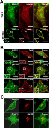
Figure 2. SNAP29 is localized on the plasma membrane and in endocytic compartments.
(A) RBL-2H3 cells were fixed, permeabilized, labeled with anti-SNAP29/anti-Rabbit AlexaFluor488 Ab and anti-SNAP23/anti-Mouse AlexaFluor594 Ab and analyzed by TIRF microscopy (top panels) or confocal microscopy (lower panels). To visualize the plasma membrane in confocal, a high focal plane was used. Thus, cytoplasmic SNAP29 is not visible. (B) RBL-2H3 cells were fixed, permeabilized, labeled with anti-SNAP29/anti-Rabbit AlexaFluor488 Abs and anti-Rab4 (top panels), anti-Rab5 (middle panels), or anti-VAMP8/anti-Mouse AlexaFluor594 Abs (lower panels) and analyzed by confocal microscopy. (C) RBL-2H3 cells were fixed, permeabilized, labeled with anti-SNAP29/anti-Rabbit AlexaFluor488 Abs and anti-Syntaxin6 (top panels), or anti-GM130/anti-Mouse AlexaFluor594 Abs (lower panels) and analyzed by confocal microscopy. All scale bars = 10 µm. Arrowheads indicate areas of colocalization.
Figure 3
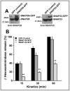
Figure 3. SNAP29 is not involved in mast cell degranulation.
(A) RBL-2H3 cells were stably transfected with SNAP29-GFP, SNAP23-GFP or GFP. Homogenates for each transfected population were immunoblotted with anti-SNAP29 and anti-SNAP23 Abs. 1.25×10⁶ cells were loaded in each lane. For each transfected population, two bands corresponding to the endogenous SNARE (lower band) and the SNARE-GFP (higher band) were observed. (B) Transfected RBL-2H3 cells were sensitized with anti-DNP IgE for 2 h, stimulated with DNP-BSA for 15, 30, and 60 min and β-hexosaminidase release was assayed. The data shown are the mean ± SD of five independent experiments each performed in triplicate. The asterisks denote a significant difference (p<0.01).
Figure 4
Figure 4. Mast cells kill E. coli while SNAP29 relocates to the phagosome.
BMMCs (A) and RBL-2H3 cells (B) were infected with E. coli at a MOI of 250 for 2 h at 37°C. Extracellular bacteria were killed with gentamicin for 1 h. Cells were then washed and incubated with DMEM without antibiotics. The cells were incubated at 37°C for an additional hour, 2 h or 24 h. Cell lysates were serially diluted on agar plates at 37°C. Surviving colonies were counted 24 h later. Both graphs represent the mean ± SD of three independent experiments, each performed in duplicate. BMMCs (C) and RBL-2H3 (D) were infected with mCherry E. coli (red) for 1 h, fixed, permeabilized, and labeled with anti-SNAP29/anti-rabbit AlexaFluor488 Abs (Green). The nucleus was labeled with Hoechst (Blue). SNAP29 colocalization with E. coli was assessed using confocal microscopy. SNAP29 staining accumulates around E. coli 1 h post infection. The inset in each image shows an enlarged area of an E. coli phagosome. This experiment is representative of n = 3. Scale bar = 5 µm (C) and 20 µm (D).
Figure 5
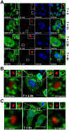
Figure 5. SNAP29 relocation to E. coli phagosomes in infected mast cells is transient.
(A) RBL-2H3 cells were infected with E. coli expressing mCherry (red) for 0 h, 1 h, 2.5 h or 4 h. Cells were then fixed, permeabilized and labeled with anti-SNAP29/anti-rabbit AlexaFluor488 Abs (green). The nucleus was labeled with Hoechst (blue). SNAP29 localization was assessed using confocal microscopy. SNAP29 staining accumulates around E. coli phagosomes at t = 1 h and t = 2.5 h. The inset in each image shows an enlarged area of E. coli phagosomes. This experiment is representative of n = 3. Scale bars = 20 µm. (B and C) RBL-2H3 cells were infected with E. coli mCherry (red) for 2.5 h (B) or 4 h (C). Cells were fixed and stained as described in A. SNAP29 localization was assessed using confocal microscopy. Two different E. coli phagosomes are shown on the left and right of the center image. Z-sections of 0.3 µm were used to reconstruct each phagosome in 3D (shown below the enlarged phagosomes). SNAP29-positive endosomes associate with phagosomes at 2.5 h, but are not present 4 h post-infection. Scale bars = 20 µm.
Figure 6
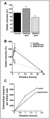
Figure 6. SNAP29 is involved in mast cell phagocytosis.
(A) RBL-2H3 cells overexpressing GFP, SNAP29-GFP, or SNAP23-GFP were infected with E. coli at a MOI of 250 for 2 h at 37°C. Extracellular bacteria were killed with gentamicin for 1 h. Cells were then lysed and serially diluted on agar plates at 37°C. Surviving colonies were counted 24 h later. (B) Transfected RBL-2H3 cells were infected as described in A. Extracellular bacteria were killed with gentamicin for 1 h. The infected cells were then washed and incubated with DMEM without antibiotics. The cells were returned to 37°C for an additional hour, 2 h or 24 h. Cell lysates were serially diluted on agar plates at 37°C. Surviving colonies were counted 24 h later. (C) After transfected RBL-2H3 cells were infected with E. coli (MOI 250), the culture medium was collected, centrifuged to concentrate the bacteria and serially diluted on agar plates at 37°C. Colonies were counted 24 h later. All the graphs represent the mean ± SD of five independent experiments, each performed in triplicate. The asterisks denote a significant difference (p<0.01).

Similar articles

Cited by

References

    1. Wasserman S (1989) Mast cell-mediated inflammation in asthma. Annals of Allergy 63: 546–550. - PubMed
    1. Pietrzak A, Wierzbicki M, Wiktorska M, Brzezińska-Błaszczyk E (2011) Surface TLR2 and TLR4 expression on mature rat mast cells can be affected by some bacterial components and proinflammatory cytokines. Mediators Inflamm 2011: 427473. - PMC - PubMed
    1. Abraham S, St John A (2010) Mast cell-orchestrated immunity to pathogens. Nat Rev Immunol 10: 440–452. - PMC - PubMed
    1. Malaviya R, Twesten N, Ross E, Abraham S, Pfeifer J (1996) Mast cells process bacterial Ags through a phagocytic route for Class I MHC presentation to T cells. J Immunol 156: 1490–1496. - PubMed
    1. Abraham S, Malaviya R (1997) Mast cells in infection and immunity. Infection and Immunity 65: 3501–3508. - PMC - PubMed

Publication types

MeSH terms