Amygdala lesions disrupt modulation of functional MRI activity evoked by facial expression in the monkey inferior temporal cortex
- PMID: 23184972
- PMCID: PMC3535608
- DOI: 10.1073/pnas.1218406109
Amygdala lesions disrupt modulation of functional MRI activity evoked by facial expression in the monkey inferior temporal cortex
Abstract
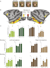
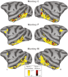
We previously showed that facial expressions modulate functional MRI activity in the face-processing regions of the macaque monkey’s amygdala and inferior temporal (IT) cortex. Specifically, we showed that faces expressing emotion yield greater activation than neutral faces; we term this difference the “valence effect.” We hypothesized that amygdala lesions would disrupt the valence effect by eliminating the modulatory feedback from the amygdala to the IT cortex. We compared the valence effects within the IT cortex in monkeys with excitotoxic amygdala lesions (n = 3) with those in intact control animals (n = 3) using contrast agent-based functional MRI at 3 T. Images of four distinct monkey facial expressions--neutral, aggressive (open mouth threat), fearful (fear grin), and appeasing (lip smack)--were presented to the subjects in a blocked design. Our results showed that in monkeys with amygdala lesions the valence effects were strongly disrupted within the IT cortex, whereas face responsivity (neutral faces > scrambled faces) and face selectivity (neutral faces > non-face objects) were unaffected. Furthermore, sparing of the anterior amygdala led to intact valence effects in the anterior IT cortex (which included the anterior face-selective regions), whereas sparing of the posterior amygdala led to intact valence effects in the posterior IT cortex (which included the posterior face-selective regions). Overall, our data demonstrate that the feedback projections from the amygdala to the IT cortex mediate the valence effect found there. Moreover, these modulatory effects are consistent with an anterior-to-posterior gradient of projections, as suggested by classical tracer studies.
Conflict of interest statement
The authors declare no conflict of interest.
Figures


Comment in
-
Facing the role of the amygdala in emotional information processing.Proc Natl Acad Sci U S A. 2012 Dec 26;109(52):21180-1. doi: 10.1073/pnas.1219167110. Epub 2012 Dec 14. Proc Natl Acad Sci U S A. 2012. PMID: 23243143 Free PMC article. No abstract available.
Similar articles
-
Perception of emotional expressions is independent of face selectivity in monkey inferior temporal cortex.Proc Natl Acad Sci U S A. 2008 Apr 8;105(14):5591-6. doi: 10.1073/pnas.0800489105. Epub 2008 Mar 28. Proc Natl Acad Sci U S A. 2008. PMID: 18375769 Free PMC article.
-
Facial Expressions Evoke Differential Neural Coupling in Macaques.Cereb Cortex. 2017 Feb 1;27(2):1524-1531. doi: 10.1093/cercor/bhv345. Cereb Cortex. 2017. PMID: 26759479 Free PMC article.
-
Facing the role of the amygdala in emotional information processing.Proc Natl Acad Sci U S A. 2012 Dec 26;109(52):21180-1. doi: 10.1073/pnas.1219167110. Epub 2012 Dec 14. Proc Natl Acad Sci U S A. 2012. PMID: 23243143 Free PMC article. No abstract available.
-
Distributed and interactive brain mechanisms during emotion face perception: evidence from functional neuroimaging.Neuropsychologia. 2007 Jan 7;45(1):174-94. doi: 10.1016/j.neuropsychologia.2006.06.003. Epub 2006 Jul 18. Neuropsychologia. 2007. PMID: 16854439 Review.
-
Functional atlas of emotional faces processing: a voxel-based meta-analysis of 105 functional magnetic resonance imaging studies.J Psychiatry Neurosci. 2009 Nov;34(6):418-32. J Psychiatry Neurosci. 2009. PMID: 19949718 Free PMC article. Review.
Cited by
-
Lesion Studies in Contemporary Neuroscience.Trends Cogn Sci. 2019 Aug;23(8):653-671. doi: 10.1016/j.tics.2019.05.009. Epub 2019 Jul 3. Trends Cogn Sci. 2019. PMID: 31279672 Free PMC article. Review.
-
Experimental evidence for a child-to-adolescent switch in human amygdala-prefrontal cortex communication: A cross-sectional pilot study.Dev Sci. 2022 Jul;25(4):e13238. doi: 10.1111/desc.13238. Epub 2022 Feb 12. Dev Sci. 2022. PMID: 35080089 Free PMC article.
-
Hierarchical Encoding of Social Cues in Primate Inferior Temporal Cortex.Cereb Cortex. 2015 Sep;25(9):3036-45. doi: 10.1093/cercor/bhu099. Epub 2014 May 16. Cereb Cortex. 2015. PMID: 24836688 Free PMC article.
-
The neurobiology of dispositional negativity and attentional biases to threat: Implications for understanding anxiety disorders in adults and youth.J Exp Psychopathol. 2016;7(3):311-342. doi: 10.5127/jep.054015. J Exp Psychopathol. 2016. PMID: 27917284 Free PMC article.
-
Oxytocin modulates fMRI responses to facial expression in macaques.Proc Natl Acad Sci U S A. 2015 Jun 16;112(24):E3123-30. doi: 10.1073/pnas.1508097112. Epub 2015 May 26. Proc Natl Acad Sci U S A. 2015. PMID: 26015576 Free PMC article.
References
-
- Adolphs R, Baron-Cohen S, Tranel D. Impaired recognition of social emotions following amygdala damage. J Cogn Neurosci. 2002;14(8):1264–1274. - PubMed
-
- Adolphs R, et al. A mechanism for impaired fear recognition after amygdala damage. Nature. 2005;433(7021):68–72. - PubMed
-
- Meunier M, Bachevalier J, Murray EA, Málková L, Mishkin M. Effects of aspiration versus neurotoxic lesions of the amygdala on emotional responses in monkeys. Eur J Neurosci. 1999;11(12):4403–4418. - PubMed
-
- Phelps EA, LeDoux JE. Contributions of the amygdala to emotion processing: From animal models to human behavior. Neuron. 2005;48(2):175–187. - PubMed
-
- Amaral DG. The amygdala, social behavior, and danger detection. Ann N Y Acad Sci. 2003;1000:337–347. - PubMed
Publication types
MeSH terms
Grants and funding
LinkOut - more resources
Full Text Sources
Other Literature Sources
Medical

