Review
. 2012 Dec;13(12):755-66.
doi: 10.1038/nrm3478.

Function and regulation of SUMO proteases

Affiliations
Review

Function and regulation of SUMO proteases

Christopher M Hickey et al. Nat Rev Mol Cell Biol. 2012 Dec.

Abstract

Covalent attachment of small ubiquitin-like modifier (SUMO) to proteins is highly dynamic, and both SUMO-protein conjugation and cleavage can be regulated. Protein desumoylation is carried out by SUMO proteases, which control cellular mechanisms ranging from transcription and cell division to ribosome biogenesis. Recent advances include the discovery of two novel classes of SUMO proteases, insights regarding SUMO protease specificity, and revelations of previously unappreciated SUMO protease functions in several key cellular pathways. These developments, together with new connections between SUMO proteases and the recently discovered SUMO-targeted ubiquitin ligases (STUbLs), make this an exciting period to study these enzymes.


Figures

Figure 1
Figure 1. SUMO conjugation and deconjugation cycle
a | SUMO (S) is translated in a precursor form (black) that must be processed by a SUMO protease before SUMO can be conjugated to other proteins. Mature SUMO is conjugated to proteins in an ATP-dependent manner in reactions catalyzed by three enzymes, E1, E2, and E3, in sequence. SUMO can be conjugated to a single lysine of a protein (monosumoylation), multiple lysines of a single protein (multisumoylation), or to SUMO that is already conjugated to a protein, forming a SUMO chain (polysumoylation). SUMO proteases deconjugate SUMO or SUMO chains from proteins. See Table 1 for a summary of the ability of each of the known SUMO protease to process and/or deconjugate the different forms of SUMO. b | The C-termini of S. cerevisiae Smt3 and the four mammalian SUMO isoforms are shown, with the di-glycine motif underlined. The site of cleavage is indicated by an arrrow. The chemical structure of the C-terminal tail of SUMO2 is also shown, with the scissile bond boxed in orange (bottom). c | The chemical structure of the isopeptide bond between the terminal glycine of mature SUMO and the lysine of a substrate is also shown, with the scissile bond boxed in orange.
Figure 2
Figure 2. Structures of SUMO proteases
The domain organizations and lengths (in amino acids) of the SUMO proteases discussed in this article are shown. Each catalytic domain (CD) is colored magenta, with key histidine (H) and cysteine (C) residues shown. Putative SUMO-interacting motifs (SIMs) are indicated by asterisks. Regions shown to be important for intracellular localization of the protease are shown in cyan. For DeSI-1 and DeSI-2, the mouse proteins are shown. For USPL1, the human protein is depicted. Representatives from each of the three known classes of SUMO proteases exist in other organisms, ; however, in some cases it is unclear whether these proteins are indeed SUMO-specific proteases. b | Crystal structure of the catalytic domain of SENP1 (magenta) in complex with SUMO2 (blue) (Protein Databank (PDB) code 2CKH) shows the orientation of SUMO2 with respect to SENP1. This complex contains a covalent thiohemiacetal linkage between the active site cysteine of SENP1 and the C-terminal glycine of SUMO2. The catalytic residues are shown in stick format, with the atoms colored according to element (carbon = cyan; nitrogen = blue; oxygen = red; sulfur = orange). c | A space-filling representation of the structure shown in part b, which highlights the intimate contacts made between SENP1 and SUMO2. d | A higher magnification view of SENP1 in complex with SUMO2 (2CKH), showing the two Trp residues of SENP1 that surround the two Gly residues of SUMO2. e | The structure of the catalytic domain of SENP7 (yellow) (PDB code 3EAY) aligned to the structure of the catalytic domain of SENP1 (magenta) highlights the similarities and differences between SENP1/2 and SENP6/7. The catalytic residues are displayed in cyan for SENP1 and red for SENP7. Loop regions, including Loop-1 (right, green), of SENP7 are unique to SENP6 and SENP7. f | The structure of a monomer of DeSI-1 (PDB code 2WP7) shows the proposed catalytic dyad (Cys-His), which differs from the catalytic triad (Cys-His-Asp) utilized by the Ulp/SENP enzymes. The catalytic residues are displayed as in b
Figure 3
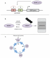
Figure 3. Regulation of SUMO proteases
a | SENP1 and SENP2 levels are known to be affected by changes in their transcription under various conditions (see text). Fittingly, the promoters of SENP1 and SENP2 contain response elements (REs) that bind specific transcription factors (TFs). Both SENP1 and SENP2 can affect their own transcription by desumoylating transcription factors or factors that affect the activity or abundance of relevant transcription factors. b | SENP3 levels are regulated by the ubiquitin-proteasome system. SENP3 is constitutively ubiquitylated under normal growth conditions, leading to its proteasome-mediated degradation. Under conditions of mild oxidative stress, SENP3 ubiquitylation is reduced and SENP3 levels increase. When the tumor suppressor p19(Arf) is expressed, it interacts with SENP3, enhancing its ubiquitylation and degradation, leading to decreased levels of SENP3. c | Yeast Ulp2 is phophorylated during the mitosis phase of the cell cycle. Phosphorylation of Ulp2 is thought to inhibit its activity.
Figure 4
Figure 4. The function of SENP1 in transcription during Wnt signaling
Activation of Wnt signaling leads to the sumoylation of TBL1 and TBLR1, which function together as a transcriptional co-activator complex. Sumoylation of TBL1-TBLR1 promotes the dissociation of TBL1-TBLR1 from the NcoR/HDAC3 corepressor complex and promotes the association of TBL1-TBLR1 with β-catenin. The TBL1SUMO-TBLR1SUMO-β-catenin complex is recruited to Wnt target genes by the sequence-specific DNA-binding protein TCF4. SENP1 is responsible for desumoylation of TBL1SUMO-TBLR1SUMO, which leads to the disassembly of the TBL1SUMO-TBLR1SUMO-β-catenin complex and decreased activation of Wnt target gene expression.
Figure 5
Figure 5. SUMO-targeted ubiquitin ligases (STUbLs)
STUbLs are enzymes that catalyze the addition of ubiquitin to proteins that have been previously sumoylated. SUMO (S), likely in the form of SUMO chains, attached to STUbL substrates is recognized by SIMs of the STUbL. The ubiquitin (U)-charged E2 enzyme (E2-U) is recruited by the dimeric RING domains of the STUbL, and ubiquitin is transferred to the substrate. Potential sites of attachment for ubiquitin on the substrate include the substrate itself and/or the attached SUMO moieties. STUbL activity results in proteins that are modified by both SUMO and ubiquitin. Ubiquitin addition can target proteins to the proteasome for destruction or non-proteolytic fates. It is currently unclear whether SUMO proteases act on STUbL substrates destined for the proteasome. If SUMO proteases act on these substrates, it could be before arrival at the proteasome (1) and/or at the proteasome (2). Alternatively, SUMO could be destroyed along with the substrate at the proteasome. Ubiquitin on STUbL substrates is likely recycled by deubiquitylating (DUB) activity of the proteasome (not shown).

Similar articles

Cited by

References

    1. Hochstrasser M. Origin and function of ubiquitin-like proteins. Nature. 2009;458:422–9. - PMC - PubMed
    1. Panse VG, Hardeland U, Werner T, Kuster B, Hurt E. A proteome-wide approach identifies sumoylated substrate proteins in yeast. J Biol Chem. 2004;279:41346–51. - PubMed
    1. Wohlschlegel JA, Johnson ES, Reed SI, Yates JR., 3rd Global analysis of protein sumoylation in Saccharomyces cerevisiae. J Biol Chem. 2004;279:45662–8. - PubMed
    1. Hannich JT, et al. Defining the SUMO-modified proteome by multiple approaches in Saccharomyces cerevisiae. J Biol Chem. 2005;280:4102–10. - PubMed
    1. Wykoff DD, O'Shea EK. Identification of sumoylated proteins by systematic immunoprecipitation of the budding yeast proteome. Mol Cell Proteomics. 2005;4:73–83. - PubMed

Publication types

MeSH terms

Substances

LinkOut - more resources