Replication kinetics of duck enteritis virus UL16 gene in vitro
- PMID: 23171438
- PMCID: PMC3560188
- DOI: 10.1186/1743-422X-9-281
Replication kinetics of duck enteritis virus UL16 gene in vitro
Abstract
Background: The function and kinetics of some herpsvirus UL16 gene have been reported. But there was no any report of duck enteritis virus (DEV) UL16 gene.
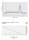
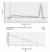
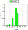
Findings: The kinetics of DEV UL16 gene was examined in DEV CHv infected duck embryo fibroblasts (DEFs) by establishment of real-time quantitative reverse transcription polymerase chain reaction assay (qRT-PCR) and western-blotting. In this study, UL16 mRNA was transcript at a low level from 0-18 h post-infection (p.i), and peaked at 36 h p.i. It can't be detected in the presence of acyclovir (ACV). Besides, western-blotting analysis showed that UL16 gene expressed as an apparent 40-KDa in DEV infected cell lysate from 12 h p.i, and rose to peak level at 48 h p.i consistent with the qRT-PCR result.
Conclusions: These results provided the first evidence of the kinetics of DEV UL16 gene. DEV UL16 gene was a late gene and dependent on viral DNA synthesis.
Figures


Similar articles
-
Expression and intracellular localization of duck enteritis virus pUL38 protein.Virol J. 2010 Jul 17;7:162. doi: 10.1186/1743-422X-7-162. Virol J. 2010. PMID: 20637115 Free PMC article.
-
Duck enteritis virus UL21 is a late gene encoding a protein that interacts with pUL16.BMC Vet Res. 2020 Jan 8;16(1):8. doi: 10.1186/s12917-019-2228-7. BMC Vet Res. 2020. PMID: 31915010 Free PMC article.
-
Establishment of real-time quantitative reverse transcription polymerase chain reaction assay for transcriptional analysis of duck enteritis virus UL55 gene.Virol J. 2011 Jun 1;8:266. doi: 10.1186/1743-422X-8-266. Virol J. 2011. PMID: 21631934 Free PMC article.
-
Molecular characterization of the duck enteritis virus US10 protein.Virol J. 2017 Sep 20;14(1):183. doi: 10.1186/s12985-017-0841-2. Virol J. 2017. PMID: 28931412 Free PMC article.
-
Adaptation and growth kinetics study of an Indian isolate of virulent duck enteritis virus in Vero cells.Microb Pathog. 2015 Jan;78:14-9. doi: 10.1016/j.micpath.2014.11.008. Epub 2014 Nov 13. Microb Pathog. 2015. PMID: 25450886
Cited by
-
Duck enteritis virus (DEV) UL54 protein, a novel partner, interacts with DEV UL24 protein.Virol J. 2017 Aug 29;14(1):166. doi: 10.1186/s12985-017-0830-5. Virol J. 2017. PMID: 28851454 Free PMC article.
-
Duck Enteritis Virus VP16 Antagonizes IFN-β-Mediated Antiviral Innate Immunity.J Immunol Res. 2020 May 15;2020:9630452. doi: 10.1155/2020/9630452. eCollection 2020. J Immunol Res. 2020. PMID: 32537474 Free PMC article.
-
Duck plague virus Glycoprotein J is functional but slightly impaired in viral replication and cell-to-cell spread.Sci Rep. 2018 Mar 6;8(1):4069. doi: 10.1038/s41598-018-22447-x. Sci Rep. 2018. PMID: 29511274 Free PMC article.
-
US10 Protein Is Crucial but not Indispensable for Duck Enteritis Virus Infection in Vitro.Sci Rep. 2018 Nov 7;8(1):16510. doi: 10.1038/s41598-018-34503-7. Sci Rep. 2018. PMID: 30405139 Free PMC article.
-
UL11 Protein Is a Key Participant of the Duck Plague Virus in Its Life Cycle.Front Microbiol. 2022 Jan 4;12:792361. doi: 10.3389/fmicb.2021.792361. eCollection 2021. Front Microbiol. 2022. PMID: 35058907 Free PMC article.
References
-
- Fadly AM, Glisson JR, McDougald LR, Nolan L, Swayne DE. Duck Virus Enteritis. Wiley-BlackwellSaif YM, Diseases of Poultry American; 2008. pp. 384–393.
-
- King A, Lefkowita E, Adams MJ. Virus taxonomy: Ninth report of the International Committee on Taxonomy of Viruses. Elsevier. 2011. pp. 111–114.
Publication types
MeSH terms
Substances
LinkOut - more resources
Full Text Sources

